Solitary Osteocartilaginous Exostosis in a Developing Community
Wilson IB Onuigbo*
Department of Pathology, Medical Foundation & Clinic, Nigeria
Submission: March 09, 2018 Published: March 19, 2018
*Corresponding author: Wilson IB Onuigbo, Department of Pathology, Medical Foundation & Clinic, 8 Nsukka Lane, Enugu 400001, Nigeria, Email: wilson.onuigbo@gmail.coms
How to cite this article: Wilson IB Onuigbo. Solitary Osteocartilaginous Exostosis in a Developing Community. Ortho & Rheum Open Access 2018;11(1): 555805. DOI: 10.19080/OROAJ.2018.11.555805
Abstract
Jaffe’s famous book on tumors defines the solitary osteocartilaginous exostosis simply as "a more or less cartilage-capped osseous projection.” He went on to generalize in terms of sex, age and localization. Therefore, this paper considers its epidemiological data among the Ibo or Igbos, an ethnic group living in the South-Eastern Region of Nigeria. The world literature was also searched on a comparative basis. Thus, 2 cases were encountered locally, although a 1982 survey claimed that "Tubular exostosis from the anterior iliac spine has not been reported in the literature.”
Keywords: Bone tumor; Osteocartilaginous; Exostosis; Solitary; Developing community
Introduction
The famous book of Jaffe [1] defines the solitary osteocartilaginous exostosis as "a more or less cartilage-capped osseous projection.” Since he generalized on its epidemiological features, this particular step is being taken here as regards age, sex, and location. In particular, following the plea of a Birmingham (UK) group that the establishment of a histopathology data pool facilitates epidemiological analysis [2], I have used the one established at Enugu, erstwhile capital city of South Eastern Nigeria, to carefully collect data on the Ibo or Igbo ethnic group [3].
Investigation
As the pioneer pathologist at the Regional Pathology Laboratory at Enugu, I encouraged the submission of specimens provided they were accompanied by epidemiological data.
Results
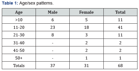
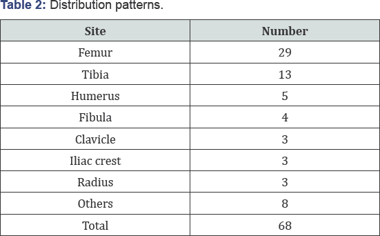
(Table 1) At a glance, the peak age incidence is 11-20 while the sexes are almost equal. Table 2 lists the involved bones.


Discussion
Searching the recent world literature revealed some similarities. Thus, the local mandible case is like the ones seen in both India [4] and Egypt [5]. In contrast, the subungual lesion found in USA [6] and elsewhere [7] did not feature locally. At the other end is the single German fibula case [8], whereas there were four of them in this developing community. With reference to X-Ray appearances, Jaffe [1] was sure that "In fact, it can hardly be mistaken for anything else.” This was true of as many as 12 of the local patients. Curiously, one case was different in that the picture exhibited pathological fracture.
References
- Jaffe HL (1958) Tumors and tumorous conditions of the bones and joints. Pa. Lea & Febiger, Philadelphia, USA.
- Macartney JC, Rollaston TP, Codling BW (1980) Use of a histopathology data pool for epidemiological analysis. J Clin Pathol 33(4): 351-353.?
- Basden GT (1966) Niger Ibos. Cass, London, UK.
- Singh SK, Bhadauria AS, Shukla AK, Srivastava R (2015) Osteocartilagenous exostosis of mandibular condyle: A rarity that is explored!!: Case report. Intl J Pre Clin Dent Res 2(4): 84-88.
- Wang-Norderud R, Ragab RR (1975) Osteocartilaginous exostosis of the mandibular condyle. Scand J Plast Reconstruct Surg 9(2): 165-169.
- Woo TY, Rasmussen JE (1985) Subungual osteocartilaginous exostosis. Dermatol Surg 11(5): 534-536.
- Bonifazi E (2009) Subungual osteo-cartilaginous exostosis. Eur J Pediat Dermatol 19(4): 252.
- Paprottka FJ, Machens HG, Lohmeyer JA (2012) Partially irreversible paresis of the deep peroneal nerve caused by osteocartilaginous exostosis of the fibula without affecting the tibialis anterior muscle. Plast Reconstruct Aesthet Surg 65(8): e223-e225.


