Bone Turnover and Bone Mass in High-Risk Neonates
Hesham H1, Abdulaziz A. Mustafa1, Ibrahim Mansour2, Gamal A3 and Hanan Amin4
1Department of Pediatrics, Egypt
2Department of Pediatrics, Egypt
3Department of Clinical Pathology, Egypt
4Department of AlAzhar And Zagazigo, Egypt
Submission: April 11, 2017; Published: April 17, 2017
*Corresponding author: Hesham H, 24 Mohamad Tawfik Diab, Makram Ebid Nasr City, Department of Rheumatology, Cairo Egypt, Tel: 201100000129; Email: hamoud.hesham@yahoo.com
How to cite this article:Hesham H, Abdulaziz A. Mustafa, Ibrahim M, Gamal A, Hanan A. Bone Turnover and Bone Mass in High-Risk Neonates . Ortho & Rheum Open Access 2017; 6(2): 555682. DOI: 10.19080/OROAJ.2017.06.555682
Abstract
Introduction: Dual energy X-ray absorptiometry DXA was used to follow postnatal changes in the bone mineral content BMC of 20 premature newborn infants with gestational age (32.2 ± 0.90) weeks and birth weight (1.981 ± 0.41)g. BMC was measured once at birth and another when these infants reached maturity by age (38.5 ± 0.17) weeks. Also BMC of an equal number of full term newborn infants was studied at birth.
Results: of this study showed that: BMC and body weight of premature infants increased significantly at term (45.15 ± 0.31g and 3.250 ±0.367g respectively) compared with BMC and body weight measured at birth (16.0 ± 0.73 g and 1.981 ± 0.41 g respectively), P <0.007. Also, at term the premature newborn BMC and body weight are still significantly low compared with BMC and body weight of full term newborn infants (56.32 ± 0.39 g and 3.492 ± 0.365 g) respectively for full terms, P<0.001. Serum calcium, phosphorus, alkaline phosphatase and osteocalcin concentrations were not significantly different between the preterm and full term newborn infants at any time point.
Conclusion: That BMC of preterm newborn infants increased at expected term but is still low compared with BMC of full term infants. This difference may be related to the difference in body size between full term and preterm at expected term.
Introduction
Preterm Infants are known to be at risk of developing metabolic bone disease, and this risk is negatively associated with birth weight and age of newborn infants. Over the past decade it has become clear that events operating during f et al life, infancy and childhood may affect peak bone mass and, therefore potentially influence the development of Osteoporosis [1]. Similarly other investigators speculated that, even silent early bone disease may interfere with the control of subsequent linear growth and emphasize the potential importance of providing preterm infants, especially those fed human milk, with adequate substrate for bone mineralization [2].
The use of simple biochemical indicators of bone mineralization such as serum alkaline phosphate (AIP), and to some extend serum phosphorus, calcium or Osteocalcin has been suggested to be an easy way of identifying preterm infants with metabolic bone disease. Some investigators report on association, although weak, between these biochemical markers and bone mineral content (BMC), where as others do not [3].
X-Ray examination of the skeleton has been used and provides direct access to measure bone mineralization. Using either conventional radiographs, quantitative ultrasounds or computed tomography is associated with poor precision [4]. Dual energy X-ray absorptiometry "DXA" is currently the gold standard for non-invasive determination of BMC because of its high precision, accuracy, speed of scanning and lower radiation exposure [1]. The aim of this study was to assess bone status of preterm and term infants by dual energy X-ray absorptiometry.
Patients and Methods
Forty healthy newborn infants were selected to be the subject of this prospective study. They were recruited from neonatal intensive care unit of Bab El Sharya University hospital. The infants were studied in two groups; preterm, the study and full term, the control group. The preterm group included 20 newborn infants (8 males and 12 females) of gestational age 32.2 (0.90) weeks and birth weight 1.981 (0.41) g.
These infants were studied twice; once at birth (average 2 days) and another when they reached the term at 38.5 (0.17) weeks. The term group included 20 full term infants (10 males and 11 females), of gestational age 38.6 (0.2) weeks and birth weight 3.492 (0.365) g. Exclusion criteria: All infants were medically stable and none had any significant respiratory, neurologic, renal, cardiovascular, hepatic, gastrointestinal, genetic or systemic infection. None of them used medicine like steroids or diuretics. All infants were fed their own human milk and the volume of feeding was checked through a proper regular weight gain. All infants were subjected to the following; anthropometeric measurements (body weight, length and head circumference), blood tests (serum calcium, phosphorus, alkaline phosphatase and osteocalcin), and BMC using DXA scan.
Anthropometric measurements
Body weight was measured by a calibrated infant scale to the nearest 10g. Crown-heal length and head circumference (at the largest occipitofrontal) width by a non stretchable tape to the next succeeding millimeter.
Blood tests
It was done on a blood obtained by venipunctures. An enzymatic colorimetric test (Boehringer Mannheim - Mannheim, Germany) using Hitachi 911 auto analyzer (Rosalki et al, 1993) was used to measure serum calcium, phosphorus and alkaline phosphatase, serum osteocalcin was measured using the immunoassay ELISA test (DSL-10-7600 kit, Diagnostic System Laboratories Inc, supplied by Clini Lab comp).
Bone mineral content (BMC)
It was measured by DXA scan with whole body single beam scanner (Hologic QDR 1000 W Hologic, Inc., Waltham, MA). The scanning procedure was performed when the naked infant placed in supine position on a pediatric scan platform and environmental temperature was maintained satisfactorily using a heat lamp. The measurement was performed after a feed, when the infant was quiet and in most cases asleep to avoid movement artifacts. No sedatives were used. The average duration of each measurement was 10min. BMC was expressed as total 1g as g/ kg body weight as total body BMC is closely associated with body weight 3.
Results
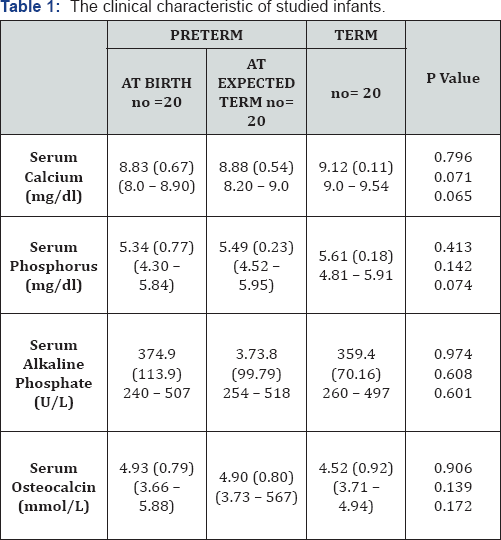
Clinical characteristics (Table 1)
It shows that a time of experted term, There was no significant difference noted comparing the preterm infants with the term control group regarding the age, the length and the head circumference (P>0.05). Only the body weight at term of preterm infants was merely significantly low (p=0.043) compared with term control infants.

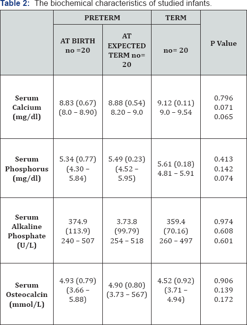
Biochemical Characteristics of the studied infants (Table 2)

Compared with the term control infants, the preterm infants did not differs significantly in mean serum concentrations of calcium, phosphorus, alkaline phosphatase or osteocalcin either at birth or at the time of expected term (P>0.05) for all.
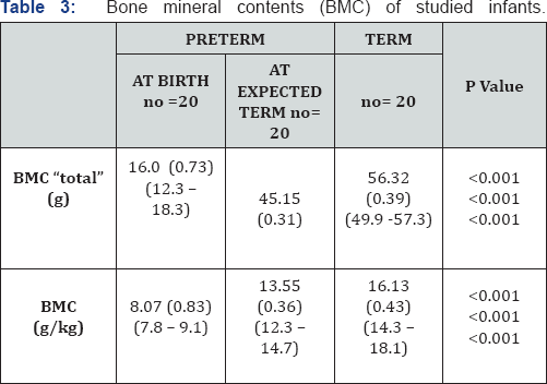
Bone mineral content of studied infants (Table 3)

Compared with its level at birth, BMC increased significantly at expected term of preterm infants (P<0.001), but is still significantly low compared with BMC of term infants (P<0.001).
Discussion
It is clear from this study that within a few weeks after birth, premature infants can absorb and retain calcium and phosphorus in sufficient amounts to approximate the estimated in utero accretion rate. This is clear from the finding in our study of no significant difference between the preterm and term infants in the levels of serum calcium, phosphorus, ALP and Osteocalcin. These results do agree KOO and Tsang [5], who found that premature infants can absorb calcium and phosphorus at the same level as mature infants.
The previous finding of no difference in the biochemical variables in face of the finding of significant changes of BMC between preterm and term infants in our study, suggests that the difference in BMC was probably not due to the difference in Vit-D status, because Vit-D is not a major determinant of calcium absorption in premature infants [6,7]. This explanation also agree the view of Rigo [8], Heaney [9], and Faerk [3], they suggested that nutrients other than calcium and phosphorus may have stimulated bone growth, and the higher protein intake of supplemented infant formulas may have had an additional effect on bone mineralization, as it is positively influence growth in premature infants. They found that BMC is highly significantly associated with body weight and to much less extent with dietary minerals or Vit-D intake, and that prevention of metabolic bone disease should focus on promotion of adequate growth. Moreover, they conclude that routine measurement of serum Alkaline phosphatase and bone minerals is therefore of no use in predicting bone mineralization in premature infants. Bronner suggested that one more important factor explaining the low rate of mineralization after birth in premature infants is that, in utero the fetus is parentally fed whereas the premature infants is enterally fed and the mineral retention of enteral feeding seems to be limited [7].
So, its seem from these previous studies that BMC is dependent more on the infant's whole growth and weight rather that on the elemental food supplementation. These finding do agree our study in that BMC was low in premature infants by time their weights were significantly low at term. Lastly, in our study we use only breast milk for feeding, and still there's a debate regarding feeding the premature infants their own human milk or supplemented infant formulas. One study by Alpay et al. [10] found that premature infants fed breast milk had a lower BMC than their formula-fed peers, and fortifying premature human milk will improve mineralization in these infants.
On the other hand many studies as those by Gross [11] and Faerk [12], investigated two groups of preterm infants feeding either human milk or fortified human or formula milk. They found a tendency to minimal increase of BMC in the fortified group, however when they correcting for non dietary factors (as the body size) they found no significant difference. This indicates that the difference was related to the difference in body size, as the weight of human milk fed infants was lower at term compared with fortified formula group. We can conclude from this study that BMC increased, but still low at term in premature newborn compared with mature newborn infants and this difference may be related to body size and that promotion of growth may be of value for bone mineralization of premature infants.
References
- Javaid MK, Cooper C (2002) Prenatal and Childhood Influence on Osteoporosis. Best practice Res Endocrinol Metab 16(2): 349-367.
- Salle B L, Braillon P, Polaniato, Covero E (1992) Bone Mineral content of lumbar spine in preterm infants. A longitudinal study during the first year of life. Pediatric Res 31: 294-297.
- Faerk J, Peitersen B, Petersen S, Michaelsen F (2002) Bone mineralization in premature infants cannot be predicted from serum alkaline phosphate or serum phosphate. Arch Dis Child Fet al Neonatal Ed 87(2): 133-136.
- Leonardo MB, Shults J, Zemel BS (2002) Interpretation of whole body dual X-ray absorptiometry measures in children. Comparison with peripheral C.T. Bone 34(6): 1044-1052.
- Koo WW, Tsang RC (1993) Calcium, Magnesium, Phosphorus and Vit D. In: Tsang R.C., Lucas A. eds Nutritional needs of preterm infants. Baltimore: Williams & Wilkins 135-155.
- Stenterre J (1991) Osteopenia versus rickets in premature infants. IN: Glorieux FH, ed. Rickets. Nestle Nutrition Workshop series 21. New York: Raven Press, USA.
- Bronner F, Salle BL, Rigo J, Stenterre J (1992) Net calcium absorption in premature infants: results 103 metabolic balance studies. Am J Clin Nutr 56(6): 1037-1044.
- Rigo J (2000) Bone mineral metabolism in the micropremie. Clin Perinatol 27:147-170.
- Heaney RP (2001) protein intake and bone health: The influence of belief systems on the conduct of nutritional science. AMJ Clin Nutr 73: 5-6.
- Alpay F, Unay B, Narin Y, Akin R (1998) Measurement of bone mineral density by DXA in preterm infants fed human milk or formula. Eur J Pediatr 157: 505-507.
- Gross SJ (1987) Bone Mineralization in preterm infants fed human milk with and without supplementation. J Pediatr III(3) : 450-458.
- Faerk J, Petersen S, Peitersen B, Michaelsen K (2000) Diet and bone mineral content at term in premature infants. Pediatr res 47(1):148- 156.


