Waste Not, Want Not: A Case of a Terrible Triad Injury of the Elbow Where a Non Salvageable Radial Head Was Used as a Source of Bone Graft for a Contra Lateral Intra-Articular Distal Radius Fracture
SJ Lewis1 and Alun Yewlett2*
1Orthopaedic Department, Prince Charles Hospital, England
2Orthopaedic Department, Grantham and District Hospital, England
Submission: April 04, 2017; Published: April 12, 2017
*Corresponding author: Alun Yewlett, Orthopaedic Department, United Lincolnshire Hospitals NHS Trust, Grantham and District Hospital, 101 Manthorpe Road, Grantham, Lines NG31 8DG, Tel: 01476 464439, Email: alunyewlett@gmail.com
How to cite this article: S Lewis, Alun Y. Waste Not, Want Not: A Case of a Terrible Triad Injury of the Elbow Where a Non Salvageable Radial Head Was Used as a Source of Bone Graft for a Contra Lateral Intra-Articular Distal Radius Fracture. Ortho & Rheum Open Access 2017; 6(1): 555680. DOI: 10.19080/OROAJ.2017.06.555680
Abstract
We present a case of a 37 year old male who sustained bilateral upper limb injuries following a mountain biking accident. He sustained a terrible triad injury of the elbow on the left side and an intra-articular distal radius fracture on the right side. The elbow was reduced emergently and placed in a backslab on the day of injury. There was a two week delay before definitive treatment could be carried out. The terrible triad injury was treated with a suture lasso technique to the coronoid fragment, a radial head excision and arthroplasty and a lateral collateral ligament reconstruction. The excised radial head was used as a source of bone graft and used to fill the intra-articular defect on the contra-lateral side in the distal radius using a K wire and plaster immobilisation. Although by no means unique we believe this case highlights an important point to remind surgeons that in multiply injured patients where some of their own bone needs to be excised it can be used as a valuable source of autograft for other injuries.
Keywords: Terrible Triad Elbow; Radial Head Arthroplasty; Bone Graft; Intra-articular Distal Radius
Case Report
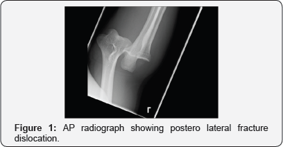
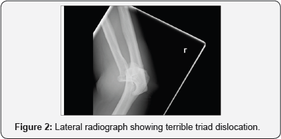
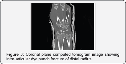
A 37 year old right hand dominant male sustained an injury when he came off a mountain bike at approximately 35 mph. His primary survey revealed no life threatening injuries but he had sustained bilateral closed upper limb injuries. On the left side he sustained a terrible triad injury of the elbow -i.e. a fracture dislocation of the ulnohumeral joint with an associated coronoid and radial head fracture (Figures 1 & 2). In addition he sustained an injury to the right distal radius which showed a dye-punch type intra-articular fracture. He was neurovascular intact throughout.
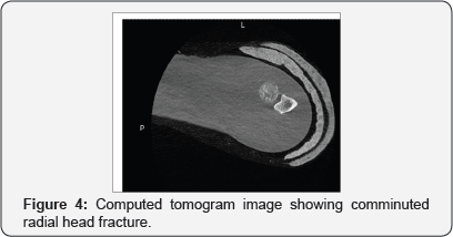
His elbow dislocation was reduced emergently in the operating room under general anaesthesia and a backslab in flexion was applied to his elbow that was documented to be very unstable by the treating surgeon. He was then admitted for observation and a specialist opinion was sought from a fellowship trained shoulder and elbow surgeon. In the interim CT scans of the elbow and wrist were performed for surgical planning purposes which illustrate the injuries in more detail (Figures 3 & 4).
Following an informed discussion about treatment options and likely outcomes with both conservative and surgical treatment with the patient, he elected to pursue a surgical solution which was carried out a fortnight after the initial injury. At the time of surgery the patient was positioned supine under general anaesthesia with intra-operative image intensification available. The decision was taken to address the right elbow first and a laterally based incision was utilised to approach the radial head, a Kocher interval was planned however on opening there was a traumatic rent in the common extensors and therefore this was utilised.
The radial head was more comminuted than expected from the preoperative imaging and was not salvageable and was therefore excised using a reciprocating saw and the articular cartilage removed to be use as a source of bone graft. A separate small dorsal incision using an ACL guide was used to drill two bone tunnels through the ulna to allow for a suture lasso technique to be used to capture the brachial is and a small bony fragment of the coronoid. Sutures were passed but not tied until the radial head had been replaced using an uncemented prosthesis (Acumed Anatomic radial head system) and bony tunnels in the lateral epicondyle drilled and sutures passed to allow for a Krakow type repair of the lateral collateral ligament complex (LCL) as described by the Mayo group. At the end of the procedure following radial head replacement, coronoid fixation and LCL reconstruction intraoperative stability was assessed. The elbow was stable to full flexion and to within 20 degrees short of full extension with gentle pressure and therefore a separate medial incision was not felt to be needed to address the medial structures and the repair was protected with a hinged elbow brace to allow a graduated return to full extension.
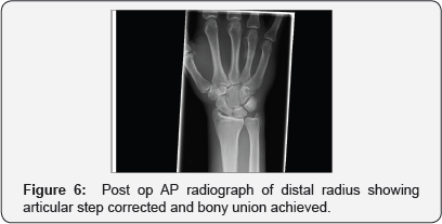
Attention was then turned to the contra lateral side and a dorsal incision was made to approach the intra-articular defect and a segment of decorticated bone from the excised radial head was shaped to fit the defect and placed into position and held with a 1.6mm K -wire using a percutaneous technique. Postoperatively, at the two week mark, his sutures were removed uneventfully and his elbow wounds had healed well. He was felt to have a superficial infection around his pin site and was started on a course of antibiotics as a precaution until the pin was removed uneventfully two weeks later. Physiotherapy was instigated immediately postoperatively following a graduated return to full extension in a hinged elbow brace to protect the LCL repair for 8 weeks but with full active range of motion permitted within the brace from the 3rd post-operative week.
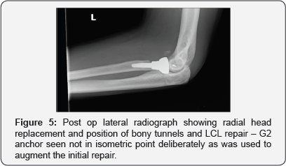
His check radiographs at 3 months show that the distal radius has gone on to unite well and that his elbow remains in joint. (Figures 5 & 6) At 6 month follow up he is back to his recreational activities and his elbow range of motion documents full pronation and supination, full flexion to 160 degrees and he lacks the terminal 10 degrees of extension and had a “functional arc” range of 150 degrees.
Discussion
Terrible triad injuries of the elbow were originally described by Hotchkiss [1] meaning fractures to the coronoid process and radial head with a resulting poster lateral elbow dislocation and refractory instability. The usual traumatic mechanism leading to this injury is a fall on to the outstretched, supinated arm with a valgus stress through the elbow. This condition accounted for 4% of all adult radial head fractures and 31% of elbow dislocations in a study by van Riet and Morrey [2]. Terrible triad injuries are more commonly seen in men and a peak occurrence in the fourth decade of life [3].
Treatment aims for terrible triad injuries is to restore the bony anatomy and reconstruct the ligamentous restraints of the elbow to provide enough stability for early elbow range of motion and prevent stiffness. With increasing understanding of elbow biomechanics and advancements in surgical techniques and implants there is no doubt that the outcome of these injuries are not as bad as they once were as evidenced by this case. We believe a surgeon should have a low threshold for replacing the radial head primarily to improve stability to allow for early mobilisation for these complex injuries and this is a view that is echoed by several recent studies [4,5]. Using a patient's own bone as a source of auto graft is not new, however we feel there is some merit in emphasising that if a patient sustains multiple injuries and has a bony defect at one injury site and requires a bony excision as part of their treatment at another anatomical site, then the excised bone could be used as a bone graft in preference to an off the shelf option to deal with defects elsewhere.
This avoids donor site morbidity from an unnecessary incision elsewhere and avoids any theoretical risk of prion disease transmission using demineralised bone matrix as a source of allograft for structural defects. Although this is seemingly common sense, we were unable to find any documented case reports in the orthopaedic literature describing this principle and would therefore urge others to "waste not, want not".
Acknowledgement
The authors would like to acknowledge I, Joseph for his help with the preparation of this manuscript.
Conflict of Interest
The authors declare that they have no ethical or financial conflicts of interest arising from the possible publication of this manuscript. The patient described in the case report has given written permission for his case to be published.
References
- Hotchkiss RN (1996) Fractures and dislocations of the elbow. In: Court-Brown C, Heckman JD, Koval KJ, Wirth MA, Tornetta P, Bucholz RW, (eds), Rockwood and Greens Fractures in Adults, Lippincott- Raven, Philadelphia, USA.
- Van Riet RP, Morrey BF (2008) Documentation of associated injuries occurring with radial head fracture. Clin Orthop Relat Res 466(1): ISO- 134.
- Chemama B, Bonnevialle N, Peter O, Mansat P, Bonnevialle P (2010) Terrible triad injury of the elbow: how to improve outcomes? Orthop Traumatol Surg Res 96 (2): 147-154.
- Watters TS, Garrigues GE, Ring D, Ruch DS (2014) Fixation versus replacement of radial head in terrible triad: is there a difference in elbow stability and prognosis? Clin Orthop Relat Res 472 (7): 21282135.
- Yan M, Ni J, Song D, Ding M, Liu T, et al. (2015) Radial head replacement or repair for the terrible triad of the elbow: which procedure is better? ANZ Journal of Surgery 85(9): 644-648.


