




 OMCIJ Home




Abstract
Filamentous actin has its own ATPase activity. That is, the splitting of ATP and the release of Pi accompany the polymerization of actin
monomers, which include the ATP molecule. There is reason to believe the same property of actin is realized when interacting with the myosin
head, which has a split ATP molecule, as occurs during contraction of muscle fibers. It is quite possible that actin attracts the inorganic phosphate
to itself and then releases it into solution. The tropomyosin, which rotates around its axis on the surface of the actin filament, after the activation
of muscle contraction by Ca2+, not only removes the obstacle to the interaction of actin with myosin, but also seems specifically activates myosin
ATPase, bringing the positively charged amino acid closer to the site of interaction between actin and Pi of myosin.
The troponin more likely serves as a Ca-sensitive lever that rotates the tropomyosin properly: in the presence of Ca2+, apparently turning
counterclockwise brings the positively charged amino acid closer to the site of the transition of Pi to actin, accelerating the speed of ATPase. In the
absence of Ca2+, troponin one can assume turns tropomyosin clockwise to its original position, inhibiting ATPase activity.
Keywords:The Regulation of Muscle Contraction; Atasi Activity of Myosin; Actin; Tropomyosin; Troponin
Why does Actin Activate?
To date the mechanism of actin-myosin interaction during contraction of skeletal muscle is generally understood. Schematically, this is the cyclic interaction of the myosin crossbridges with the actin filaments, as was described [1]. Moreover, the combination in silico computational chemistry and cryoelectron microscopy of reconstituted F-actin-myosin complexes allow to offer detailed models of such interaction [2-8]. There is a consensus that the step of removing Pi from the active site of the myosin motor domain is essential for understanding actinactivated myosin ATPase activity. The details of conformational changes in the motor domain that accompany the release of inorganic phosphate (back door) are discussed in several reviews [9-12]. The problem is that this escape route may be sterically blocked when myosin head is bound to actin [9].
This difficulty can be circumvented by assuming that inorganic phosphate is released towards actin rather than away from it. Incidentally, one of the two pathways for the release of Pi, namely, backdoor I path guides the Pi into the cleft between the U50 and L50 subdomains [13]. That is, it directly indicates that phosphate moves towards the actin surface. Again, the release of Pi through the back door towards actin leads to a situation where the release of inorganic phosphate is directly related to the actin surface, which serves as the main arena for the regulation of activity by troponin-tropomyosin complex of striated muscle. This means that now there is a connection between the release of Pi and the activating effect of actin on myosin.
As is known, the actin filament is assembled during polymerization from actin monomers containing the ATP with Mg2+ or Ca2+ ions. During polymerization, actin monomers hydrolyze the ATP molecules to ADP·Pi, followed by the release of Pi into the surrounding solution. It is highly likely that this ability of actin underlies the activation of myosin ATPase during muscle contraction. The process might look like this. After myosin head in the weak-binding state (M*.ADP. Pi state) only weakly binds to actin by L50 (lower 50kDa) subdomain [14], further mutual attraction of the two molecules occurs due to electrostatic interactions. In the course of this attraction, the 50kDa myosin head cleft starts to close and the distance between the surfaces of two molecules decreases, increasing the strength of the interaction [3].
At this moment, the actin monomer likely changes from the stable A-state to the unstable R-state. The R-state is characterized by opening the cleft because of the propeller-like rotation of the outer actin domain with respect to the inner domain by 12°-13° [15]. All this can create conditions for the movement of inorganic phosphate from the active center of the myosin head to the cleft between the large and small subdomains of the actin monomer. The bottom of this cleft is made up of charged amino acids such as R183, K336, H73, which together create a positively charged part. One can call this part of the actin monomer, a positive actin center (PAC). Inorganic phosphate leaves the active center of the myosin head apparently enters to PAC and releases into the surrounding solution as occurs during the polymerization of the monomeric actin-ATP complexes [16,4]. In this case, the actin monomer from the R-state returns obviously to the stable A-state, at which the cleft closes.
Transition of actin from R-state to A-state and release of Pi occurs obviously much faster than during actin polymerization, facilitated by conformational changes in the myosin head left without Pi in the active site. Thus, a change in the conformation of the myosin head contributes to a change in the conformation of actin, and thus myosin helps itself to be released from phosphate. G-actions have ATPase activity, but the rate of the actin ATPase is enhanced by about 7,000-fold by the polymerization process. Thus, F-actin is an effective tool for removing Pi that has fallen into its zone of influence. In such a situation, what are the chances that Nature will come up with a new way to remove phosphate from the myosin head, when the problem has already been solved by the F-actin structure itself? The answer is no chance. Everything suggests that F-actin snatches phosphate from myosin when it enters its sphere of influence.
A number of facts confirm the importance of the interdomain space on actin influencing the ATPase activity of myosin. The replacement of ADP with Br8ADP in the actin molecule significantly decreased both Vmax and Km of myosin S1 ATPase [18] probably due to the introduction of a negatively charged bromine atom into the PAC region of actin. FRET assays reveal shifts in the position of subdomain 2 in ATP vs. ADP in actin [19]. Case reports the fatal hypertrophic cardiomyopathy and nemaline myopathy associated with ACTA1 K336E mutation, amino acid lysine, which is part of the positively charged PAC network [20]. Thus, it is the creation of a local positive charge at the actin center PAC, which attracts the released by myosin inorganic phosphate, that is the mechanism of activation of myosin ATPase. Now actin transfers the arena of actions to regulate the contraction to itself.
Today the existing models of actin-myosin interaction do not reflect this state of affairs. It seems that myosin phosphate in nucleotide binding site is located too far from the acceptors on the actin surface. Our current knowledge of actin-myosin interaction is based on X-ray crystallography and on electron microscopy. Both of these techniques determine a static structure of actinmyosin complex of the strong-binding actomyosin interface. In addition, the events associated with the transition of Pi from the myosin molecule to the actin molecule occur in the time interval between the weakly bound and strongly bound states of actomyosin. Moreover, such a transition is associated with conformational transformations of both molecules, which cannot be traced by currently existing means. Only the consequences of such a transition are available in the form of existence a static structure of actin-myosin complex. It is known that actin is a very dynamic molecule and allosteric interactions with actin binding proteins are decisive to most of its functions [21].
There is evidence that during the interaction of actin with the myosin head, the latter comes into much closer contact with the actin surface, so that myosin phosphate may well be under the influence of F-actin ATPase. Direct contact known of the light chains A1 and A2 of the myosin head with the outer domain of actin [22]. Here is evidence that the light chains A1 binds to the C-terminal part of actin at residues 360-363 [23,24]. Even if the neck of myosin head contacts the actin surface (which is not reflected in modern models of actin-myosin interaction), why not contact his active center, which would just fall in the center of the actin monomer? Until now, the “active” or “inactive” state of the actin monomer in the regulation of muscle contraction was considered only indirectly, through the activity of the myosin ATPase. This model allows characterizing this state by the protein conformation. The inactive state of actin (“OFF” state) in the thin filament can be viewed as a state in which the PAC is impaired either by introducing a negative charge or by steric shielding. Accordingly, the active state (“ON” state) occurs when the PAC is restored. In conclusion, it should be noted that acceptance of the concept of the presence of PAС and its active participation in the release of cleaved phosphate from the myosin head creates the basis for understanding the regulation of muscle contraction by tropomyosin and troponin molecules, which will be discussed further.
How does Tropomyosin Regulate the Actomyosin Cycle?
Tropomyosin (Tm) is a coiled-coil dimer, which binds end-toend along the actin filament and covers seven actin monomers. The most widely cited model for regulation of actin-myosin activity mediated by Tm is one, which allows the existence of thin filaments in three biochemically distinguishable states: the “blocked” state, the “closed” state and “open” state [25-28]. It is known that Tm has a low affinity of binding to actin and therefore is easily displace by the myosin head from its position on the actin filament. Ca2+-free troponin (Tn) increases the affinity of the interaction of Tm with the actin by about a hundred times.
Nevertheless, even this strengthening of the bond does not allow Tm to keep the onslaught of myosin, and at a high concentration of the myosin heads, the rate of the ATPase becomes almost the same as that of an unregulated actin filament [29]. In other words, the blocking properties of Tm in conjunction with Tn, become apparent only in the absence of Ca2+- and only at a low concentration of myosin heads, as is the case in a state of relaxation, when the degree of overlap of actin and myosin filaments is minimal. Experiments show that myosin subfragment 1 does bind to thin filaments in the absence of Ca2+ but unable to complete of interaction [30].
Suggested model assume that with the flux of Ca2+ into the muscle fiber, Тn rotates Tm in such a way that tropomyosin rolls by an azimuthal rotation of about 16° and brings the positively charged amino acids closer to the actin PAC, whereas before its rotation Tm creates a negatively charged or non-polar field next to the PAC of the actin. Due to this, tropomyosin accelerates the ATPase of actin- myosin during muscle contraction. Further, in this section, evidence will be given to confirm the correctness of this assumption. [31], originally propose the idea that Tm “rolls”, not slides across the actin surface. In this model the actin surface interact with Tm’s α-band residues in the absence of Ca 2+ but in the presence of calcium the actin surface interact with β-band residues. The data of other authors are consistent with this model [32, 33].
Now, if we return to the problem of regulating the ATPase cycle, the process of the actin-myosin interaction will look like this. In the relaxed state (without Ca2+) the M-ADP-Pi weak state rebinding to actin in inactive state through the positively charged loop 2, interacting with the negative charge of the N-terminus of actin. Тm located near outer domain of actin with the support of Tn, does not allow (either sterically, or allosterically, or by a combination of these two ways), myosin head to crawl over the actin monomer. In such prepowerstroke state, the myosin head remains until Ca2+ begins to flow. Under the influence of calcium on Tn, the latter begins to shift Tm towards the center of the actin axis, and myosin is able to complete its attachment to the actin. The first stage is characterized by the formation of stereo-specific hydrophobic interactions between actin and the L50 domain. The second step is followed by 50 kDa cleft closure provides additional interactions between actin and the U50 domain with the formation of the complete actin-myosin contact surface. Simultaneously with the advancement of the motor domain towards the center of the actin filament, Tm turns over and now when the actin PAC approaches the nucleotide-binding site of myosin as much as possible; Tm completes its turn and strengthens the positively charged field near the PAC. Phosphate is released. The loss of Pi is associated with relieving the strain in the upper 50K β -sheet and the swing of the converter and lever – arm. These events are likely to have equivalent processes in the pre-powerstroke to rigor transition. Thus, this interpretation of events is more suitable for the concept of a two-state model for Tm-mediated regulation of actin-myosin activity: inactive state at which the ATPase is significantly slowed down and active at which the ATPase works at a suitable speed [34,35].
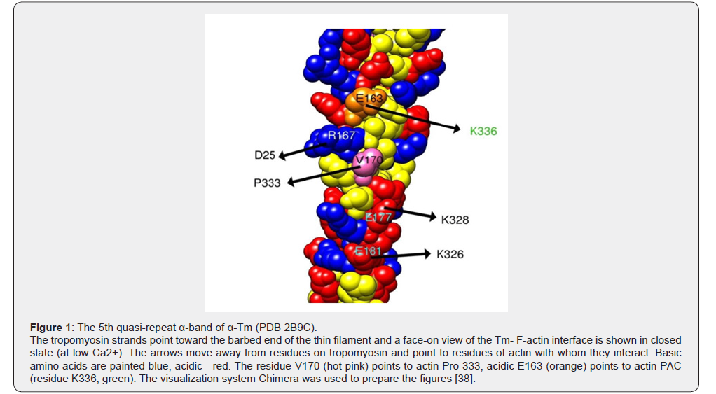
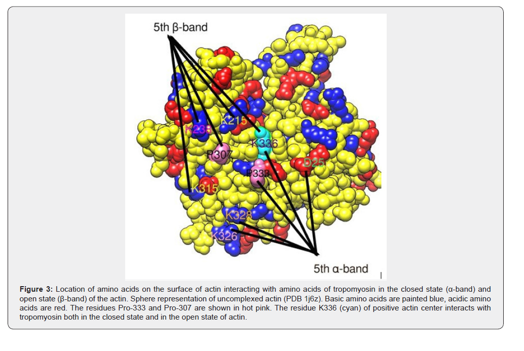
Recently, the exact position of the tropomyosin molecules on the actin filaments has become known [36,37]. Now we have the opportunity to compare which amino acids on the actin surface will interact with amino acids on the tropomyosin surface when tropomyosin moves from a closed state to an open one using the example of the 5th quasi-repeat of α-tropomyosin. As follows from studies reported [37], in closed state the α-band of the 5th quasirepeat of α-Tm covers outer domain of the actin. Wherein the K326 and K328 residues of actin approach E181 and E177 residues of tropomyosin, respectively, actin’s P333 residue comes to V170 residue on tropomyosin, actin D25 residue goes closer to R167 on tropomyosin and K336 residue (PAC) on actin approaches the E163 residue on tropomyosin Figure 1 & 3. Thus, the negatively charged tropomyosin’s E163 residue is located above the region of the PAC in actin, inhibiting the work of the ATPase in the closed state of tropomyosin. In the 4th quasi-repeat α-band of Tm, the PAC region of actin is in contact with E124, and in the 6th with T199, for example [39], using a direct electron detector and drift correction have determined the complex of F-actin-tropomyosin. From their data, it follows that rolling of Tm leads to not only a shift in azimuthal direction by ~ 12 Å, but also a shift in Tm of a half-tropomyosin repeat along the F-actin filament. In other words, upon the transition of Tm to the open state, next to the actin PAC, the 5th quasi-repeat β-band of tropomyosin, covering the inner actin domain, will appear.
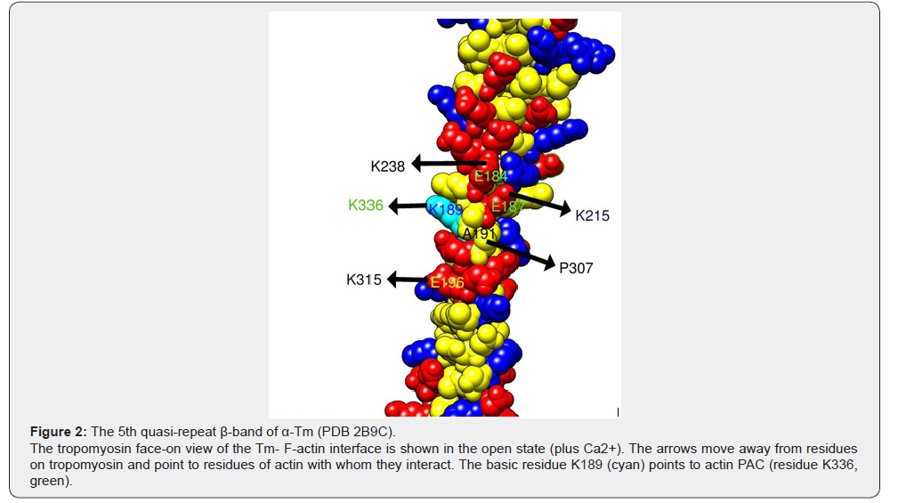
Wherein K315 residue of actin approaching E196 on tropomyosin, P307 on actin approaching A191 on tropomyosin; actin’s residues K215 and K238 are close to E187 and E184 on tropomyosin, respectively and K336 (PAC) on actin is close to K189 on tropomyosin Figure 2 & 3. Thus, the positively charged K189 tropomyosin residue is located nearby the actin region of the PAC, accelerating the work of ATPase in the open state of tropomyosin. In the third quasi-repeat β-band of α-tropomyosin, the PAC region of actin is in contact with K112 and in the 4th with K152, for example. It follows that Tm in striated muscle plays a more active role than the steric blocking theory suggests. Tropomyosin not only blocks and then moves to allow actin to interact with myosin, but also activates actin-myosin ATPase itself when calcium ions enter during muscle contraction.
It has long been noted that Tm is able to accelerate myosin ATPase but this property was attributed to the cooperative effect. The concept of a cooperative effect appeared in connection with the study of the kinetics of actin binding to tropomyosin. It turned out that at first the process is slow, but as the first attached molecules appear, the rate of attachment of the remaining molecules increases. The cooperative effect of ATPase upregulation was explained by the fact that binding of the first Tm molecules to the actin filament facilitates the attachment of subsequent. It is right. However, how can one explain the fact that in the presence of Tm in thin filaments, the ATPase rate exceeds the ATPase rate of unregulated actin-myosin, where all myosin-binding sites of actin are initially open? In addition, there is no need to open and activate neighboring actin molecules? The assumption that Tm enhances the positively charged field and thereby accelerates the ATPase, it is thus easy to imagine. If we assume that Tm touched actin with a “negative” surface before rotation, then at the end of rotation on 70 degree, Tm contacts actin with a “positive” surface. If the main function of tropomyosin in the muscle fiber is to prevent actin from interacting with myosin, as the steric blocking hypothesis insists, then any “log” would be suitable for this purpose.
However, tropomyosin has a unique composition that not repeated in any other protein; at each turn, an area saturated with negatively charged amino acids (“negative” surface) alternates with a “positive” surface area [31]. Moreover, why did tropomyosin need to roll over at all? For blocking, a simple displacement of the molecule to the center of the actin filament would be suitable. This model of interactions also makes it possible to explain the S-shaped dependence of the calcium sensitivity of actin-myosin ATPase on the concentration of free calcium. With an increase in low Ca2+ concentrations (pCa 9–7.5), ATPase activity is low and the dependence curve runs almost parallel to the x-axis. In the range from 7.5 to 6, the dependence graph rises almost in direct proportion to the maximum values. Above pCa 6, the ATPase values do not change and the graph again runs parallel to the x-axis.
This phenomenon is explained by the rotation of tropomyosin around its axis, in which a positively charged amino acid approaches the PAC on actin. At low Ca2+ concentrations, tropomyosin rotates through a small angle and the amino acid does not yet entered into the electrostatic field of the PAC, and therefore the ATPase values do not change. In the range of pCa from 7.5 to 6, the amino acid already enters this field, and even a slight rotation of tropomyosin already noticeably increases the positively charged PAC field, which is expressed in an increase in the ATPase rate. The maximum approach of the amino acid with the PAC occurs at the completion of the turn, when the maximum ATPase rate is noted. A further increase in Ca2+ concentration from pCa 6 and above does not lead to further closing in, and the ATPase rate remains maximum throughout this interval.
Role of Troponin
Troponin, together with tropomyosin, forms a system that regulates skeletal muscle contraction and relaxation at the molecular level. Troponin consists of three subunits: TnC, TnI and TnT. Troponin at low Ca2+ operates as an inhibitor, while at high Ca2+ it acts as an initiator of the contraction. The inhibitory role of troponin is carried out by the C-terminus of TnI (residues 163 to 210) [40]. The activating role of troponin is imparted by TnC. Binding of Ca2+ to the N terminal domain of TnC would induce the widening of two EF hands (AB and CD) [41]. Ca2+ binding induce opening of the hydrophobic pocket in the N-terminus of TnC and joining the switch segment (residues 149 to 164) of TnI to it [41]. At the same time, the TnI inhibitory segment and central helix of TnC are significantly stabilized [42].
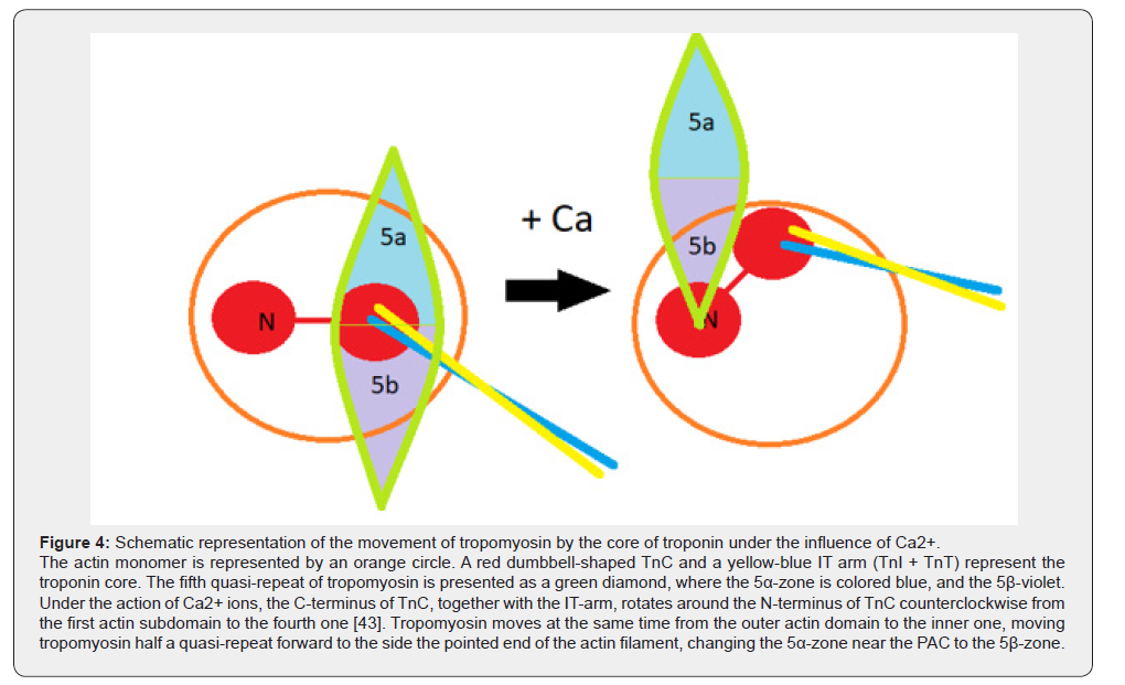
In work [43], the authors determine the three-dimensional structure of thin filaments by cryo-microscopy. At high Ca2+, the peak of the electron density of the V block was located above the inner actin domain but at low Ca2+ shifted by 28 Å towards the outer domain. At low Ca2+ the C-terminal domain of TnC shifted by 50 Å to the outer domain of actin, whereas its N-terminal domain practically did not move much. Therefore, TnC acted as a lever arm, and N-TnC was its fulcrum. In general, the Ca2+ induces movement of TnC from the first actin subdomain towards the fourth one [43]. This is exactly the direction of movement of the core part of troponin, which ensures the rotation and advancement of the tropomyosin cord by half of the quasi-repeat (from the α-zone to the β-zone) towards the pointed end of the actin thin filament Figure 4 described in the second section of this article about the role of tropomyosin.
Thus, according to this model, the activating effect of troponin on the cyclic work of cross-bridges consists in the precise and coordinated movement of Tm (rotation plus move), the purpose of which is to bring the positive charge closer to the PAC of actin. Considering the contraction and relaxation of the muscle, one must not forget that two cyclic processes occur in this case: the cyclic movement of actomyosin cross-bridges and the cyclic movement of the Tn-Tm complex, while the first is preceded by the second with an interval of 12-17msec [44]. At the same time, it should be borne in mind that the cyclic nature of actomyosin bridges includes not only the alternating attachment-detachment of myosin-actin, but also the cyclic nature of the movement of the myosin head over the actin surface. The myosin head moves cyclically from the outer edge of the first domain to the border between the outer and inner actin domains, as shown in [4]. During this movement, the myosin head is forced to push the Tn-Tm complex in front of it if there is no Ca2+ in the medium. This inhibitory effect of the passive Tn- Tm complex is expressed in the slowing down of the ATPase rate, which is clearly seen from work [29].
Mutations in genes encoding sarcomeric proteins including β-MyHC, myosin light chains, myosin-binding protein C, titin, actin, Tm and Tn will affect Ca2+-sensitivity if they cause replacement of amino acids essential for these two cyclic processes. All mutations that facilitate these cycles will increase Ca2+ - sensitivity and those that make it difficult - reduce the sensitivity. This hypothesis allows us to explain not only the activation of myosin ATPase, but also for the first time to separate it from Ca2+-sensitivity. It is no coincidence that the number of mutations leading to an increase in Ca2+-sensitivity significantly exceeds the number of mutations that increase the rate of ATPase. For the latter case, it is necessary that a strong positively charged field approach the PAC of the actin, and for the first case, any mutations leading to facilitation of the course of these two cycles.
Because of the shift in focus from the inorganic phosphate exit through the «back door» to the active uptake of the inorganic phosphate by PAC on actin, the active role of actin is asserted. Instead of passive displacement of tropomyosin for the release of myosin-binding sites of actin, offered targeted and precise movement, leading to the activation of the ATPase. Instead of opening a hydrophobic pocket on N-TnC for TnI attachment and thus eliminating the inhibitory effect of TnI as the main event in troponin activity, a dynamic picture of the movement of the core part of troponin is proposed, which ensures the movement of tropomyosin along the actin filament, aimed at regulating the ATPase cycle.
Conclusion
We can summarize all of the above by listing the advantages that follow the adoption of the concept of the PAC on actin. First, the activation of myosin ATPase becomes simple and clear: actin returns to its active role. The fact that in in vitro experiments Pi can leave the nucleotide site of myosin spontaneously without the participation of actin suggests that the force of this process is not conformational changes in actin, but the acceleration of this release with increasing ionic strength of the solution (0.6M KCl) indicates participation of electrostatic forces. However, the process of Pi release must be structured in a living cell, so actin uses electrostatic forces to regulate the activity of myosin ATPase. For the first time, the state of actin (on, off) is expressed through the conformation of the protein itself, and is not derived from another protein (myosin).
A logical continuation of this regulation is the activation of actin-myosin ATPase by tropomyosin by increasing the positive charge in the region of the actin PAC. It becomes clear why tropomyosin turns over and not just shifts, why a positively charged one replaces a negatively charged area on a protein [31] when rotated, why protein has pseudo repeats and is divide into α- and β-zones. Finally, the question of the ability of tropomyosin to cause cooperative enhancement of ATPase (S-shaped dependence of ATPase on Ca2+ concentration) is resolved. In the old paradigm, which looked murky and mystical, this question now has a simple and obvious answer. By the way, an attempt to explain such cooperativity by overlapping the N- and C-termini of successive molecules on the actin filament failed [45]. Finally, the work of troponin in regulating the thin filament ATPase, despite the complexity of this protein, comes down to a simple counterclockwise turn in the presence of Ca2+, and in the opposite direction in the absence of the latter. This causes the same rotation of tropomyosin in order to influence the state of the actin PAC center, and therefore the myosin ATPase.
References
- Lymn RW, EW Taylor (1971) Mechanism of adenosine triphosphate hydrolysis by actomyosin. Biochemistry 10(25): 4617-4624.
- Reubold TFS, Eschenburg A, Becker FJ, Kull, DJ Manstein (2003) A structural model for actin-induced nucleotide release in myosin. Nature Structural Biology 10(10): 826-830.
- Lorenz M, KC Holmes (2010) The actin-myosin interface. Proceedings of the Proc Natl Acad Sci USA 107(28): 12529-12534.
- Behrmann EM, Muller PA, Penczek HG, Mannherz DJ, Manstein, et al. (2012) Structure of the Rigor Actin-Tropomyosin-Myosin Complex. Cell 150(2): 327-338.
- Varkuti BH, ZH Yang, A Malnasi Csizmadia (2015) Structural Model of Weak Binding Actomyosin in the Prepowerstroke State. Journal of Biological Chemistry 290(3): 1679-1688.
- Houdusse A, HL Sweeney (2016) How Myosin Generates Force on Actin Filaments. Trends in Biochemical Sciences 41(12): 989-997.
- Vonder Ecken J, SM Heissler, S Pathan Chhatbar, DJ Manstein, S Raunser (2016) Cryo-EM structure of a human cytoplasmic actomyosin complex at near-atomic resolution. Nature 534(7609): 724-728.
- Mentes A, A Huehn, XQ Liu, A Zwolak, R Dominguez, et al. (2018) High-resolution cryo-EM structures of actin-bound myosin states reveal the mechanism of myosin force sensing. Proc Natl Acad Sci USA 115(6): 1292-1297.
- Yount RGD, Lawson, I Rayment (1995) Is myosin a back door enzyme. Biophysical Journal 68: S44-S49.
- Lawson JDE, Pate I, Rayment, RG Yount (2004) Molecular dynamics analysis of structural factors influencing back door P-i release in myosin. Biophysical Journal 86(3): 3794-3803.
- Geeves MA, KC Holmes (2005) The molecular mechanism of muscle contraction. Fibrous Proteins: Muscle and Molecular Motors 71: 161-193.
- Sweeney HL, A Houdusse (2010) Structural and Functional Insights into the Myosin Motor Mechanism. In: DC Rees, KA Dill, JR Williamson (Eds.), Annual Review of Biophysics, Vol 39. Annual Reviews Palo Alto, pp. 539-557.
- Cecchini M, Alexeev Y, Karplus M (2010) Pi release from myosin: a simulation analysis of possible pathways. Structure 18(4): 458-470.
- Onishi H, Mikhailenko SV, Morales MF (2006) Toward understanding actin activation of myosin ATPase: the role of myosin surface loops. Proc Natl Acad Sci USA 103(16): 6136-6141.
- Oda T, Iwasa M, Aihara T, Maéda Y, Narita A (2009) The nature of the globular- to fibrous- actin transition. Nature 457(7228): 441-445.
- Iwasa MK, Maeda A, Narita Y, Maeda, T Oda (2008) Dual roles of Gln(137) of actin revealed by recombinant human cardiac muscle alpha-actin mutants. Journal of Biological Chemistry 283(30): 21045-21053.
- Pollard TD, AG Weeds (1984) The rate constant for ATP hydrolysis by polymerized actin. FEBS Lett 170(1): 94-98.
- Hegyi G, Szilágyi L, Belágyi J (1988) Influence of the bound nucleotide on the molecular dynamics of actin. Eur J Biochem 175(2): 271-274.
- Moraczewska J, Strzelecka Gołaszewska, H Moens, PD dos Remedios (1996) Structural changes in subdomain 2 of G-actin observed by fluorescence spectroscopy. Biochem J 317: 605-611.
- D'Amico A, Graziano C, Pacileo G, Petrini S, Nowak K, et al. (2006) Fatal hypertrophic cardiomyopathy and nemaline myopathy associated with ACTA1 K336E mutation. Neuromuscul Disord16(9-10): 548-552.
- Egelman EH (2001) Actin allostery again? Nat Struct Biol 8(9): 735-736.
- Milligan RAM, Whittaker, D Safer (1990) Molecular-structure of f-actin and location of surface binding-sites. Nature 348(6298): 217-221.
- Sutoh K (1982) Identification of Myosin-Binding Sites on the Actin Sequence Biochemistry 21(15): 3654-3661.
- Trayer IP, HR Trayer, BA Levine (1987) Evidence that the n-terminal region of a1-light chain of myosin interacts directly with the c-terminal region of actin - a proton magnetic-resonance study. Eur J Biochem 164(1): 259-266.
- McKillop DFA, MA Geeves (1993) Regulation of the interaction between actin and myosin subfragment-1 - evidence for 3 states of the thin filament. Biophysical Journal 65(2): 693-701.
- Vibert PR, Craig, W Lehman (1997) Steric-model for activation of muscle thin filaments. Journal of Molecular Biology 266(1): 8-14.
- Lehman W (2017) Switching Muscles on and Off in Steps: The McKillop-Geeves three-state model of muscle regulation. Biophysical Journal 112(12): 2459-2466.
- Geeves MA, SS Lehrer, W Lehman (2019) The mechanism of thin filament regulation: Models in conflict? Journal of General Physiology 151(11): 1265-1271.
- Robaszkiewicz KE, Dudek AA Kasprzak, J Moraczewska (2012) Functional effects of congenital myopathy-related mutations in gamma-tropomyosin gene. Biochimica Et Biophysica Acta-Molecular Basis of Disease 1822(10): 1562-1569.
- Chalovich JM, E Eisenberg (1982) Inhibition of actomyosin atpase activity by troponin-tropomyosin without blocking the binding of myosin to actin. Journal of Biological Chemistry 257(5): 2432-2437.
- McLachlan AD, M Stewart (1976) 14-fold periodicity in alpha-tropomyosin and interaction with actin. Journal of Molecular Biology 103(2): 271-298.
- Bacchiocchi C, SS Lehrer (2002) Ca2+-induced movement of tropomyosin in skeletal muscle thin filaments observed by multi-site FRET. Biophysical Journal 82(3): 1524-1536.
- Holthauzen LMF, F Correa, CS Farah (2004) Ca2+-induced rolling of tropomyosin in muscle thin filaments - The alpha- and beta-band hypothesis revisited. Journal of Biological Chemistry 279(15): 15204-15213.
- Hill TL, E Eisenberg, LE Greene (1983) Alternate model for the cooperative equilibrium binding of myosin subfragment-1-nucleotide complex to actin troponin tropomyosin. Proceedings of the National Academy of Sciences of the United States of America-Biological Sciences 80(1): 60-64.
- Heeley DH, HD White, EW Tayor (2019) Investigation into the mechanism of thin filament regulation by transient kinetics and equilibrium binding: Is there a conflict? Journal of General Physiology 151(5): 628-634.
- Lehman WA, Galinska Rakoczy, V Hatch LS, Tobacman, R Craig (2009) Structural Basis for the Activation of Muscle Contraction by Troponin and Tropomyosin. Journal of Molecular Biology 388(4): 673-681.
- Li XC, LS Tobacman, JY Mun, R Craig, S Fischer, W Lehman (2011) Tropomyosin Position on F-Actin Revealed by EM Reconstruction and Computational Chemistry. Biophysical Journal 100(4): 1005-1013.
- Pettersen EF, TD Goddard, CC Huang, GS Couch, DM Greenblatt, et al. (2004) UCSF chimera - A visualization system for exploratory research and analysis. Journal of Computational Chemistry 25(13): 1605-1612.
- von der Ecken, J M Muller, W Lehman, DJ Manstein PA, Penczek, et al. (2015) Structure of the F-actin-tropomyosin complex. Nature 519(7541): 114-117.
- Galinska Rakoczy AP, Engel C, Xu H, Jung R, Craig LS (2008) Tobacman, and W. Lehman. 2008. Structural basis for the regulation of muscle contraction by troponin and tropomyosin. Journal of Molecular Biology 379(5): 929-935.
- Marston S, JE Zamora (2020) Troponin structure and function: a view of recent progress. Journal of Muscle Research and Cell Motility 41(1): 71-89.
- Vinogradova MV, DB Stone, GG Malanina, C Karatzaferi, R Cooke, et al. (2005) Ca2+-regulated structural changes in troponin. Proc Natl Acad Sci USA 102(14): 5038-5043.
- Narita A, Yasunaga T, Ishikawa T, Mayanagi K, Wakabayashi T (2001) Ca2+ -induced switching of troponin and tropomyosin on actin filaments as revealed by electron cryo-microscopy. J Mol Biol 308(2): 241-261.
- Kress M, HE Huxley, AR Faruqi, J Hendrix (1986) structural-changes during activation of frog-muscle studied by time-resolved x-ray-diffraction. Journal of Molecular Biology 188(3): 325-342.
- Walsh TP, Trueblood CE, Evans R, Weber A (1985) Removal of tropomyosin overlap and the co-operative response to increasing calcium concentrations of the acto-subfragment-1 ATPase. Journal of Molecular Biology 182(2): 265–269.

























