Prevention of Crystallization in Stones Infection by Chemical Inhibitors
Mohamed Beghalia 2*, Hocine Allali1 and Saïd Ghalem1
1Laboratory of Naturals Products and Bio Actives, Lasnabio Department of Chemistry, Faculty of Sciences, Aboubakr Belkaid University, Tlemcen, Algeria
2Laboratory of agronomy and ecology , University Centre of Tissemsilt Algeria
Submission: July 26, 2019; Published: August 19, 2019
*Corresponding author: Mohamed Beghalia, Laboratory of agronomy and ecology, University Centre of Tissemsilt, Algeria
How to cite this article: Mohamed Beghalia, Hocine Allali, Saïd Ghalem. Prevention of Crystallization in Stones Infection by Chemical Inhibitors. Organic & Medicinal Chem IJ. 2019; 8(5): 555746 DOI: 10.19080/OMCIJ.2019.08.555746
Abstract
Struvite or magnesium ammonium phosphate hexahydrate (MAP) is one of the components of urinary stone. It forms in human beings as a result of urinary tract infection with urea splitting organisms. These stones can grow rapidly forming “staghorn-calculi”, which is a painful urological disorder. Therefore, it is of prime importance to study the growth and inhibition of struvite crystals. In the present work, we have exanimated the interactions between the enzyme urease and two inhibitors (aluminum and citrate acid) by molecular modeling methods. The results show that the presence of aluminum or citrate acid significantly affects struvite crystal growth. It suggested that our inhibitors induced morphology changes that reflect the interactions between the inhibitor and the crystal surfaces at a molecular level. Similar changes were observed in the presence of identical concentrations of citrate acid, and aluminum, emphasizing the unique interaction of phosphocitrate with the struvite crystal. .
Keywords: Inhibitors Struvite Modeling Interactions
Introduction
Infection lithiasis are composed primarily of magnesium ammonium phosphate hexahydrate (MgNH4PO46H2O) but may, in addition, contain calcium phosphate in the form of carbonate apatite (Ca10[PO4]6CO3) [1,2]. Infection of the urinary tract with urease-producing organisms is required for the formation of infection stones in humans. Although the species Enterobacteriaceae comprises most urease-producing pathogens, a variety of gram-positive and gram-negative bacteria and some yeasts and mycoplasma species have the capacity to synthesize urease [3].
An elevated urinary pH reduces the solubility of magnesium ammonium phosphate and favors precipitation of Struvite crystals. Higher intake of phosphate (from Proteins) and magnesium-based food and lower intake of water gives rise to the PO4 3- and Mg2+ ions in the supersaturated urine, which leads to the conditions of formation of Struvite [4]. Probably, the negative charged points of the external side of the cellular structures could reduce the metastability field of struvite and calcite, acting as heterogeneous nuclei of crystallization [5].
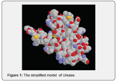
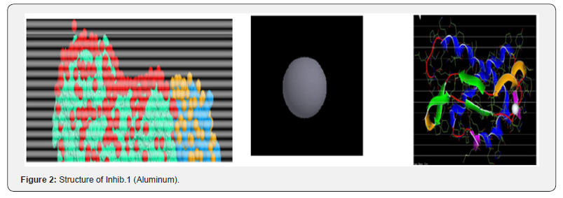
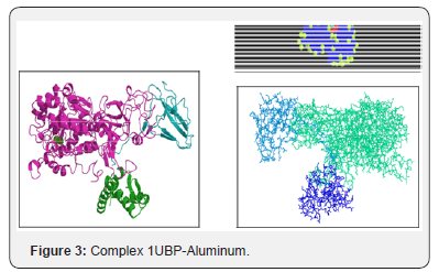
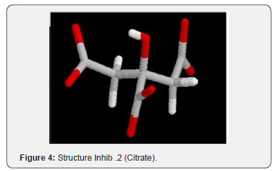
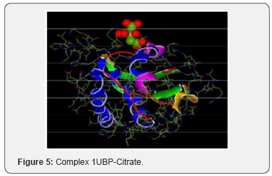
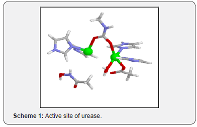
For example, during digestion, are enzymes that accelerate the decomposition and food processing. An enzyme catalyzes only one reaction in general, because it can bind efficiently thana single substrate. This specificity results from the fact that when two molecules meet, their association may be stabilized by the establishment of many weak bonds (hydrogen bonding, ionic bonding, Van Der Waals forces) these bonds are about 100 times weaker than a covalent bond. Modeling and simulation of struvite growth, incorporating solution chemistry and thermodynamics, growth kinetic and process description of the recovery system. An ensemble of experimental data is combined with the dynamic model to estimate struvite growth kinetics [6] (Figures 1-5) (Schemes 1 & 2).

Experimentation
Struvite (MgNH4PO46H2O) crystals were produced by infection associated with urea generating organisms. The aim of this study is to examine the interactions between the enzyme urease and two inhibitors, the first is an inhibitor monoatomic: Aluminum and the second is a polyatomic: Citrate by the methods of molecular modeling: molecular mechanics, molecular dynamics (MM+, AMBER) and molecular docking (FleX). Supersaturated solutions induce crystallization by nucleation and subsequent crystal growth .The mechanisms for the formation of calcium phosphate urinary stones are still not understood.
Chemicals product has been studied extensively as inhibitors and has been observed in the attachment of crystals to in vitrostudy. As a complement we have using an electron microscope Hitachi TM1000, we examined specimens of crystals struvite. The various figures show a set of grains of sizes of the order of 20 μm. Most these particles’ present regular forms. This suggests the crystal growing. This result to an alteration in the expression of these faces and the development of a characteristic architectural struvite morphology. Similar changes were observed in the presence of identical concentrations of citrate acid, and Aluminum, emphasizing the unique interaction of phosphocitrate with the struvite crystal.
Results
The results of the bibliography. Ions Al (III) exert their action directly on the crystals blocking the growth sites located on their surface and prevent their dissolution process, when present in low concentrations .Comparing the two complexes formed (their energy),we can say that the complex polyatomic inhibitor is more stable than the complex of the inhibitor because the interactions between the inhibitor polyatomic (Citrate) which comprises several pairs donors (electron pairs) and receiver (active site) of the enzyme is much more than the monatomic inhibitor (Al) which is one lone pair.
Using an electron microscope Hitachi TM1000, we examined specimens of a renal lithiasis. The various figures show a set of grains of sizes of the order of 20 μm . Most these particles’ present regular forms. This suggests the crystal growing. Frequently a certain number of differently directed crystalline grains from to each other and joined to the interface by grain boundaries. Qualitatively, we can say that the crystals present an axis of rotation of order 2, from where the possibility that crystals belong to the monoclinic system. Some crystals of size 20 μm are in the center of a concretion which seems of amorphous structure on all the scales and the crucial role of which is to stop the growth of the initial crystal [7].
Discussion
This result, proved by the absence of crystals detected by optical microscope in polarized light, is important. It confirms the inhibitory effect of the solution of aluminum at low concentration of nucleation, in agreement with the results of the bibliography. Ions Al (III) exert their action directly on the crystals blocking the growth sites located on their surface and prevent their dissolution process, when present in low concentrations .Comparing the two complexes formed (their energy),we can say that the complex polyatomic inhibitor is more stable than the complex of the inhibitor because the interactions between the inhibitor polyatomic (Citrate) which comprises several pairsdonors (electron pairs) and receiver (active site) of the enzyme is much more than the monatomic inhibitor (Al) which is one lone pair.
Using an electron microscope Hitachi TM1000, we examined specimens of a renal lithiasis. The various figures show a set of grains of sizes of the order of 20μm. Most these particles’ present regular forms. This suggests the crystal growing. Frequently a certain number of differently directed crystalline grains from to each other and joined to the interface by grain boundaries. Qualitatively, we can say that the crystals present an axis of rotation of order 2, from where the possibility that crystals belong to the monoclinic system. Some crystals of size 20 μm are in the center of a concretion which seems of amorphous structure on all the scales and the crucial role of which is to stop the growth of the initial crystal.
Conclusion
The aim of this study was to examine the possible effects of some metals on the inhibition of phosphate calcium crystallization. These results obtained confirmed the beneficial effect of chemical inhibitors and may justify its use as a preventive agent against the formation of struvite urinary stones. The inhibition capacity of aluminium and citrate, ion was important on the aggregation and size of struvite crystals.
References
- Griffith DP (1978) Struvite stones. Kidney Int 13(5): 372-382.
- Griffith DP, Osborne CA (1987) Infection (urease) stones. Miner Electrolyte Metab 13(4): 278-285.
- Gleeson MJ, Kobashi K, Griffith DP (1992) Non calcium nephrolithiasis. Disorders of Bone and Mineral Metabo-lism, (Coe FI, Favus MJ, eds.). Raven, New York, NY, pp. 801.
- Chauhan C K, MJ Joshi, A D B Vaidya (2008) Growth inhibition of struvite crystals in the presence of herbal extract Commiphora wightii. J Mater Sci Mater Med.
- Ma Teresa González Muñoz, Nabil Ben Omar, Magdalena Martínez Cañamero, Manuel Rodríguez Gallego, Alberto López Galindo, et al. (1996) Stru-vite and calcite crystallization induced by cellular mem-branes of Myxococcus xanthus. Journal of Crystal Growth 163(4): 434-439.
- Md Imtiaj Ali, Philip (2008) An approach of estimating struvite growth kinetic incorporating thermodynamic and solution chemistry, kinetic and process descry. Chemical Engineering Science Volume 63(13): 3514-3525.
- Mohamed Beghalia, Hocine Allali, Saïd Ghalem, Aïssa Belouatek, Abdelhamid Sari (2011) Molecular Modeling of Chemicals Products Inhibitors of Growth Struvite Crystal. Advances in Molecular Imaging 1(03): 33-42.


