Chemokine Ligand 2 – Chemokines Receptor 2 As A Noval Drug Target
Ms. Kinjal Gamit1* , Ms. Vishwa M Patel2, Dr. Jimik B patel3 and Dr. Gaurang Shah4
C. K. Pithawalla Institute of Pharmaceutical Science and Research, Opposite Surat Airport, Behind DPS School,Near Malvan Mandir, Dumas Road, Surat, India
University of Texas at Austin, Department of Pharmacology, Austin-78712, Texas, US
Thomas Jefferson University, 901 Walnut St, Philadelphia, PA 19107, USA
L.M College of pharmacy, Ahmedabad- 380009, Gujarat, India
Submission:August 30, 2022; Published: November 02, 2022
*Corresponding author: Ms. Kinjal Gamit, C. K. Pithawalla Institute of Pharmaceutical Science and Research, Opposite Surat Airport, Surat - 395007
How to cite this article: Ms. Kinjal G , Ms. Vishwa M P, Dr. Jimik B p, Dr. Gaurang S. Chemokine Ligand 2 – Chemokines Receptor 2 As A Noval Drug Target. Open Acc J of Toxicol. 2022; 5(3):555665. DOI: 10.19080/OAJT.2022.05.555665.
Abstract
In the last couple of years special attention paid to the role of chemokines, cytokine, and their receptors in controlling several pathophysiological processes by attracting chemokines. Our goal is to understand regulating mechanism and activation of the molecular mechanism by chemokines and their receptors. There are many numbers of chemokines that combine with their reporter and produced chemokine ligand and receptor interaction. The chemokine Ligand 2 (CCL2) and its main chemokine receptor 2 (CCR2) have been playing an important role in various inflammatory diseases. Chemokines mediate normal host defense and tissue repair. However, CCR2 has dual roles and has both pro-inflammatory and anti-inflammatory actions. CCR2 may be involved in the pathology of diseases such as neurodegenerative disorders, chronic obstructive pulmonary disease (COPD), obesity, atherosclerosis, and cancer. CCL2-CCR2 is therefore used as a therapeutic target for various neurological and pain-related disorders.
Keywords:Adhesion molecules; Pro-inflammatory cytokines; Matrix metalloproteinase; Chemotactic cytokines; Chemokine ligands; Glycosaminoglycans
Abbreviations:GPCRs: G protein-coupled receptors; HIV: Human Immunodeficiency Virus; GAGs: Glycosaminoglycans; TAMs: Tumor-associated macrophage; MCP-1: Monocyte chemo-attracting proteins-1; COPD: Chronic obstructive pulmonary disease; HDL: high-density lipoprotein; CNS: central nervous system; TRPV1: Transient receptor potential vanilloid receptor subtype 1d
Introduction
The inflammatory response can be defined as the physiological reaction of vascularised tissues to injury. For the inflammatory response generation, the migration and recruitment of leukocytes to an endothelial cell into the inflamed tissue is necessary. That is a complex process in which several adhesion molecules and the chemokines play a key role [1]. Leukocyte recruitment is a well-orchestrated process that involves several protein families. That includes adhesion molecules, pro-inflammatory cytokines, matrix metalloproteinase, and the large cytokine subfamily of chemotactic cytokines, the chemokines [2]. Chemokines are defined by a common structure that is the chemokine fold. Chemokines form a large family composed primarily of 40 small, secreted cytokine proteins, which is secreted by the different cell [3]. The variety of cells includes leukocytes and structural cell types of the immune system and inflammatory cytokines. Chemokines bind to their specific 7- transmembrane (7-TM) G protein-coupled receptors (GPCRs). The varieties of downstream signals are activated by them, which notably modulate polymerization of the actin cytoskeleton, and drive cellular motility [3]. To control cell migration during the inflammation and immune surveillance host defense depends on the ability of chemokines. The lymphocyte development, homing, and maturation are regulated by the chemokine, and also, regulate lymphoid and other organs’ development. However, chemokines also contain a darker side [4]. Chemokines mediate normal host defense and tissue repair, and they may also support pathological immune responses, including chronic inflammation, autoimmunity, and cancer.
The chemokine system is also a major target for immune system evasion or exploitation by pathogens e.g., human immunodeficiency virus [HIV] and Plasmodium vivax. The non-immunological functions can be beneficial as in embryogenesis or harmful as in cancer. An inappropriate inflammatory response is a characteristic of many human diseases which include multiple sclerosis (MS), metabolic syndromes, and also atherosclerosis. This may lead to much attention to molecules involved in the inflammatory response, including chemo-attractants and their receptors, as potential drug targets for the future management of such disorders [1].
Chemokine Structure
There are 50 chemokine ligands and 22 CKRs in humans, not including splice variants or isoforms [5,6]. Usually ligands are proteins with 7–12 kDa. They are classified into 4 subfamilies based on their terminative cysteine amino residues patten. The subfamilies are C, CC, CXC, and CX3C. (where X = any amino acid) [7]. Generally, most chemokines bind to their own class of receptors. The many chemokines bind with the multiple receptors and vice versa. Chemokines contain 3 β-sheets in Greek key shape arrangement. Chemokines have a C-terminal α-helical domain and an N-terminal domain that lacks order. All chemokines contain at least 2 cysteines and all but 2 have at least 4. In the 4-cysteine group, the first 2 are either adjacent (CC motif, n = 24) or separated by either 1 amino acid (CXC motif, n = 16) or 3 amino acids (CX3C motif, n = 1). C-1 to C-3 and C-2 are linked by the Disulfide bonds. Corresponding to C-2 and C-4 in the other groups C chemokines (n = 2) contain only 2 cysteines. The group names of chemokines are followed by the letter “L” and with the number (e.g., CL1, CCL1, CXCL1, CX3L1) in a systematic nomenclature to resolve competing for aliases [8]. Each chemokine contains a transmembrane domain, mucin-like stalk, C- terminal cytoplasmic module, and classic chemokine domain. Each of them exists as either a shed form or a membrane-bound form and enabling as chemotaxis or direct cell-cell adhesion. Chemokine has different forms such as monomer, dimer, and tetramer structures and complex quaternary structures. And on the cell surface, they bind with glycosaminoglycans (GAGs). This may be important for invivo functions [4], (Figure 1).

Chemokine Receptor
They are the mediators upon binding with chemokines they activate cellular responses. There are 23 subtypes of chemokine receptors that have been identified in humans. All chemokine receptors are members of the 7-TM (seven-transmembrane) domain superfamily of receptors [4].
CCRs are divided into two main groups:
a) The G protein-coupled chemotactic chemokine receptors (n = 19) and
b) The atypical chemokine receptors (n = 4).
Chemokine binding, membrane anchoring, and signaling domains for receptors from both groups come from a single polypeptide chain, and by the structural and biochemical evidence, these receptors form homodimers, and heterodimers. Some of them pair alone with their chemokine ligands and some are promiscuous but restricted to one chemokine structural group. Each receptor and chemokine have a unique specificity profile for each. Mostly all chemokines are chemotactic agonists. Few of them may be agonists at 1 G protein-coupled chemokine receptor and antagonists at another, in addition to binding to atypical receptors.
Atypical chemokine system components contain three classes
a) The atypical 7TM chemokine receptors: bind chemokines without signaling or with atypical signaling. These proteins are thought to function as chemokine scavengers but may also facilitate chemokine transcytosis across endothelial barriers. They are structurally unique chemokine-binding proteins (scavengers) [9].
b) Endogenous non-chemokine agonists: act as chemokine receptors. (Agonists or antagonists) [10].
c) Virally encoded chemokines: Viral chemokine avoids recruiting new target cell proliferation, gene expression, and angiogenesis, lastly inhibits target cell entry by inhibiting inhibits immunes system [11].
Immunological classification of the chemokine system
i. Homeostatic system: They play an important role in hematopoiesis and the immune system and expressed ligand and receptors for it.
ii. Inflammatory system: inflammatory system play role in adaptive and innate immunity. In both immune systems, they expressed receptor and inducible ligands [12].
Chemokine receptors can be signaled by the various Gα- protein families, and it leads to various transduction pathways and produces biological effects. At the leading edge of a migrating cell, the coupling of Gαi regulates the gradient sensing and polymerization of F-actin. The “backness” signaling depends on chemo-attractant induce activation of Gα12/13 and this results in contraction of actin-myosin. The coupling of the chemokine receptor with Gαq/11 will cause an increase in cell adhesion and activation [13].
By diverse factors, CCL and its receptor expression can be negatively/positively regulated at the transcriptional level. That includes pro-inflammatory cytokines, antigen uptake, bacterial products (e.g., lipopolysaccharide), Oxidative stress, T-cell costimulation, cell adhesion, and diverse transcription factors (e.g., NF-κB) (Table 1 & 2).
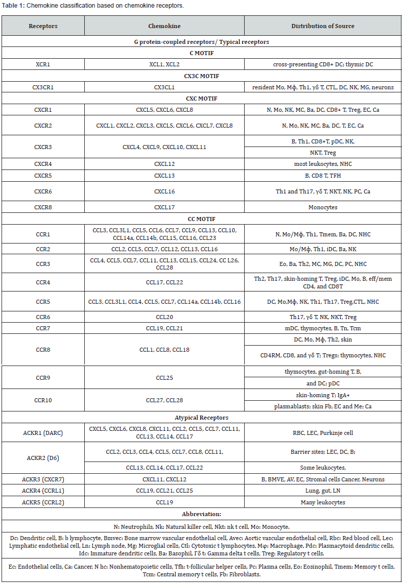
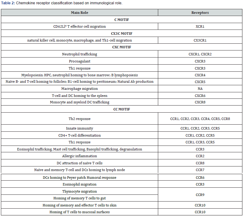
CCR2-CCL2: CCR2-CCL2 (MCP-1) is a CC chemokine that activates the CCR2 on T cells and monocytes [14]. CCL2 is first discovered in human CC chemokine, and it is localized on chromosome 17. Its size is 13 kDa and is composed of 76 amino acids polypeptide and is a member of the C–C chemokine family characterized by adjacent cysteine residues [15,16]. CCL2 recognizes the CCR2, all chemokines interact with Chemokines receptor 2. Chemokine receptor is present on the surface of several leukocytes that are dendritic cells, monocytes, basophils, and natural killer cells and activated T lymphocytes. The main function of chemokine was inducing granule release, chemotaxis, histamine and cytokines releases, and respiratory burst. CCR2 has 2 different forms of the receptor such as CCR2A and CCR2B and these different forms of receptor differ from each other by C-terminal region.
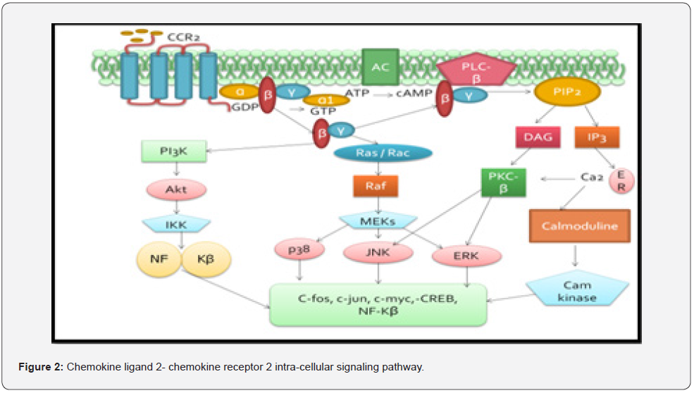
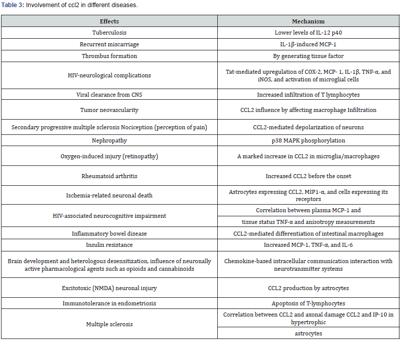
CCR2A is the major isoform expressed by mononuclear cells and vascular smooth muscle cells, and the CCR2B isoform expresses predominantly by the monocytes and activated NK cells. There is the possibility of activation of various signaling pathways by CCR2A and CCR2B and this produces the various actions. Chemokine receptor 2 has dual roles as pro-inflammatory and anti-inflammatory like both actions. The sequence homology between Chemokine ligand 2 and other family members is high and varies between 61% for CCL8 and CCL4, and 71% for CCL7. Several studies have shown that the CCR2 can bind with the 5 proinflammatory Chemokine Ligands i.e., CCL2, CCL7, CCL8, CCL12 and CCL13 [17]. Several chemokines like CCL2, CCL7, CCL8, CCL13, and CCL16, act as CCR2 agonists and other like CCL11 and CCL26 act as antagonists. Effects of the various chemokine ligands 2 on ß-arrestin recruitment to the receptor are qualitatively different from one another. There are differences found in the efficacy and identification of CCL7, CCL8, and CCL13 as partial agonists. The study result supports the validity of models for receptorligand interactions in which different ligands stabilize different receptor conformations also for endogenous receptor ligands, with corresponding implications for drug development targeting CCR2 [18]. CCR2 may be involved in the signaling pathology of diseases such as chronic obstructive pulmonary disease, obesity, atherosclerosis, cancer metastasis, neurodegeneration, and multiple sclerosis (Figure 2), (Table 3).
The role of CCL2-CCR2 in disease pathology
CCL2-CCR2 signaling in immunity and inflammation: Inflammation is a complex mechanism, it is characterized by the activation and migration of inflammatory cells, and displacement of immune cells through lymphoid organs to the site of inflammation and initiate immune responses. But such movement of inflammatory cells required chemokines receptor to trigger the inflammatory cell expression on target sites. The interaction between immune cells and chemoattractant triggers a cascade of cellular events. CCL2- CCR2 stimulates monocytes, macrophages, and several cellular chemotaxis events, including integrin expression and calcium influx. It is also an inducer of cytokine and chemokines expression in monocytes and microphage, at high concentrations, and expedites the generation of reactive oxygen species by doing respiratory bust.
CCL2 is rapidly produced by immune and stromal cells after toll-like receptor (TLR) activation. As the activation of TLR, CCL2 undergo dimerizes of ligand and binds to their extracellular matrix glycosaminoglycans, thus establishing a stable gradient for CCR2+ inflammatory monocytes. CCL2 expression has been identified as upregulated in certain cell types, such as macrophages/ foam cells and smooth muscle cells in atherosclerosis, delayed type of hypersensitivity reactions, endothelial cells, cancer, and inflammatory diseases. Monocyte chemo-attracting proteins (MCP-1), includes CCL2, CCL7, CCL8, and CCL13. In the inflamed tissue CCL2 and receptors of it show a non-redundant role in the monocyte trafficking. And also, in the regulation of specific aspects of the response of adaptive immune and involvement in mobilization of monocyte precursors of bone marrow [19,20].
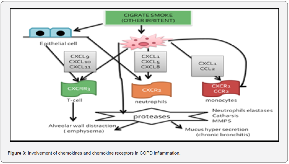
CCL2-CCR2 signaling in chronic obstructive pulmonary fibrosis: Subject with Chronic obstructive pulmonary disease (COPD), CCR2, has a role and they were markedly increased level of chemokines and neutrophils [21]. The concentration of chemokines more acclimated in the BAL (bronchoalveolar lavage) fluid, sputum, and lungs of COPD patients compared to non-COPD [22]. Furthermore, CCL2 is also expressed by T cells, alveolar macrophages, type 2 pneumocytes, and epithelial cells in culture followed by stimulation with lipopolysaccharide. The CCR2 is down-regulated by Toll-like receptor-2 (TLR2), TLR4, and other inflammatory receptors, and the mechanism for terminating chemotactic response is provided by it, which leads to monocyte accumulation. Macrophages have a major role in chronic obstructive pulmonary disease by blocking CCR2, it may help in therapy [23], (Figure 3).
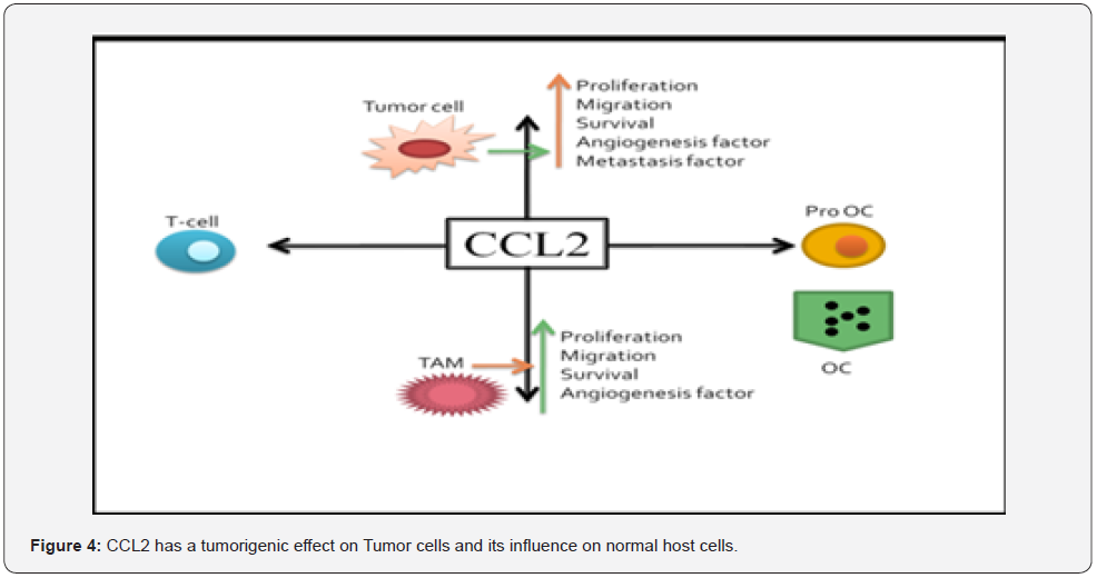
The role of CCL2-CCR2 in cancer: The chemokines and growth factors are responsible for recruitment of cytokines and activation microenvironment for development of tumors in the host [24] Alteration of chemokines and their receptor function affects the function of cells and accelerates progression of cancer in cells. Although altering the function of chemokines and cytokines does not participate not only in progression of cancer but also in metastasis and invasion of cancer. However, there is distinct mechanism of chemokines, such as the endocrine and the paracrine actions play role in metastasis and progression of cancer [25]. Chemokines and their receptor signaling has recently been identified as a player in promoting tumorigenesis and metastasis [26], (Figure 4).
CCL2-CCR2 signaling in the metastatic cascade: Metastasis of cancers is a multistep process that includes invasion of cancer cells into surrounding environment of tissues and followed by intravasation and extravasation of cells into blood vessels and secondary sites of tissue. Now cancer cells have to adapt their new environment at secondary sites for proliferation of cells and which promote outgrowth of cells at sites and form colonization at letter stage of cancer. Chemokines ligand 2 and their receptor play an important role in the metastasis of cancer. Several experimental shreds of evidence suggest that CCL2 is involved in the both early as well as letter stage of metastasis. Chemokines are involved in metastasis by affecting activities of pro-tumorigenic mediators in cancer cell microenvironment and they excite direct effect on function of cancer cells [27].
Firstly, in the early phase of metastasis, cancer cells have to achieve migration to surrounding environment by breaking extracellular matrix of neighboring tissue and move it toward the blood vessels for invasion. Chemokine ligand 2 plays a role in increasing the expression of matrix metalloproteinases such as MMP9 and MMP2 in cancer cells, which break down the extracellular matrix of tissue and helps in migration of cancer cells into blood vessels and increases invasion [28]. Intravasation of Cancer cells permitted into the circulation is needed for metastatic dissemination. Wyckoff et al. demonstrated that intravasation of cancers cell is an important process to interact with tumor-associated macrophages (TAMs) [29]. Extravasation of cancer cells to secondary sites is also dependent on TAMs and bone marrow endothelial cells. CCL2 is a potent chemoattractant for TAMs, it may indirectly promote the intravasation and extravasation process of cancer cells [30].
The most common challenging step for cancer cells to series in a secondary site, the cancer cell enters to secondary site they have to adopt homeostasis mechanism for cell growth and apoptosis, but sometimes they cannot be able to adopt that and it goes to quiescent phase. This state of dormancy is in keeping with clinical observations that many patients with prostate and breast cancer develop metastatic relapse after the initial diagnosis or treatment [31]. Now this kind of cell dormancy occurs in tumor cells called as delayed cell adaptation to the new environment. For example, in an adaptation to new environment cancer cell needs to adopt immune dormancy, cellular dormancy, and mass dormancy for successful cancer formation at secondary sites which means metastatic proliferation of cells [32]. Although there has certain evidence that CCL2 play role in cell cycle arrest, by initiating recruit TAMs and encouraging an angiogenesis control and switch level of myeloid-derived suppression cells to suppress immunemediated death [33,34].
In later stage of metastasis CCL2 play an additional role in cancer by increasing attraction of leucocytes at secondary site. Leucocyte trafficking attracts the cancer cell at secondary site which prompts metastasis of cancer. However, the trafficking and migration process is quite complex in nature, and it may require other immense cell involvement for this process [35]. In addition to that CCL2 play role in cell survival and stimulate cancer cell proliferation at secondary site by facilitating environment by promoting migration, recruitment, and proliferation of endothelial cells. Endothelial cells of both humans, as well as animals, have a receptor of CCL2 that is CCR2 that expresses CCL2 and recruit microenvironment of metastasis of cancer cells. In addition to that CCL2 and CCR2 signaling play role in angiogenesis, there are several in vitro and in vivo models demonstrate that CCL2 and CCR2 help in neovascularization and its facilities angiogenesis.
Apart from direct action of CCL2 on tumor and endothelial cells, CCL2 also have an indirect effect via recruiting various immune cells, such as monocytes and macrophages at site of metastasis and medicating polarization and differentiation of various cell [36]. Polarization of immune cell play role in function property of immune cell and altimetry shift balance between tumor growth and inhibition. For example, polarized macrophages shift balance towards an M2 phenotype, they are responsible for immunosuppressive and may lead to tumor cell survival by dampening immune attack [37,38]. Similarly, CCL2 is also involved in T cell differentiation and polarization and has been shown to regulate Th2 polarization towards a more immunosuppressive T regulatory phenotype in vitro and in mouse models. It is evident that CCL2-CCR2 signaling has multiple key functions, in both metastatic development and progression [39]. Inhibition of CCL2-CCR2 signaling by using an antagonist of CCR2 helps us to inhibit cancer progression and metastasis.
CCL2-CCR2 signaling in cardiovascular disease and metabolic syndrome
i. Metabolic syndromes
Obesity, hypertension, insulin resistance, and dyslipidemia all are factors of metabolic syndrome, which often lead to cardiovascular diseases and endocrine diseases. Many tissues in the body, such as the skeletal muscle, liver, pancreas, heart, and vessels are adversely influenced by metabolic syndrome. Although recent data suggest that complex molecular mechanism responsible for inflammation and migration of macrophages/ inflammatory marker in tissues which is responsible for development of obesity-related complication and play a central role in Pathological role in obesity-related disorders [40]. Obesity is characterized by activation and infiltration of inflammatory and immune cells into metabolic organs such as adipose tissue, skeletal muscles, liver and leading to chronic inflammation in tissues and generates a signal that attracts chemokines and cytokines which regulated the immune response into tissues [41]. Adipocytes are major source of CCL2 in obesity. In addition, adipocytes trigger the production of CCL2 during high fatty acid and inflammatory conditions. Recently one article suggests that microRNA193b and 126 have been implicated in the regulation of chemokines ligand in obesity [42]. In addition, other cells such as vascular smooth muscle, skeletal muscle cells, hepatocytes, and endothelial cells are capable to secrete chemokines in obesity. CCL2 role in obesity is not well studied but some evidence found that higher levels of circulating CCL2 in obese adults as well as children [43,44]. This circulatory CCL2 level was directly correlated with obesity inflammatory markers like high-density lipoprotein (HDL) interleukins and creative protein as well as body mass index (BMI) in obesity. But some conflicting evidence found that CCL2 administration causes insulin resistance in mice and humans [45,46]. Some research articles suggest that reduction in weight by doing exercise reduced CCL2 levels and improved insulin sensitivity [47].
ii. Atherosclerosis
Atherosclerosis is a chronic metabolic inflammatory disease; cause of atherosclerosis leads to some heart disease as well as stock. The accumulation of inflammatory and immune cells and fatty acids in the intima is an essential step in the development of atherosclerosis. The development of atherosclerosis is primarily triggered by section of chemokines and cytokines from endothelial and epithelial cells. Various members of the CC chemokine family play crucial role in progression and development of atherosclerotic plaque [48]. CCL2 have primarily been involved in inflammation due to their role in the recruitment of leukocyte migration as well they also up-regulate some inflammatory and fibrotic cells [49,50]. In patients at risk for developing atherosclerosis, it has been seen that there is a higher level of serum CCL2 is direct correlate with the inflammatory activity,[51] and therefore CCL2 serves as a marker for cardiovascular risk.
Study evidence suggests that deficiency in CCL2 leads to reduction of atherosclerosis lesions in mice LDLR-/-.[52]. In support of the theory, some more evidence suggests that the chemokine receptor axis plays an important role in the development of atherosclerotic lesions. Some studies have manifested that a deficiency in receptor CCR2, which is binding receptor of CCL2, ultimately represents reduction in the progression of atherosclerosis in ApoE-/- mice [53,54]. Administration of an inactive N-terminal deletion mutant of CCL2 that binds CCR2 and thus blocks CCL2 mediated chemotaxis of immune and inflammatory cells and has shown positive modulatory effects in ApoE-/- mice, thus positive modulatory effects play vital role in development of atherosclerosis and related several consequences [55]. The above decibels all findings suggest the importance of the CCL2–CCR2 axis as a target in the development of future clinical therapies.
iii. CCL2-CCR2 signaling in the central nervous system
CCL2-CCR2 signaling also has important functions in the central nervous system. The central nervous system (CNS) has resident immune cells such as microglia and it is phylogenetically related to monocytes [56]. During neuron infection or inflammation activated astrocytes secret CCL2 and attract microglia at site of inflammation and where they phagocytose microbes or cellular debris [57]. However, endothelial cells and neurons under both basal and neuroinflammatory conditions macrophage/ microglia produced CCL2. During neuroinflammatory conditions, peripheral microphage has migrated into CNS. In this condition, CCR2 increases expression of brain microvascular endothelial cell response to the CCL2, and it increases permeability of the bloodbrain barrier by activating RhoA signaling and reorganization of actin cytoskeletal [58]. Also, at the site of neuronal damage and inflammation neuronal progenitor cells (NPCs) are attracted [59]. CCL2 and CCR2-dependent migration of NPCs in the CNS has been reported during many brain conditions, such as glial tumors, ischemia and stroke, epilepsy, and striatal cell loss. CCR2 expression in neurons and astrocytes has also been reported. Activation of astrocytes which enhances CCR2. Survival of astrocytes via NF-κB and Akt signaling pathways promotes the production of neurotrophic factors [60,61].
iv. The role of CCL2-CCR2 in neurodegenerative diseases
CCl2-CCR2 axis play role in neurodegenerative diseases. Alzheimer’s disease (AD) is one of the most common neurodegenerative diseases. AD is characterized by deposition of the β-amyloid peptide, which is a degenerative product of the β-amyloid precursor protein (β-APP). B-APP is a peptide and has capacity to aggregate and form neurotoxins intracellular as well as extracellularly. This is responsible for formation of fibrils and senile plaques in neurons. The accumulation of beta-amyloid peptides triggers neurodegeneration. Amyloidal peptides more affect the cholinergic neurons and lead to a loss of memory function which is common symptom of AD. During this process activated immune cells are a hallmark for AD. In the affected area of brain, a large number of inflammatory cytokine and chemokines was found including CCL2.
The patient suffering from mild cognitive impairment and examine the level of CCL2 in CSF, which was found to be high in AD suffering patients, so higher level of CCL2 in CSF was an indicator of early pathogenesis of AD [62]. In studies, they found that higher CCL2 expression leads to microgliosis and plague formation in biogenic mice. In addition to that, this mouse model was originally established as beta amyloidal mice model with overexpression of CCL2 via under regulation of glial acidic fibril protein promoter. Some research on biogenic mice has proven that involvement of CCL2 were enhance plaque formation and Aβ accumulation in neurons [63]. CCL2 also play role in accumulation of amyloidal peptides and cognitive dysfunction has been found in a murine model of beta-amyloidosis. From the study, they support that CCL2 plays role as a co-factor in amyloidal peptide-induced cognitive impairment in APP mice. However, the exact mechanism behind oligomerization is not elucidated. But it has been revealed that beta-amyloid uptake by microglia, which is significantly enhanced CCL2 and its leads to an oligomerized Aβ species in intracellular as well as extracellularly. Therefore, CCL2 is a potent chemokine chemoattractant for microglia and may activate cytoskeleton dynamics as a part of its chemoattractant signaling and thereby affect Aβ uptake. This research evidence indicates a possible role of CCL2/CCR2 system in the pathogenesis of AD where CCL2 levels in CSF and plasma are augmented.
Multiple sclerosis (MS)
Multiple sclerosis (MS) is a chronic autoimmune disease causing severe neurologic impairment. It is characterized by immeasurable demyelination and inflammation of neurons. The activated macrophages and infiltration of lymphocytes, reactive astrocytes, and microglia, are the majorly associated with Multiple sclerosis [64]. In MS, inflammatory process is controlled by cytokines and chemokines, and which generate chemotaxis and attract the leukocytes [65]. The presence of Chemokine ligand 2 has been detected in autopsy tissue samples collected from patients with active and chronic MS [66]. More specifically, the expression of CCL2 and CCR2 in MS have been described in the cerebrospinal fluid, brain, and blood [67]. In a clinical study, the higher expression of chemokine ligand 2 has been found in acute as well as chronic MS plaques. Also, it has been found that chemokine ligand 2 is expressed by astrocytes and macrophages within demyelinating MS plaques. Many studies have indicated the involvement of CCR2 in MS. One study was done by utilizing CNS tissue and revealed that chemokine receptor 2 is highly expressed by microphage and microglia and perivascular cells in chronic MS conditions [68]. Some other studies support that mice lacking CCR2 cannot undergo encephalopathy-like conditions however the mice show overexpression of chemokine ligand 2.
Thus, the study concludes that CCR2 signaling is essential for CCL2-produced effects [69]. This research article agreed with the above finding involvement of CCL2 signaling pathways, plays an impotent role in the progression of EAE induced by a peptide derived from myelin oligodendrocytes glycoprotein peptide 35- 55.72 However, In this study mice lacking immunized and CCR2 with myelin oligodendrocyte glycoprotein peptide, 35–55 and cannot produce inflammatory cell infiltrates in the CNS and prevent elevation of Chemokine levels in CNS. Accordingly, it has been concluded that CCR2 and CCL2 are essential for the production of active lesions, which is mediated via the production of inflammatory chemokines that amplify the local inflammatory response [70,71].
CCL2-CCR2 signaling in Pain: Expression of the Chemokine receptor is done by monocytes, and they act in the presence of high level of membrane Ly6C. For entry of the monocyte in different inflammatory sites the CCR2 is required and it also includes CNS. CCL2 is the ligand for trafficking of CCR2+ ‘‘inflammatory’’ monocytes into the nervous system. Initial evaluated affected tissues lead to a CCR2 up-regulation in injured nerve, in dorsal root ganglia, and at the site of inflamed injection. In presence of CCR2+ inflammatory monocytes, the pain pathways are modulated. And the loss of Chemokines signaling ameliorates the pain by dampening macrophage-mediated inflammation. In the context of chronic nerve root damage, the CCL2-CCR2 is up-regulated by the DRG neurons and becomes a more complex situation. Furthermore, in synaptic-like vesicles, CCL2 is packed and the action of it at CCR2 leads to the pain-promoting response. That includes the transient receptor potential vanilloid receptor subtype 1 (TRPV1) ion channel sensitization. They prove that the Chemokine receptor plays a complex role inmonocytes and by its startling and novel properties as an inducible neuromodulatory receptor on DRG neurons [72].
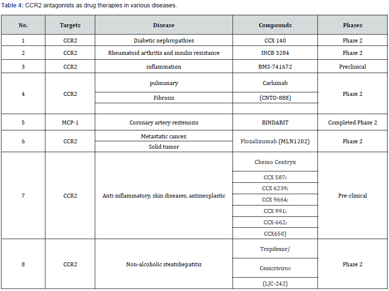
Clinical significance of chemokine receptor antagonists: Chemokines are highly conserved small signaling proteins, which are secreted by a variety of cell types such as B cells, T cells, stem cells innate lymphocytes, dendritic cells, myeloid cells, and stromal cells. Chemokines are known for their multifactorial functions such as chemotaxis, migration, and inflammatory. They are important not only for their dual role in protection and/or distortive property but also in development and mentioning of immune system homeostasis in humans [73]. Due to this chemokine have important roles in several pathophysiological conditions such as cancer, pain, immunity, and inflammation, and several more. As above we show the role of chemokines and chemokines reporter are therapeutic targets of antagonists intended for the blockade of chemokine receptor-ligand interactions and helps to prevent disease progression. Several antagonists of CCL2 and CCR2 in preclinical and clinical trials have been listed in [74], (Table 4).
Conclusion
Cellular and molecular mechanism of CCL2-CCR2 has been made in the last couple of years it plays an important role in health and various diseases. Some small molecule of CCR2 antagonists are translated in the progress from basic research is still in the starting phase, although initial clinical trials are running, currently launched, or upcoming. Still, a substantial amount of research work is needed to establish these targets which can help in the management of healthcare systems.
References
- Feria M, Díaz-González F (2006) The CCR2 receptor as a therapeutic target. Expert Opin Ther Patents 16(1): 49-57.
- Proudfoot AEI ( 2002) Chemokine receptors: multifaceted therapeutic targets. Nat Rev Immunol 2(2): 106-115.
- Solari R, Pease JE, Begg M (2015) Chemokine receptors as therapeutic targets: Why aren’t there more drugs? Eur J Pharmacol 746: 363-367.
- Kufareva I, Salanga CL, Handel TM (2015) Chemokine and chemokine receptor structure and interactions : implications for therapeutic strategies. Immunol Cell Bio 93(4): 372-383.
- Scholten DJ, Canals M, Maussang D (2012) Pharmacological modulation of chemokine receptor function. Br J Pharmacol 165(6): 1617-1643.
- Bot I, Zacarías NVO, De Witte WEA, Vries HD, Santbrink PJV, et al. (2017) A novel CCR2 antagonist inhibits atherogenesis in apoE deficient mice by achieving high receptor occupancy. Sci Rep 7(1): 52.
- Bachelerie F, Ben-Baruch A, Burkhardt AM, Combadiere C, Farber JM, et al. (2013) International Union of Pharmacology. LXXXIX. Update on the Extended Family of Chemokine Receptors and Introducing a New Nomenclature for Atypical Chemokine Receptors. Pharmacol Rev 66(1): 1-79.
- Zlotnik A, Yoshie O, Nomiyama H (2006) The chemokine and chemokine receptor superfamilies and their molecular evolution. Genome Biol 7(12): 243.
- Ulvmar MH, Hub E, Rot A (2011) Atypical chemokine receptors. Exp Cell Res 317(5): 556-568.
- Slinger E, Langemeijer E, Siderius M, Vischer HF, Smit MJ (2011) Herpesvirus-encoded GPCRs rewire cellular signaling. Mol Cell Endocrinol 331(2): 179-184.
- Berger EA, Murphy PM, Farber JM (1999) Chemokine receptors as HIV-1 coreceptors : Roles in Viral Entry, Tropism, and Disease 17: 657-700.
- Locati M, Murphy PM (1999) Chemokines and chemokine receptors: Biology and Clinical Relevance in Inflammation and AIDS. Annu Rev Med 50: 425-440.
- Griffith JW, Sokol CL, Luster AD (2014) Chemokines and Chemokine Receptors : Positioning Cells for Host Defense and Immunity. Annu Rev Immunol 32: 659-702.
- Donnelly LE, Barnes PJ (2006) Chemokine receptors as therapeutic targets in chronic obstructive pulmonary disease. Trends Pharmacol Sci 27(10): 546-553.
- Zhang J, Patel L, Pienta KJ (2010) CC chemokine ligand 2 (CCL2) promotes prostate cancer tumorigenesis and metastasis. Cytokine Growth Factor Rev 21(1): 41-48.
- Deshmane SL, Kremlev S, Amini S, Sawaya BE (2009) Monocyte Chemoattractant Protein-1 (MCP-1): An Overview. J Interf Cytokine Res 29(6): 313-326.
- Bose S, Cho J (2013) Role of chemokine CCL2 and it receptor CCR2 in neurodegenerative diseases. Arch Pharm Res 36(9): 1039-1050.
- Berchiche YA, Gravel S, Pelletier M-E, St-Onge G, Heveker N (2011) Different Effects of the Different Natural CC Chemokine Receptor 2b Ligands on -Arrestin Recruitment, G i Signaling, and Receptor Internalization. Mol Pharmacol 79(3): 488-498.
- Viola A, Luster AD (2008) Chemokines and Their Receptors : Drug Targets in Immunity and Inflammation. Anu Rev Pharmacol Toxicol 48: 171-197.
- Zimmerman KA, Hopp K, Mrug M (2020) Role of chemokines, innate and adaptive immunity. Cell Signal 73: 109647.
- Soler N, Ewig S, Torres A, Filella X, Gonzalez J, Zaubet A (1999) Airway inflammation and bronchial microbial patterns in patients with stable chronic obstructive pulmonary disease. Eur Respir J 14(5): 1015-1022.
- Traves SL, Culpitt S V, Russell REK, Barnes PJ, Donnelly LE (2002) Increased levels of the chemokines GRO α and MCP-1 in sputum samples from patients with COPD. Thorax 57(7): 590-595.
- Witherden IR, Bon EJ Vanden, Goldstraw P Effects of Cigarette Smoke and Neutrophil Elastase.
- Borsig L, Wolf MJ, Roblek M, Lorentzen A, Heikenwalder M (2014) Inflammatory chemokines and metastasis-tracing the accessory.Oncogene 33(25): 3217-3224.
- A Ben-Baruch (2006) The multifaceted roles of chemokines in malignancy. cancer Metastasis rev 25(3): 357-371.
- Markers P (2020) Chemokines and their Receptors : Multifaceted Roles in Cancer Progression and Potential Value as Cancer.
- Chaffer C L , Weinberg R A (2011) A Perspective on Cancer cell metastasis. Science 331(6024): 1559-1565.
- Tang CH, Tsai CC (2012) CCL2 increases MMP-9 expression and cell motility in human chondrosarcoma. Biochem Pharmacol 83(3): 335-344.
- Wyckoff JB, Wang Y, Lin EY, Li JF, Goswami S, et al. (2007) Direct Visualization of Macrophage-Assisted Tumor Cell Intravasation in Mammary Tumors Direct Visualization of Macrophage-Assisted Tumor Cell Intravasation in Mammary Tumors. Cancer Res 67(6): 2649-2656.
- Qian B, Deng Y, Im JH, Muschel RJ, Zou Y (2009) A Distinct Macrophage Population Mediates Metastatic Breast Cancer Cell Extravasation, Establishment and Growth. PLoS One 4(8): e6562.
- Kleffel S, Schatton T (2013) Tumor Dormancy and Cancer Stem Cells : Two Sides of the Same Coin ? Adv Exp Med Biol 734: 145-179.
- Giancotti FG (2015) Mechanisms Governing Metastatic Dormancy and Reactivation. Cell 155(4): 750-764.
- Janine M Low-Marchelli, Veronica C Ardi, Edward A Vizcarra, Nico van Rooijen, James P, et al. (2014) Twist1 induces CCL2 and recruits macrophages to promote angiogenesis. Cancer Res 73(2): 662-671.
- Huang B, Lei Z, Zhao J, Gong W, Liu J, et al. (2007) CCL2 / CCR2 pathway mediates recruitment of myeloid suppressor cells to cancers. Cancer Lett 252(1): 86-92.
- Witz IP (2008) Tumor - Microenvironment Interactions : Dangerous Liaisons. Adv Cancer Res 100: 203-239.
- Qian B, Li J, Zhang H (2012) 475(7355): 222-225.
- Jaillon B, Galdiero MR, Garlanda C, Marone G, Mantovani A (2012) Tumor-Associated Macrophages and Neutrophils in Tumor Progression. J Cell Physiol 228(7): 1404-1412.
- Liu T, Guo Z, Song X (2020) High-fat diet-induced dysbiosis mediates MCP-1 / CCR2 axis-dependent M2 macrophage polarization and promotes intestinal adenoma-adenocarcinoma sequence. J cell Mol Med 24(4): 2648-2662.
- Matsukawa A, Lukacs NW, Theodore J, Chensue SW, Kunkel SL (2015) Adenoviral-Mediated Overexpression of Monocyte Chemoattractant Protein-1 Differentially Alters the Development of Th1 and Th2 Type Responses In Vivo. J Immunol 164(4): 1699-1704.
- Weisberg SP, Mccann D, Desai M, Rosenbaum M, Leibel RL, et al. (2003) Obesity is associated with macrophage accumulation. J Clin Invest 112(12): 1796-1808.
- Rakotoarivelo V, Variya B, Langlois M, Ramanathan S (2020) Cytokine Chemokines in human obesity. Cytokine 127: 154953.
- Arner E, Mejhert N, Kulyté A, Balwierz PJ, Pachkov M, et al. (2012) Adipose Tissue MicroRNAs as Regulators of CCL2 Production in Human Obesity. Diabetes 61(8): 1986-1993.
- Kim C, Park H, Kawada T, Kim JH, D Lim , et al. (2006) Circulating levels of MCP-1 and IL-8 are elevated in human obese subjects and associated with obesity-related parameters. Int J Obes (Lond) 30(9): 1347-1355.
- Roth CL, Kratz M, Ralston MM, Reinehr T (2011) Changes in adipose-derived inflammatory cytokines and chemokines after successful lifestyle intervention in obese children. Metabolism 60(4): 445-452.
- Kamei N, Tobe K, Suzuki R, Ohsugi M, Watanabe T (2006) Overexpression of Monocyte Chemoattractant Protein-1 in Adipose Tissues Causes Macrophage Recruitment and Insulin Resistance. J Biol Chem 281(36): 26602-26614.
- Ezenwaka CE, Nwagbara E, Seales D, Okali F, Sell H, et al. (2009) Insulin resistance, leptin and monocyte chemotactic protein-1 levels in diabetic and non-diabetic Afro- Caribbean subjects. Arch Physiol Biochem 115(1): 22-27.
- Chen Y, Hubal MJ, Hoffman EP, Thompson PD, Clarkson PM (2015) Molecular responses of human muscle to eccentric exercise Molecular responses of human muscle to eccentric exercise. Clinical Trial 95(6): 2485-2494.
- Charo IF, Peters W (2003) Chemokine Receptor 2 ( CCR2 ) in Atherosclerosis, Infectious Diseases, and Regulation of T-Cell Polarization. Microcirculation 10(3-4): 259-264.
- Inoshima I, Kuwano K, Hamada N, Hagimoto N, Yoshimi M, et al. (2004) Anti-monocyte chemoattractant protein-1 gene therapy attenuates pulmonary fibrosis in mice. Am J Physiol Lung Cell Mol Physiol 286(5): 1038-1044.
- Barrett TJ (2020) Macrophages in Atherosclerosis Regression. Arterioscler Thromb Vasc Biol 40(1): 20-33.
- Martinovic I, Abegunewardene N, Seul M, Vosseler M, Horstick G (2005) Elevated Monocyte Chemoattractant Protein-1 Serum Levels in Patients at Risk for Coronary Artery Disease. Circ J 69(12): 1484-1489.
- Gu L, Okada Y, Clinton SK, Gerard C, Sukhova GK (1998) Absence of Monocyte Chemoattractant Protein-1 Reduces Atherosclerosis in Low Density Lipoprotein Receptor - Deficient Mice. Mol Cell 2(2): 275-281.
- Boring L, Gosling J, Cleary M, Charo IF (1998) Decreased lesion formation inCCR2−/− mice reveals a role forchemokinesintheinitiation of atherosclerosis. Nature 394(6696): 894-897.
- Veillard NR, Steffens S, Pelli G, B Lu, Kwak BR, et al. (2005) Differential Influence of Chemokine Receptors CCR2 and CXCR3 in Development of Atherosclerosis In Vivo. Circulation 112(6): 870-879.
- Inoue S, Egashira K, Ni W, Kitamoto S, Usui M (2002) Basic Science Reports Limits Progression and Destabilization of Established Atherosclerosis in Apolipoprotein E - Knockout Mice pp. 2700-2707.
- Williams JL, Holman DW, Klein RS (2014) Chemokines in the balance : maintenance of homeostasis and protection at CNS barriers. Front Cell Neurosci 8: 154.
- Biber K, de Jong EK, van Weering HRJ, Boddeke HWGM (2006) Chemokines and their receptors in central nervous system disease. Curr Drug Targets 7(1): 29-46.
- Dzenko KA, Song L, Ge S, Kuziel WA, Pachter JS (2005) CCR2 expression by brain microvascular endothelial cells is critical for macrophage transendothelial migration in response to CCL2. Microvasc Res 70(1-2): 53-64.
- Belmadani A, Tran PB, Ren D, Miller RJ (2006) Chemokines Regulate the Migration of Neural Progenitors to Sites of Neuroinflammation. J Neurosci 26(12): 3182-3191.
- Kalehua AN, Nagel JE, Whelchel LM, Gides JJ, Pyle RS, et al. (2004) Monocyte chemoattractant protein-1 and macrophage inflammatory protein-2 are involved in both excitotoxin-induced neurodegeneration and regeneration. Exp Cell Res 297(1): 97-211.
- Quinones MP, Kalkonde Y, Estrada CA, Jimenez F, Ramirez R, et al. (2008) Role of astrocytes and chemokine systems in acute TNF α induced demyelinating syndrome : CCR2-dependent signals promote astrocyte activation and survival via NF- κ B and Akt. Mol Cell Neurosci 37(1): 96-109.
- Galimberti D, Fenoglio C, Lovati C (2006) Serum MCP-1 levels are increased in mild cognitive impairment and mild Alzheimer’s disease. Neurobiol Aging 27(12): 1763-1768.
- Kiyota T, Yamamoto M, Xiong H, Lambert M P, Klein W L, et al. (2009) CCL2 Accelerates Microglia-Mediated A b Oligomer Formation and Progression of Neurocognitive Dysfunction. PLoS One 4(7): e6197.
- Baecher-allan C, Kaskow BJ, Weiner HL (2018) Review Multiple Sclerosis : Mechanisms and Immunotherapy. Neuron 97(4): 742-768.
- Conductier G, Blondeau N, Guyon A, Nahon J, Rovère C (2010) The role of monocyte chemoattractant protein MCP1/CCL2 in neuroinflammatory diseases. J Neuroimmunol 224(1-2): 93-100.
- Semple BD, Kossmann T, Morganti-kossmann MC (2009) Role of chemokines in CNS health and pathology : a focus on the CCL2 / CCR2 and CXCL8 / CXCR2 networks. J Cereb Blood Flow & Metab 30(3): 459-473.
- Mahad DJ, Ransohoff RM (2003) The role of MCP-1 (CCL2) and CCR2 in multiple sclerosis and experimental autoimmune encephalomyelitis (EAE) Semin Immunol 15(1): 23-32.
- Simpson J, Rezaie P, Newcombe J, Cuzner ML, Male D, et al. (2000) Expression of the b -chemokine receptors CCR2, CCR3, and CCR5 in multiple sclerosis central nervous system tissue 108: 192-200.
- Huang D, Wujek J, Kidd G (2005) Chronic expression of monocyte chemoattractant protein-1 in the central nervous system causes delayed encephalopathy and impaired microglial function in mice. FASEB J 19(7): 761-772.
- Izikson BL, Klein RS, Charo IF, Weiner HL, Luster AD (2000) Resistance to Experimental Autoimmune Encephalomyelitis in Mice Lacking the CC Chemokine Receptor (CCR) 2. J Exp Med 192(7): 1075-1080.
- Cui L, Chu S, Chen N (2020) International Immunopharmacology The role of chemokines and chemokine receptors in multiple sclerosis. Int Immunopharmacol 83: 106314.
- Ransohoff RM (2009) Review Chemokines and Chemokine Receptors : Standing at the Crossroads of Immunobiology and Neurobiology. Immunity 31(5): 711-721.
- Colotta F, Allavena P, Sica A, Garlanda C, Mantovani A (2009) Cancer-related inflammation, the seventh hallmark of cancer : links to genetic instability. Carcinogenesis 30(7): 1073-1081.
- Chiu H, Sun K, Chen S, Wang H, Tang S, et al. (2012) Cytokine Autocrine CCL2 promotes cell migration and invasion via PKC activation and tyrosine phosphorylation of paxillin in bladder cancer cells. Cytokine 59(2): 423-432.


