A Rare Entity of Clear Cell Carcinoma from Sinonasal Location: Sinonasal Renal-Cell Like Adenocarcinoma SNRCLA a Case Report and Literature Reviews
Nyein Nyein Htun*, Daniel Nguyen, Beverly Wang Y and Behdokht Nowroozizadeh
Department of Pathology, University of California Irvine Medical Center, UC Irvine School of Medicine, USA
Submission: August 26, 2023; Published:September 12, 2023
*Corresponding author:Nyein Nyein Htun, Department of Pathology, University of California Irvine Medical Center, UC Irvine School of Medicine, USA
How to cite this article:Nyein Nyein H, Daniel N, Beverly Wang Y, Behdokht N. A Rare Entity of Clear Cell Carcinoma from Sinonasal Location: Sinonasal Renal-Cell Like Adenocarcinoma SNRCLA a Case Report and Literature Reviews J Tumor Med Prev. 2023; 4(2): 555633. DOI: 10.19080/ JTMP.2023.07.555633
Abstract
Background
Very few Sinonasal malignancies can express clear cell histomorphology. Primary sinonasal tumors that can appear as clear cell malignancies include Sinonasal clear cell carcinoma SCCC and Sinonasal renal cell like carcinoma SNRCLA. Metastatic renal clear cell carcinoma is one of the differentials of sinonasal clear cell malignancy. Primary SNRCLA exclusively occurred in sinonasal tract.
Results
Histologically, the tumor was identical to clear cell renal cell carcinoma. No evidence of renal malignancy on abdominal computed tomographic scan or gadolinium-enhanced magnetic resonance imaging and negative urinary symptoms like hematuria, pain and mass. Histochemistry confirmed no mucin with negative mucicarmine stain. Immunohistochemistry confirmed robust expression CAIX, keratin AE1/AE3 and patchy CK7 and patchy inhibin.
Keywords: Neoplasms; Morphology; Metastatic Renal Cell Carcinoma; Tumor; Hematoma; Kidneys; Oncocytoid; Radiotherapy; Abdominal Pain; Hematuria
Introduction
The sinonasal tract has been an anatomic region in which a remarkable diversity of neoplasms can develop. Sinonasal malignancy is a rare entity and accounting for less than 3% of all head and neck malignancies and the incidence has been estimated to be on the order of 0.5–1.0 per 100,000 people. In addition to rarity and diverse nature of neoplasms due to various histologic origin, sinonasal malignancy are still challenges to Pathologists. Among them, sinonasal renal cell–like adenocarcinoma (SNRCLA) is an extremely rare neoplasm that mimics the clear cell variant of renal cell carcinoma. A few cases have been reported about SNRCLA and differential include tumor with clear cell morphology such as salivary clear cell carcinoma SCCC, mucoepidermoid carcinoma and metastatic renal cell carcinoma [1]. The descriptive term, “renal cell like,” was originally proposed by Zur et al. The authors acknowledged that the major differential diagnosis of was a metastatic clear cell RCC because the two neoplastic entities looked very similar [2]. Final diagnosis of sinonasal clear cell renal cell like carcinoma require interpretation the context of immunohistochemical features in the supportive negative pertinent imaging findings of abdominal masses possibly renal origin tumors.
Case presentation
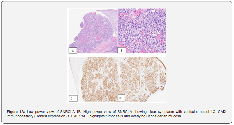
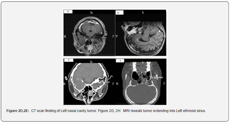
We reported an unusual case of a woman with a sinonasal tumor that mimicked, histologically, metastatic RCC. A-49-year-old women with no significant past medical history presented with 2 months history of headache and recurrent epistaxis for 2 months. She visited ER and 4.5x 4x 2cm high attenuation lesion in the left nasal cavity which is extending into the left ethmoid sinuses and bulging towards the left maxillary sinus on CT scan. The lesion raises the suspicion of polyp or hematoma, and she received treatment with nasal packing. MRI finding revealed 3.0 x 2.6 x 1.5 cm heterogeneously enhancing mass centered in the left middle turbinate and left nasal cavity, internally convoluted cerebriform pattern of enhancement. The mass obstructed the left osteomeatal unit, frontoethmoidal and sphenoethmoidal recesses and associated with mucosal thickening throughout the paranasal sinuses except right sphenoid sinus. She denies any urinary symptoms such as hematuria, abdominal pain and renal masses [3]. Negative lymphadenopathy on physical examination and MRI. CT abdomen finding discovered no suspicious masses in the kidneys or elsewhere in the abdomen or pelvis or suspicious lymphadenopathy. Biopsy of the lesion was taken, and histological findings disclosed carcinoma with clear cell features involving sinonasal submucosa, diffusely infiltrating between the seromucinous glands, and associated with rich vascular network. The tumor cells show nests to sheets of polygonal cells bearing clear to slightly eosinophilic cytoplasm and vesicular nuclei with prominent nucleoli. Extensive immunohistochemical work up showed positivity with CAIX, AE1/AE3, patchy positivity with CK7 and inhibin. The tumor cells are immunonegative for S100, HMB45, PAX-8, P63, CD68, CD163, chromogranin, synaptophysin, SMA, D2-40, ERG, beta-catenin [4]. The proliferation index is low and 10%. Mucicarmine stain is negative. Our diagnosis is clear cell carcinoma favor low-grade sinonasal renal cell like adenocarcinoma(SNRCLA). The case had been sent out for second opinion and outside Pathology agreed with our diagnosis. Wide excision with negative margins has been performed and postoperative recovery was uneventful. 2 months follow-up after wide excision is no recurrence of tumor.

Case Discussion
Salivary clear cell carcinoma SCCC is uncommon salivary gland tumor and slightly female preponderant (female to male ratio 1.6:1). SCCC usually occurs in the sixth or seventh decade of life, the palate and base of tongue being the most common primary sites of tumor [5]. Sinonasal location is very unusual location and assumed developed from minor salivary glands in locations. Sinonasal renal cell like carcinoma SNRCLA is a rare, low-grade neoplasm that bears no resemblance to any other sinonasal primary tumor. The tumor also has a female predominance of 2:1. The nasal cavity is the most common site of involvement, followed by paranasal sinuses and nasopharynx. Histologically, the tumor cells have monomorphous cuboidal to columnar glycogen-rich clear cells lacking mucin production [6]. The cellular cytoplasm may be “crystal clear” or may be slightly eosinophilic arranged in follicular/glandular structures to entirely solid and nonglandular, glandular pattern being most dominating in reported cases (Figure 1A,1B,1C,1D). Maharaj et al reported a case of SNRCLA arising from patient with Von Hippel Lindau syndrome and tumor cells are arranged in follicular pattern with focal colloid like secretions. One reported case of SNRCLA was evident for prominent foci of oncocytoid and basophilic tumor cells. SNRCLA has no necrosis with limited pleomorphism and scarce mitotic activity. Hyalinized stroma and evidence of myoepithelial differentiation are not features of SNRCLA. In SCCC, the tumor cells are arranged in nests, cords, and trabeculae. Amyloid like hyalinization can variably accompany SCCC. Our reported case has clear cytoplasmic tumor cells arranged in nests to diffuse sheets [7-10]. The epithelial markers including CK7, CK19, CK14, CAM5.2, and EMA are positive in SCCC and usually express p63, p40 and CK5/6.1 In SNRCLA, most of the reported cases have been positive for CKs7 variable expression of vimentin and S100. Iv Robust cytoplasmic and membranous CA-IX expression was seen in 6 out of 7 SNRCLAs. All SNRCLAs examined for Human melanoma black-45 (HMB45), Melan A, and smooth muscle action (SMA) are negative, thus excluding balloon cell variant of melanoma and PEComa, respectively. As our case described strong immunoreactivity with CAIX, AE1/AE3 and patchy positivity with CK7 and inhibin while negative for HMB45 and S100, the diagnosis is leaning towards the SNRCLA rather than SCCC. In addition, PAX-8 non-immunoexpression and negative imaging findings of abdominal masses and normal renal imaging entirely argue against the possibility of metastatic clear cell renal cell carcinoma (Figure 2D,2E,2G,2H).

Conclusion
We reported a rare case of sinonasal renal cell like carcinoma SNRCLA which is a low-grade malignancy. Being clear cell histomorphology, wide differential includes SCCC and metastatic renal clear cell carcinoma. Our SNRCLA case developed in the left nasal cavity extending towards ethmoid sinus and bulging towards the maxillary sinus favoring SNRCLA rather than SCCC. Despite strong and diffuse CAIX expression, negative immunoreactivity with PAX-8 and negative imaging findings for abdominal masses impede inclining towards metastatic renal clear cell carcinoma. The specific diagnosis is crucial as SNRCLA has a very low malignant potential and excision is sufficient for treatment without necessitating adjuvant radiotherapy and central neck dissection.
References
- WHO Classification of Tumours Editorial Board. Head and neck tumours [Internet; beta version ahead of print] (2022). Lyon (France): International Agency for Research on Cancer.
- Muir CS, Nectoux J (1980) Descriptive epidemiology of malignant neoplasms of nose, nasal cavities, middle ear and accessory sinuses. Clin Otolaryngol Allied Sci 5(3):195-211.
- Dulguerov P, Jacobsen MS, Allal AS, Lehmann W, Calcaterra T (2001) Nasal and paranasal sinus carcinoma: are we making progress? A series of 220 patients and a systematic review. Cancer 92(12): 3012-3029.
- Zur KB, Brandwein M, Wang B, Peter S, Ronald G, et al. (2002) Primary description of a new entity, renal cell-like carcinoma of the nasal cavity: van Meegeren in the house of Vermeer. Arch Otolaryngol Head Neck Surg 128(4):441-447.
- Daniele L, Nikolarakos D, Keenan J, Schaefer N, Lam A (2016) Clear cell carcinoma, not otherwise specified/hyalinising clear cell carcinoma of the salivary gland: the current nomenclature, clinical/pathological characteristics, and management. Crit Rev Oncol Hematol 102: 55-64.
- Wu J, Fang Q, He YJ, Chen WX, Qi YK, et al. (2019) Local recurrence of sinonasal renal cell-like adenocarcinoma: A CARE compliant case report. Medicine (Baltimore) 98(7): e14533.
- Brandwein-Gensler M, Wei S (2014) Envisioning the next WHO head and neck classification Head Neck Pathol 8 (1): 1-15.
- Maharaj S, Seegobin K, Wakeman K, Simone C, Kevin P (2022) Sinonasal renal cell-like adenocarcinoma arising in von Hippel Lindau (VHL) syndrome. Oral Oncol 125: 105705.
- Shen T, Shi Q, Velosa C, Shuting B, Lester T (2015) Sinonasal renal cell-like adenocarcinomas: robust carbonic anhydrase expression. Hum Pathol 46(11): 1598-1606.
- Storck K, Hadi UM, Simpson R, Ramer M, Brandwein-Gensler M (2008) Sinonasal renal cell–like adenocarcinoma: a report on four patients. Head Neck Pathol 2 (2): 75-80.


