Abstract
Athletes in the outdoors are at risk of being bitten by a tick. This can sometimes lead to serious illnesses. Therefore, it is necessary to remove a tick from the skin as quickly as possible. Knowledge of the anatomy of a tick (Ixodes ricinus) is helpful in avoiding complications such as the breakage of mouthparts or the squeezing of the tick’s intestinal contents into the wound during removal. The method presented here to quickly remove a tick using rotating movements with a wooden stick, the end of a match, a pencil, or something similar has been successfully performed by the authors in over 70 cases.
Keywords: Ixodes Ricinus; Mouthparts; Borrelia; Ticks bore
Introduction
The attack of ticks is not uncommon among outdoor athletes, adventurers, or mountaineers. In this case, there is sometimes a risk of infection. Tickborne diseases that affect patients in the United States include Lyme disease, Rocky Mountain spotted fever, Ehrlichiosis, Anaplasmosis, Babesiosis, Tularemia, Colorado tick fever, and tickborne relapsing fever [1]. In West Africa the presence of Rickettsia, Borrelia, Bartonella, Coxiella burnetii, Theileria, Hepatozoon and Crimean–Congo haemorrhagic fever viruses in humans, animals or ticks was reported too [2]. Molecular tools show a wider distribution of various types of Borrelia, Anaplasma, Rickettsia and Babesia on the European continent, which are responsible for causing diseases of great medical and veterinary concern [3]. Human infections with mainly Borrelia burgdorferi, Rickettsia, Anaplasmataceae, Babesia spp., Borrelia miyamotoi were found in Austria [4]. In a study of 75 patients with fever in North-Eastern Switzerland with a tick bite, bacteria were detectable in 48% (Lyme borreliosis. Tick-borne encephalitis, Granulocytic Ehrlichiosis, Babesia mikroti infection) [5]. Ixodes ricinus is one of the most important tick species in Europe because of its role as a vector for Lyme disease and tick-borne encephalitis [literature s. 6,7]. To reduce the risk of infection, the tick should be removed from the body as quickly as possible. Because the Borrelia are located in the intestine of the tick, the tick has to suck for a longer time before the pathogen is transmitted. The risk of infection increases after a sucking time of more than 12 hours [8,9]. If the tick is removed early, the risk of transmission is therefore very low. In the prevention of Lyme disease, it is of great importance to remove a tick from the skin as fast as possible without manipulating the tick [9].
How Ticks will fix their head in the skin
Ticks bore into the skin by moving the paired mouthparts (chelicerae) like a small saw.
Sample
The sample consists of the totality of the games from the FIBA Basketball World Cup 2019, held in China. National teams from around the world participated, having previously played qualifying rounds in their respective continents to earn a spot in this international tournament. The World Cup comprised 92 games, divided into 48 first-phase games, 32 second-phase games, and 12 final-phase games. A total of 32 teams were observed: 12 from FIBA Europe, 5 from FIBA Africa, 8 from FIBA Asia, and 7 from FIBA Americas. These teams, representing different regions, qualified for the 2019 World Cup. A total of 16,668 possessions were observed, along with other in-game variables, requiring nearly 184 hours of work. We chose this tournament because we believe it is the best competition for studying various performancerelated variables in high-level basketball. Focusing on a single league, such as the ACB or NBA, could lead to inaccuracies, given that each continent — and even each league - has its own unique characteristics. The FIBA World Cup 2019 featured a total of 32 teams, including 12 from FIBA Europe, 5 from FIBA Africa, 8 from FIBA Asia, and 7 from FIBA Americas. Each team played a minimum of 5 games (those that did not qualify for the knockout rounds) and a maximum of 8 games (those that reached the final stage of the tournament).
The mouthparts of a tick (Ixodes ricinus) consist of 3 anatomical parts:
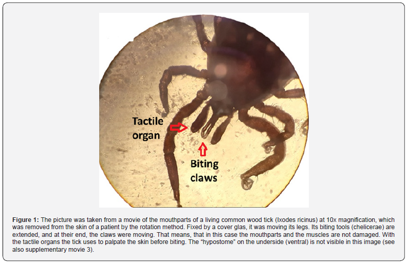
1. The two chelicerae (a pair of telescopic spear-like structures) have movable cutting tools at the end with saw-like teeth. These cheliceral teeth cut - and also firmly hold onto the skin of their hosts [10]. The intrinsic muscle in the cheliceral base (which extends into the fingers via asymmetrically arranged tendons) is responsible for their movement. With this, they saw a narrow path into the skin. Then they hook onto the sides and now pull the hypostome (s. below) into the wound. This is caused by the simultaneous activity of both chelicerae, which then fix their serrated end-claws in the skin. Then, to penetrate deeper into the skin, the telescope-like cheliceral shafts are pushed forward by hydrostatic pressure and the procedure starts again. This movement resembles pulling both arms in breaststroke swimming [7]. The retraction of the chelicerae occurs through external muscles that originate at the posterior edge of the scutum (hardened back plate of the tick) and extend into the ends of the cheliceral base [10]. Because these muscles attach above the tick`s head to the scutum, one should be careful not to injure them when removing ticks.


2. The hypostome (a toothed lower jaw) of the I. ricinus tick is firmly attached to the head of the tick on the underside. It resembles a half-tube and is fringed by rows of prominent, recurved denticles. This half-tube channels the flow of food into the oral opening of the tick and, vice versa, its saliva into the skin of the host. As mentioned, before it will be inserted into the skin. In addition, the hypostome serves as a third mouthpart to stabilize and to anchor the tick’s mouthparts in the skin of its host [10]. The trick is firmly anchored in the blood pool under the skin by these 3 mouthparts, similar to an anchor bolt in the wall. While the tick has to exert a lot of effort to penetrate the skin, it has on the other hand, only to activate a few muscles when it lets go. Therefore, it is important to find a method that allows the ticks to actively retract their mouthparts. If these muscles are responsible for causing the tick to detach from the skin, it would make sense to protect them when removing the tick. This means that one should not press hard on the mouthparts of the ticks. In particular, the pressure above the mouthparts (on the top of the head and around the lower scutum) also should be avoided. However, this is exactly where tools like pointed tweezers, tick cards, or tick levers are applied. It would be better to use such tools (like levers or tick cards) only from the underside of the tick, which can be easily recognized by its crawling legs. At the underside the (ventral) head and mouthpart (hypostome) are very stable and not so sensitive to pressure and tension. When using tweezers, it could be better to grasp the tick from both sides rather than from the top and bottom. Or even better, grasp it above the frist pair of legs. If you fix the tick with tweezers or a lever, or with a tick card, and then twist it, the mouthparts and/or muscles can be cut off. Therefore, it makes sense to use these tools only for pulling out – and not to rotate them.
Methods to remove a tick
Ticks have been accustomed for thousands of years to being pulled from animals during grooming. Therefore, they have developed a strong anchoring through their mouthparts in the skin that counteracts being pulled out. As a result, it requires quite a bit of force to pull out the tick. Thereby is a risk that the tick’s mouthparts may break - or that pressure on the abdomen or on salivary glands may lead to the release of pathogens. To remove a tick from the skin, various methods are published. The following instruments are used: Tick key, tick twister, tick tweezer, tick loop, tick card or other tick kits. The problems with these methods are that the appropriate device is not always at hand, or that strong pressure or tension must be exerted against the fixation of the mouth parts of the tick, or that there are useless because the tick is stuck in a deep fold of skin. Especially if you grasp the body of a tick with normal tweezers (that means broader tweezers) or similar device, the pressure on the abdomen of the tick can become too high, causing the mouthparts to break off - or the tick could empty its stomach contents into the wound. Sharp tweezers with pointed tips do not squeeze the body of the tick, if you use it from the side and not from above, but nevertheless the head and the mouthparts may remain spread in the skin and break off. Therefore, it is necessary to use this special tweezer, tick cards, or tick levers with caution and some patience. The pressure or pull on the tick should not be too abrupt and too strong. With small, rocking movements, you often can slowly loosen the tick. In contrast to different opinions [11], under no circumstances should the tick be sprinkled with oil, alcohol, disinfection or glue before removal [12]. This would irritate the animal unnecessarily and could lead to it releasing its saliva or stomach contents - and thus possible infectious agents. This can increase the chance of infection
The rotation method: A simple and safe method to remove a tick from the skin
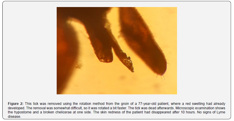
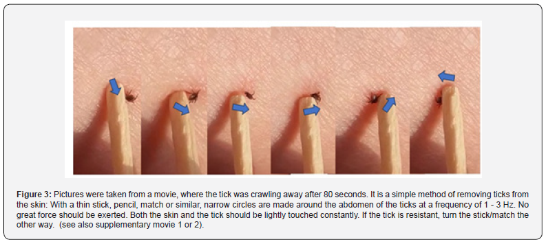
The better way is to convince the tick to leave its feeding place in the skin. A simple method for removing a tick will be presented here, which the author has used successfully for 25 years on more than 70 patients. In most cases no part of the tick (mouthparts or head) remains in the skin and the tick moves away. It is a painless method with only a few complications: Only 2 patients subsequently showed skin redness some days later that corresponded to Lyme disease. In this method, the abdomen of the tick is constantly rotated with a wooden stick or the end of a match or a pencil or similar. In almost all patients the corresponding tick removed alive within 20 seconds until to 2 minutes from his feeding place in the skin. Even in difficult skin folds - or when treating small ticks - this method was successful. The stick/match should be held slightly inclined so that there is as much and often contact as possible with the abdomen of the animal, while this is twisted to one side multiple times. This must be done repeatedly and continuously. It may happen that one accidentally presses on the abdomen. Therefore, the pressure on the skin should not be too strong and the rotation speed should be between 1-3 Hz. After about several seconds of rotation, the tick gives up and crawls away. The reason for this behavior might be that the mouth tools of the tick are paired and a constant rotation is unpleasant for the animal. A similar method using cotton swabs was recently published on the internet [13]. Some of these crawling ticks were then examined alive under a Zeiss microscope by placing them on a glass carrier and fixing them with a cover glass. With a tenfold magnification you could then see how they move their legs and mouthparts. There were no signs that any part of the mouth was defective. Here is also presented one of the few dead ticks that were removed using this method. It shows a broken chelicerae under microscopic examination.
How to use the rotation method to remove a tick smoothly:
The first requirement is not to stress the tick and not to kill it,
otherwise the mouthparts will remain in the skin.
• Do not clean the area around the tick bite with alcohol or
other chemical substances before the tick is away.
• Do not kill the tick by ice, alcohol or other chemical
substances before it is out of the skin
• Take a thin stick, or the end of a match, or a pencil, or
an ear cleaner, and position it on the skin - very close to the tick.
• Now start with small circles on the skin around the tick’s
body with 1 – 3 Hz.
• Thereby the stick/match frequently should have contact
to the abdomen of the tick. In this manner the abdomen of the tick
often has to be lightly touched and rotated slightly
• Continue these rotations several times with no - or only
gentle - pressure on the skin.
• Do not stress the tick by high pressure or tension with
instruments or your finger
• If the tick is resistant, turn the stick/match the other
way round
• The tick will almost always withdraw from the skin after
20 - 120 seconds and crawl away. Then other rules are provided
for the tick
• Now the wound can be disinfected, for example with
alcohol
• If signs of another illness (reddening of the skin, fever,
etc.) occur, it is necessary to see a doctor
• In risk areas, it makes sense to see a doctor at the same
time to rule out a disease. You should also take the dead tick with
you for analysis [12].
Conclusion
Removing a tick from the skin by continuous rotation of its abdomen is a simple and safe method. The mouth parts of a tick have been trained over many thousands of years to act against external tension - but apparently not to withstand continuous rotation. If you carry out such a rotation gently and without much force, the tick itself seems to loosen its bite in the skin. This method has several advantages: it is easy and does not require any special tools, it can quickly detach the tick, no tick parts remain in the body and the stress on the tick seems to be less than with mechanical or chemical action. Thus, the risk of infection seems be lower. Another scientific investigation with microscopic examination of the mouthparts of the detached tick and a statistic on subsequent infections would be useful to accurately assess the various methods of tick removal.
References
- Emma J Pace, Matthew O`Reilly (2020) Tickborne Diseases: Diagnosis and Management. American Family Physician 101(9): 530-540.
- Adama Z, Diarra, Patrick Kelly, Bernard Davoust, Philippe Parola (2023) Tick-Borne Diseases of Humans and Animals in West Africa. Pathogens 12(11): 1276.
- Alberto Moraga-Fernandez, Clara Munoz-Hernandez, Marta Sanchez Sanchez, Isabel G Fernandez de Mera, Jose de la Fuente (2023) Exploring the diversity of tick-borne pathogens: The case of bacteria (Anaplasma, Rickettsia, Coxiella and Borrelia) protozoa (Babesia and Theileria) and viruses (Orthonairovirus, tick-borne encephalitis virus and louping ill virus) in the European continente. 2023.Veterinary Microbiology journal 286: 109892.
- Mateusz Markowicz, Anna-Margarita Schötta, Dieter Höss, Michael Kundi, Christina Schray, et al. (2021) Infections with Tickborne. Pathogens after Tick Bite. Austria, 2015-2018. (April 2021). Emerging Infectious Diseases 27(4): 1048-1056.
- D Baumann, N Pusterla, O Peter, F Grimm, PE Fournier, et al. (2003) Fever after a tick bite: Clinical manifestations and diagnostic after acute tick-born infections in North-Eastern Switzerland. (2003). Dtsch Med Wochenschr 128(19): 1042-1048.
- Arda Cem Kuyucu, Olcay Hekimoglu (2024) Predicting the distribution of Ixodes ricinus in Europe: integrating microclimatic factors into ecological niche models. Published online by Cambridge University Press.
- Marie Vancová, Tomáš Bílý, Ladislav Šimo, Jan touš, Petr Horodyský, et al. (2020) Sonenshine, Libor Grubhoffer & Jana Nebesářová: Three-dimensional reconstruction of the feeding apparatus of the tick Ixodes ricinus (Acari: ixodidae): a new insight into the mechanism of blood-feeding. Scientific RepoRtS.
- Sibylle Rahlenbeck, Volker Fingerle (2014) Medizinreport. Ixodes ricinus: Wie man sich vor Zecken schützt. Aerzteblatt.
- Robert Koch Institut Berlin.
- Dania Richter, Franz-Rainer Matuschka, Andrew Spielman, L Mahadevan (2013) How ticks get under your skin: insertion mechanics of the feeding apparatus of Ixodes ricinus ticks. Proc Biol Sci 280(1773): 20131758.
- Tick removal – WikEM.
- Government of Canada: Lyme disease: Monitoring.
- David Priest (2025) Tick Season Has Arrived. Try This Trick to Safely Remove a Tick Without Tweezers 12: 30.


