Matriderm and Split-Thickness Skin Graft for Burn Contractures of the Hands
Cezar Buzea1*, Ileana Boiangiu1, Cristina Brezeanu1, Manuela Popa2, Cristina Huian1 and Maria Dinu1
1 The Clinical Emergency Hospital for Plastic Reconstructive Surgery and Burns, Bucharest, Romania
2University of Medicine and Pharmacy “Carol Davila”, Bucharest, Romania
Submission: August 08, 2020;Published: August 17, 2020
*Corresponding author: Cezar Buzea, The Clinical Emergency Hospital for Plastic Reconstructive Surgery and Burns, Bucharest, Romania
How to cite this article: Cezar B, Ileana B, Cristina B, Manuela P Maria D.Matriderm and Split-Thickness Skin Graft for Burn Contractures of the Hands. JOJ Orthoped Ortho Surg. 2020; 2(5): 555596. DOI: 10.19080/JOJOOS.2020.02.555596
Abstract
Scar contracture and hypertrophic scar of the hands are common late complications encountered in burn patients which result in imperfect wound healing. Burns often cause full-thickness skin defects which should be ideally reconstructed with full-thickness skin grafts or flaps. In burn victims with large TBSA these options are limited due to the lack of sufficient donor site. Nonetheless, the dermis can be partially reconstructed using split-thickness grafts which have become the mainstay of treatment in extensive burns. Although the split-thickness skin grafts are versatile, their use on the face or on the hands has undesirable outcomes such as poor color match, poor skin elasticity, graft contracture with limitation of joint movement and hypertrophic scars, due to a discontinuous healthy dermal layer. To compensate for this, numerous artificial dermal substitutes have been developed such as Matriderm.
In this paper we report the case of a patient with severe hypertrophic scars following burn injury on the dorsal aspect of the hands with important functional limitation in which we resurfaced the entire anatomical region using Matriderm and split-thickness skin graft in a single stage. Each hand was operated on separately with good functional and aesthetical results with a maximum follow-up of 12 months.
Keywords: ADM; Burn contracture; Hypertrophic scars
Introduction
Wound healing is a complex process involving many mechanisms with the goal to restore the skin barrier. Deep partial thickness or full-thickness burns cause a loss of the dermis which will be replaced by granulation tissue and then epithelized from skin grafts or from the wound edges. However, a healthy dermal layer for the wound cannot be provided by the patchy dermal remnants of the dermal papillae supplied by split-thickness skin graft or by the scar tissue. Since the function and quality of the scar tissue depends largely on orientation and the amount of the elastic fibers, burn scars tend to be of poor quality, often unstable, hypertrophic, or contracted limiting function.
The gold standard for reconstructing the full thickness defects both in the acute setting of burns and in scar resurfacing remains the full-thickness skin grafts and the flaps, but their scarce availability remains an issue [1-2]. In order to overcome these shortcomings and to improve the quality of the dermal wound bed, acellular dermal matrix has been developed. The acellular dermal matrix provides a stable scaffold for partial-thickness skin graft in which the fibroblasts, macrophages, lymphocytes and capillaries from the wound bed infiltrate and vascularize the matrix and the skin graft.
There are many types of acellular dermal matrix commercially available. One of the most popular is AlloDerm which contains both collagen and elastin fibrils which prevents wound contraction and provides good elasticity for the new skin. However, this is a human-derived product and has its limitations. Matriderm is an artificial acellular dermal matrix designed to overcome the drawbacks of the human-derived products . It consist of 3D pore structure naturally cross-linked, with a pore size that encourages cell migration. The bovine collagen supports guided regeneration and the bovine solubilized elastin stimulates early elastin synthesis and neoangiogenesis. Once the fibroblasts get activated inside the matrix they start producing the components of the extracellular matrix: new collagen, elastin and hyaluronic acid. Their activity is modulated by Matriderm, the elastin synthesis being up-regulated while the collagen synthesis slows down. Also, elastin stimulates the angiogenic phenotype of the endothelial cells which start to build up new capillaries [3-4]. Moreover, due to its hemostatic properties, Matriderm reduces the risk of hematoma and it can be used in a single or double stage procedure.
Case Report
A 26-year old male patient was admitted to the ICU in our clinic with 20 % TBSA deep partial-thickness and full-thickness burn injuries involving the face, the upper limbs, the trunk and the ankles sustained after an indoor explosion. He underwent staged excisions and grafting. The lesions on the face were grafted using full-thickness grafts. Most of the dorsal aspect of the hand healed by secondary intention and in about 4 weeks some small granular areas were grafted with split-thickness skin graft. Twelve months later, the patient presented to our clinic with extensive hypertrophic scars.
The examination of the left hand revealed the following: extensive hypertrophic scarring on the dorsal aspect extending from the web spaces to the wrist joint with small areas of integrated pliable skin graft; normal capillary refill and sensation; nails deformity secondary to contraction of the eponichium; irreducible boutonniere deformity of the fifth finger; limited abduction of the second to the fifth finger with normal adduction; both passive and active motion of the wrist and thumb within normal limits; active and passive flexion and extension of the PIP and DIP joints of the second to forth fingers within normal limits; normal active and passive extension of the MP joints with important limitation of MP flexion (45˚). The findings on the examination of the right were similar except that the right fifth finger did not have a boutonniere deformity. Staged resurfacing of the entire dorsal aspect of the hands was decided [5-6]. Firstly, the left hand was operated on. We performed excision of the scar from the wrist joint to the level of the PIP joint on the second to the fifth finger and to the level of the MP joint on the thumb with preservation of the dorsal veins and release of MP and web space contractures. On the fifth finger we performed cup and cone arthrodesis of the PIP joint in an angle of 45˚. After meticulous hemostasis a 1 mm Matriderm sheet was applied and a thin split-thickness skin graft including in the web spaces. The wound was dressed with paraffin gauze and the dressing was removed 5 days postoperatively. Graft take at day 5 was about 95%.
Three months later, resurfacing on the entire dorsal aspect of the right hand was performed. We excised the scar from the wrist joint up to the PIP joints on the second and third fingers and to the level of the MP joints on the thumb, the fourth and the fifth finger also preserving the dorsal veins and releasing the MP and web space contractures. One mm Matriderm and thin split-thickness graft were applied. The wound was dressed in the same way and at day 5 graft take was 99%.
At the time of the second surgery, the scar was excised and a biopsy was taken from the area previously grafted using Matriderm. In both instances, a thin split-thickness graft harvested from the thigh was used, the donor site healing within 10 days. Histopathological aspects of the initial scar tissue show thick, dense, collagen bundles with zonal verticalization and disordered disposition, whereas graft biopsy exposes more orderly, thinner and parallel distribution of collagen in the dermis ((Figure 1)–The microscopic examination reveals dermal architecture improvement).

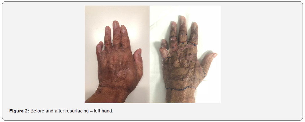
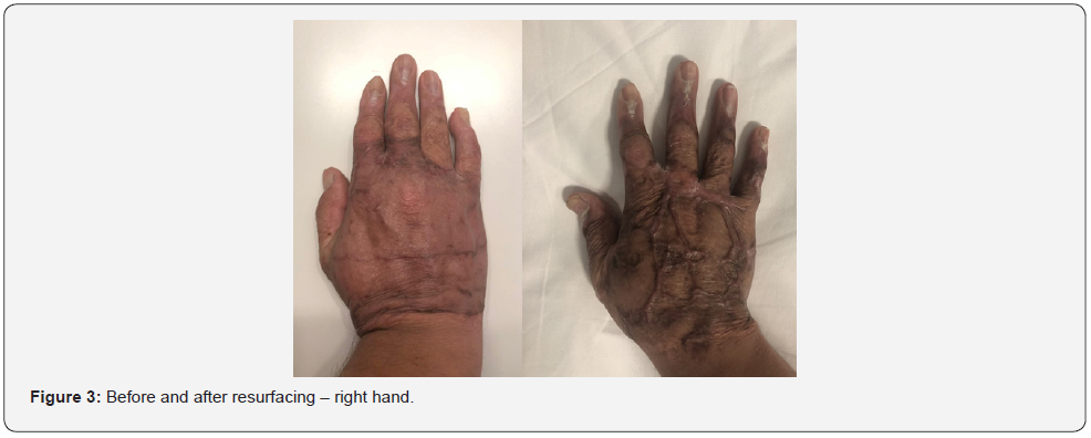
Scar quality was evaluated clinically at 12 months postoperatively on the left hand respectively at 9 months postoperatively on the right hand using both the Vancouver Scar Scale and the range of motion of the joints. The modified Vancouver Scar Scale, usually used to evaluate the burn scars, was employed to assess subjectively the quality of the scars. The old hypertrophic scar scored 12 points out of 13 while the scar obtained using Matriderm scored only 4 points ((Figure 2 & Figure 3) – After surgery, the patient regained good hand function with good scar quality). Also, the contractures of the web spaces were completely released the patient regaining the ability to fully abduct the finger and about 85˚ of MP joint flexion. The donor site scar was inconspicuous. It is also worth mentioning that the patient strictly adhered to the recommendation of wearing pressure garments both after healing and after scar revision.
Discussion
The use of acellular dermal matrix such as Matriderm both in burns and scar revisions reduces donor site morbidity since it requires a thin split-thickness graft and leads to a stable and pliable scar by providing a dermal scaffold for wound healing.


We believe that acellular dermal matrix like Matriderm might be a cost-effective alternative in the treatment of full-thickness burns. Its relatively high cost can be compensated for by the reduction in hospitalization time considering that it can be used in a single stage procedure, it has a graft take rate and prevents scar contracture which would warrant further surgeries. Moreover, it is also worth using it in scar revisions since the final scar does not diminish the patient’s work capacity.
There are, however, some limitations of our observations. We assessed the quality of the scar mostly subjectively using the VSS and objectively using the ROM of the joints. There are other devices such as Cutometer, Corneometer, Tewameter, and Mexameter which are not available in our clinic and might add some objective data in the assessment of scar quality. Also, it might be interesting to compare the results of different brands of acellular dermal matrix.
Compliance with Ethical Standard
The authors received no financial support for the research, authorship, and/or publication of this article. All procedures performed in studies involving human participants were in accordance with the ethical standards of the institutional and/or national research committee and with the 1964 Helsinki declaration and its later amendments or comparable ethical standards. All procedures performed in studies involving human participants were in accordance with the ethical standards of the institutional and/or national research committee and with the 1964 Helsinki declaration and its later amendments or comparable ethical standards. Informed consent was obtained from all individual participants included in the study.
References
- Yannas IV, Burke JF, Gordon PL et al. (1980) Design of an artificial skin II Control of chemical composition. J Biomed Mater Res 14: 65-81.
- Juhasz I, Kiss B, Lukacs L et al. (2010) Long-term followup of dermal substitution with acellular dermal implant in burns and postburn scar corrections. Dermatol Res Pract :210150.
- Lamme EN, de Vries HJ, van Veen H, Gabbiani G, Westerhof W, Middelkoop E (1996) Extracellular matrix characterization during healing of full-thickness wounds treated with a collagen/elastin dermal substitute shows improved skin regeneration in pigs. J Histochem Cytochem 44:1311-1322.
- Cervelli V, Brinci L, Spallone D et al. (2011) The use of MatriDerm (R) and skin grafting in post-traumatic wounds. Int Wound J 8(4): 400-405.
- Oliveira GV, Chinkes D, Mitchell C, Oliveras G, Hawkins HK (2005) Objective assessment of burn scar vascularity, erythema, pliability, thickness, and planimetry. Dermatologic Surgery 31(1): 48-58.
- Ryssel H, Gazyakan E, Germann G et al. (2008) The use of MatriDerm in early excision and simultaneous autologous skin grafting in burns: a pilot study. Burns 34(1): 93-97.


