Chondromyxoid Fibroma of The Distal Clavicle: Report of An Additional Case at Very Unusual Anatomic Location
Jean Marie Vianney Hope*1, Jean Claude Sane2, Souleymane Diao1, Francis Mugabo3, Jean Paul Bitega4, Anselme Noaga Nikiema1, Amadou Ndiassé Kassé1, El Hadji Souleymane Camara1, Mouhamadou Habib Sy1
1Orthopedics and Trauma Surgery, Department of Grand-Yoff General Hospital, Cheikh Anta Diop University of Dakar, Senegal
2Gaston Berger University of Saint Louis, Senegal
3Rwanda Military Hospital, University of Rwanda, East Africa
4PLA National Defence University, Beijing, China
Submission: September 26, 2018; Published: October 09, 2018
*Corresponding author: Jean Marie Vianney Hope, Orthopedic Surgeon Orthopedics and Trauma Surgery Department Grand-Yoff General Hospital, Dakar, Senegal, Tel: +221705899741; Email: hopejmv@gmail.com
How to cite this article: Hope J M V, Sane JC, Souleymane D, Mugabo F, Bitega JP, Nikiema AN, Kassé AN, El Hadji S C, Sy M H. Chondromyxoid Fibroma of The Distal Clavicle: Report of An Additional Case at Very Unusual Anatomic Location. JOJ Orthoped Ortho Surg. 2018; 2(2): 555581. DOI: 10.19080/JOJOOS.2018.02.555581
Abstract
Chondromyxoid fibroma is a relatively rare, benign cartilaginous bone tumor accounting for <1% of primary bone neoplasms, usually involving bones of the lower extremity during the second or third decades of life. We report one such case occurring in one-third distal end of the right clavicle of a 7-year-old schoolgirl. After the definitive diagnosis made by histological examination, the patient underwent curettage, followed by phenolization and synthetic processed bone grafting. The patient made an uneventful post-operative recovery. She has been followed up for 5 years to date, with no evidence of recurrence.
Keywords: Chondromyxoid fibroma; Clavicle; Histological examination; Curettage.
Abbreviations: CMF: Chondromyxoid Fibroma; CT: Computed Tomography; OPD: Out Patient Department; WHO: World Health Organization
Introduction
Chondromyxoid fibroma (CMF) is a rare benign cartilaginous tumor accounting for <1% of primary bone neoplasms [1-4]. It was first described as a distinctive clinical entity by Jaffe and Lichtenstein in 1948; formerly it was classified as myxoma or a myxomatous variant of giant-cell tumour, or mistaken for a malignant lesion, especially chondrosarcoma, chondromyxosarcoma or myxosarcoma [5]. The 2002 World Health Organization (WHO) classification of bone and soft tissue tumors [6] defines CMF as ‘‘benign tumor characterized by lobules of spindle or stellate shaped cells with abundant myxoid or chondroid intercellular material’’. It usually presents during the second or third decades of life [7-10]. Most CMF cases occur in the bones of the lower extremities and has a tendency for the metaphyseal region of the distal femur and proximal tibia, followed by the foot [11-13]. Other frequent sites are the pelvis, spine and sternum [14,15]. In general, the clavicle is a rare site for primary bone tumors, approximately 80% of which are malignant [16]. CMF of the clavicle is extremely rare. To our knowledge, only seven cases have been previously mentioned in the English language literature [13,16-21]. We report the clinical presentation, imaging and pathological findings of an additional patient with CMF arising from one-third distal end of the right clavicle.
Case Presentation
The parents of a 7-year-old schoolgirl noticed swelling over one-third distal end of the right clavicle that had slowly increased in size over 5 months. The swelling was associated with a constant slight dull pain. Her parents initially attributed this pain to carrying of a school bag that was often slung over her right shoulder. She was previously healthy without trauma history of the shoulder. She was then brought to the hospital outpatient department (OPD) for evaluation of her condition.
On physical examination, a slightly tender, fixed, bony, hard mass measuring 2.9 cm x 1.3 cm was palpable over the acromioclavicular joint of the right shoulder. The overlying skin was normal. The range of motion of shoulder was slightly restricted due to pain. No other abnormalities were revealed by the full systemic review. All laboratory tests (including full blood count, electrolytes, erythrocyte sedimentation rate, C-reactive protein) were within normal limits.
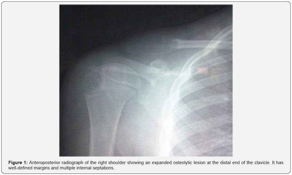
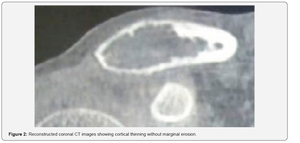
Conventional radiographs (Figure 1) showed an expanded radiolucent osteolytic lesion at the distal end of right clavicle. The lesion had well defined margins with internal septa. No periosteal reaction or associated soft tissue mass was present. Reconstructed coronal Computed Tomography (CT) image (Figure 2) showed that the lesion had a thin sclerotic rim and endosteal scalloping measuring 2.9 cm x 1.3 cm. The margins of the lesion were preserved without erosion. The acromioclavicular joint was not involved. No calcification or associated soft tissue mass were present.


Open biopsy was then carried out. On inspection during the operation, the cortex surrounding the tumor was thinned and expanded, rendering a diagnosis of a chondroid lesion without overt malignancy but deferring the final decision to paraffin histology. The histological sections showed mitochondrion lobules with increased cellularity in the peripheral area. The tumors were characterized by components of chondroid, myxoid and fibrous tissues in various proportions. Multiple stellate cells with compact nuclei were seen within the chondromyxoid components. Giant cells and osteoid formation were also observed. Secondary changes, such as necrosis or hemorrhage, were not seen. The histological diagnosis was chondromyxoid fibroma (Figure 3).

The tumor was curetted until it was completely removed and treated with phenol to reduce possible recurrence. The cavity was then packed with synthetic processed bone graft. The patient made an uneventful post-operative recovery. She has been followed up for 5 years to date, with no evidence of recurrence (Figure 4).

Discussion
Chondromyxoid fibroma is a rare tumor which comprises less than 1% of all benign bone tumors, and it is the least common benign cartilaginous tumor of bone [22]. More than 700 cases of CMF have been previously mentioned in the modern English language literature, and only seven cases involved the clavicle [13,16-21]. This tumour occurs with an approximately equal sex ratio [10-16]. The patient age is variable, ranging from 6 to 87 years. However, most occur in the second or third decade of life with mean age of 31.1 years, with a second peak in the fifth to seventh decades [8-10,17,21,23]. A case of congenital chondromyxoid fibroma has been reported by Mendoza et al. [24] Our case is a 7-year-old female patient. The age is below the common peak incidence of CMF because of early diagnosis. The symptoms presented were pain and swelling over the affected area. It measured 2.9 cm x 1.3 cm in size at clinical presentation. This is in agreement with other investigators [21]. There may also be some restriction of movement, as was seen in our case.
The clavicle is classified as a flat bone. It has a unique development process, with almost the entire clavicle developing by intramembranous ossification. However, the sternal and acromial ends are performed in cartilage, known as endochondral ossification. Chondromyxoid fibroma arises from cells related to the epiphyseal cartilage [25]. Therefore, the location of tumors in our case may be explained by the endochondral ossification at the acromial (or distal) end.
Because they frequently have atypical pleomorphic hyperchromatic nuclei, without radiographic and clinical correlation, chondromyxoid fibroma may potentially be misinterpreted as a malignant lesion; however, mitoses are a rarity [5-7,9,10,13]. The differential diagnosis of CMF includes myxoid chondrosarcoma, CMF-like or chondroblastic osteosarcomas, fibrous dysplasia, and chondroblastomas [8]. Histologically, the three components characterizing CMF are chondroid, the myxoid and the fibrous tissue in varying proportions [13]. In our case, the well-preserved fat plane and the lack of features of an aggressive bone lesion, such as soft tissue mass or periosteal reaction, led us to an initial diagnosis of a benign rather than malignant bone tumor. Our final diagnosis depended on histological examination of the biopsy material.
CMF can be treated with curettage alone or together with bone grafting or polymethylmethacrylate. The overall rate of recurrence of chondromyxoid fibroma following curettage has been reported to be around 25%, although bone grafting or bone cement can reduce it [16]. The age of diagnosis was proposed as a factor for increased recurrence rates, with the suggestion that the reduced resistance of the pediatric thin cortices and spongiosa contributes to the aggressive behavior of the lesion [7]. The prognosis of chondromyxoid fibroma of the clavicle has not been reported owing to its rarity. Clavicular tumors can be easily discerned, and the clavicle is readily accessible for treatment. Consequently, early diagnosis and en bloc excision of the tumor can be expected, with the result that the prognosis of chondromyxoid fibroma of the clavicle can be expected to be better than the overall prognosis of chondromyxoid fibroma [20]. In our case, intraoperative histological extemporaneous reading of the biopsy sample was able to arrive at the definitive diagnosis of CMF. The management was then curettage with phenol treatment and synthetic processed bone grafting. Recurrence was not observed 5 years after the operation. However, a careful follow-up is required because of the possibility of recurrence as our patient was 7 years at the time of diagnosis.
Conclusion
We report a case of chondromyxoid fibroma arising from one-third distal end of the right clavicle of a 7-year-girl. This is an additional patient with this rare tumor in this very unusual anatomic location. The clinical and imaging features alone are non-specific, and the definitive diagnosis relies on pathological examination.
Acknowledgment
The authors thank Miss Larissa Mahoro Musaninyange for expert assistance with the manuscript.
Ethical approval
The authors thank Miss Larissa Mahoro Musaninyange for expert assistance with the manuscript.
Informed consent
Informed consent was obtained from the child’s parents to publish the information, including their photographs.
References
- Dürr HR, Lienemann A, Nerlich A, Stumpenhausen B, Refior HJ (2000) Chondromyxoid fibroma of bone. Arch Orthop Trauma Surg 120: 42- 47.
- Minasian T, Claus C, Hariri OR, Piao Z, Quadri SA, et al. (2016) Chondromyxoid fibroma of the sacrum: A case report and literature review. Surg Neurol Int 17(7): S370-S374.
- Bagewadi RM, Nerune SM, Hippargi SB (2016) Chondromyxoid fibroma of radius: a case report. Journal of Clinical and Diagnostic Research 10(5): ED01-ED02.
- Schajowicz F, Gallardo H (1971) Chondromyxoid fibroma (fibromyxoid chondroma) of bone: a clinico-pathological study of thirty-two cases. J Bone Joint Surg Br 53(2): 198-216.
- Jaffe HL, Lichtenstein L (1948) Chondromyxoid fibroma of bone: a distinctive benign tumor likely to be mistaken especially for chondrosarcoma. Arch Pathol 45(4): 541–552.
- Fletcher CDM, Unni KK, Mertens F (2002) World Health Organization Classification of Tumors. Pathology and Genetics of Tumors of Soft Tissues and Bone Lyon France IARC press.
- Bhamra JS, Al Khateeb H, Dhinsa BS, Gikas PD, Tirabosco R et al. (2014) Chondromyxoid fibroma management: a single institution experience of 22 cases. World Journal of Surgical Oncology 12:283.
- Desai SS, Jambhekar NA, Samanthray S, Merchant NH, Puri A, et al. (2005) Chondromyxoid Fibromas: A Study of 10 Cases. Journal of Surgical Oncology 89(1): 28–31.
- Rahimi A, Beabout JW, Ivins JC, Dahlin DC (1972) Chondromyxoid fibroma: a clinicopathologic study of 76 cases. Cancer 30(3): 726–736.
- Wilson AJ, Kyniakos M, Ackerman LV (1991) Chondromyxoid fibroma: radiographic appearance in 38 cases and in a review of the literature. Radiology 179(2): 513-518.
- Fahmy ML, Al Rayes M, Iskaf W, Hammouda A (1998) Chondromyxoid fibroma of the foot: case report and literature review. Foot 8(2):106- 108.
- Lersundi A, Mankin HJ, Mourikis A, Hornicek FJ (2005) Chondromyxoid fibroma: a rarely encountered and puzzling tumor. Clin Orthop Relat Res 439: 171- 175.
- Wu CT, Inwards CY, O Laughlin S, Rock MG, Beabout JW, et al. (1998) Chondromyxoid fibroma of bone: a clinicopathologic review of 278 cases. Hum Pathol 29(5): 438-446.
- Di Giorgio L, Touloupakis G, Mastantuono M, Vitullo F, Imparato L (2011) Chondromyxoid fibroma of the lateral malleolus: a case report. Journal of Orthopaedic Surgery 19(2): 247-249.
- Merine D, Fishman EK, Rosengard A, Tolo V (1989) Chondromyxoid fibroma of the fibula. J Pediatr Orthop 9(4): 468-471.
- Pattamapaspong N, Peh WCG, Tan MH, Hwang JSG, Tan PH (2006) Chondromyxoid fibroma of the distal clavicle. Pathology 38(5): 4 64- 466.
- Aggarwal A, Bachhal V, Soni A, Rangdal S (2012) Chondromyxoid fibroma of the clavicle extending to the adjacent joint: a case report. Journal of Orthopaedic Surgery 20(3): 402-405.
- Cilli F, Ozyurek S, Erler K , Kiral A (2007) Chondromyxoid fibroma of the clavicle. J Orthop Sci 12(2): 193.
- Nakazora S, Kusuzaki K, Matsumine A, Seto M, Fukutome K, et al. (2003) Case report: chondromyxoid fibroma arising at the clavicular diaphysis. Anticancer Res 23(4):3517-3522.
- Sakamoto A, Tanaka K, Matsuda S, Hosokawa A, Harimaya K et al. (2006) Chondromyxoid fibroma of the clavicle. J Orthop Sci 11(5): 533-536.
- Zillmer DA, Dorfman HD (1989) Chondromyxoid fibroma of bone: thirty-six cases with clinicopathologic correlation. Human Pathol 20(10): 952-964.
- Sami SH, Shooshtarizadeh T, Zekavat H, Bahrabadi M (2017) juxtacortical chondromyxoid fibroma of tibia. Biomed J Sci & Tech Res 1(5):1-3.
- Yaghi NK, DeMonte F (2016) Chondromyxoid fibroma of the skull base and calvarium: surgical management and literature review. J Neurol Surg Rep; 77(1): e23-e34.
- Mendoza M, González I, Aperribay M, Hermosa J R, Nogués A (1998) Congenital chondromyxoid fibroma of the ethmoid: case report. Pediatr Radiol 28(5): 339-341.
- Steiner GC (1979) Ultrastructure of benign cartilaginous tumors of intraosseous origin. Hum Pathol 10: 71-86.


