Incidence of Loose Bodies and Baker’s Cysts in the Arthritic Knee
Aatif Mahmood1*,DG Shivarathre2, Kim Howard3 and RW Parkinson4
1Wirral University Hospitals, UK
2Specialty Registrar- Trauma & Orthopaedics, Wirral University Hospitals, UK
3Advanced Arthroplasty Nurse Practitioner, Wirral University Hospitals, UK
4Consultant Orthopaedic Surgeon, Wirral University Hospitals, UK
Submission: August 08, 2018; Published: August 10, 2018
*Corresponding author: Aatif Mahmood, Wirral University Hospitals, Department of Trauma &Orthopaedics Arrowe Park Hospital, United Kingdom, Tel: 0044-7828621588; Email: aatifm@gmail.com
How to cite this article: Aatif Mahmood, DG Shivarathre, Kim Howard, RW Parkinson. Incidence of Loose Bodies and Baker’s Cysts in the Arthritic Knee. JOJ Orthoped Ortho Surg. 2018; 2(1): 555576. DOI: 10.19080/JOJOOS.2018.02.555576
Abstract
The loose bodies and Baker’s cysts are common in the arthritic knee, although the exact incidence is unknown. We performed a prospective study on 202 consecutive knee replacements performed at our institution to evaluate the incidence, shape, size and site of the loose bodies in the knee. Radiographs were examined to predict the presence of loose bodies prior to surgery. The incidence of loose bodies and Baker’s cysts was 24.3% and 46.5% respectively. The commonest site for the loose bodies was the posteromedial recess. One-thirds of the loose bodies were tethered to the soft tissues. There was no significant association of the loose body or the Baker’s cyst with respect to age, gender, BMI, degree of deformity and preoperative range of motion or previous knee surgery. The study aids the arthroscopic surgeons to locate elusive loose bodies in common sites and also appreciate the fact that a significant proportion are tethered which may result in difficult removal. Standard knee radiographs underestimate the incidence of loose bodies.
Introduction
Loose bodies and Morant Baker’s cysts in the arthritic knee are common[1-4]. The exact incidence is unknown. We have performed a prospective study to ascertain the incidence of loose bodies and Baker’s cysts in the arthritic knee. The study also recorded the morphology, site and the accuracy of radiographs in detecting loose bodies in the knee joint.
Material and Methods
The data were prospectively collected from 202 consecutive knee replacements in 198 patients performed at our institution by the senior author (RWP) between June 2008 and November 2009. The indication for knee arthroplasty was osteoarthritis (199 knees), rheumatoid arthritis (3 knees) or avascular necrosis (1 knee). 15% (31 knees) had Unicompartmental knee replacement (UKR) and the remaining 85% (171 knees) underwent Total Knee replacement (TKR). Prior to surgery, the radiographs (AP, lateral & skyline views) were examined carefully by the senior author and the presence and number of loose bodies were predicted. All patients underwent a TKR via a trivector arthrotomy and UKR via a minimally invasive medial parapatellar approach. All loose bodies were extracted and noted for number, size, shape and location. At surgery, the presence of a Baker’s cyst was recorded in addition.
Results
There were 119 female and 79 were male patients. The average age was 73.2 years (Range 49 – 95). The average BMI was 29.3 (16 -59). There were equal numbers of right and left knees. Four patients had staged bilateral knee replacements. None of the patients were judged to have symptoms specific to intra-articular loose bodies such as locking or giving way. Twenty-four patients had had previous surgical interventions in the form of arthroscopy or open meniscectomy to the knee. None of these patients had arthroscopic removal of loose body prior to knee replacement surgery.
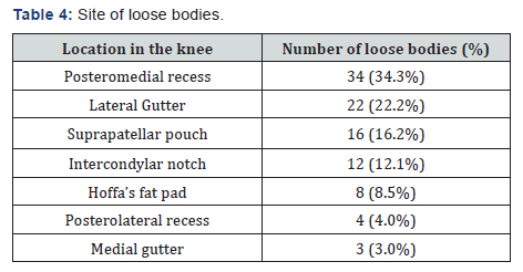
Loose bodies were found in 49 out of 202 knees giving an overall incidence of 24.3% (Table 1).The incidence of loose bodies was higher in patients who underwent TKR (26.9%) when compared to the UKR group (9.7%).Five knees were thought to contain loose bodies radiographically, but none were found intraoperatively. Furthermore, 9 patients had loose bodies which were not identified on the preoperative radiographs. The size shape and the sites of the loose bodies are shown in Tables 2-4 respectively. Majority of the loose bodies were irregular and the greatest dimension in millimetres was used to classify them. One third of the total 99 loose bodies (33.3%) retrieved were found to be tethered to the soft tissues. Twenty-one loose bodies were tethered to the capsule, 7 to Hoffa’s fat pad, 3 to the quadriceps tendon and 2 to the patellar tendon.


Baker’s cyst was noted in 94 knees (46.5%). The incidence of Baker’s cyst was higher in patients who underwent TKR (50.3%) when compared to the UKR group (25.8%)Statistical analysis using Chi-square test (SPSS version 15) revealed no significant association of the loose body or the Baker’s cyst with respect to age, gender, BMI, degree of deformity and preoperative range of motion or previous knee surgery. The presence of loose bodies and Baker’s cyst was significantly higher in patients undergoing TKR (p < 0.05).
Discussion
Although loose bodies were first described in detail by Koenig in 1888 [5], the incidence in the arthritic knee has never been formally reported to the best of our knowledge. Wroblewski et al described a 12% incidence of loose bodies in the arthritic hip joint [6]. Our study records a 24.3% incidence of loose bodies in the arthritic knee joints. Whether the loose bodies are a cause or effect of osteoarthritis remains a matter of debate. The posteromedial recess was the most common site of loose bodies and this may help arthroscopic surgeons locate elusive loose bodies in the posterior aspect of the knee when it is unclear whether a loose body is posteromedial or posterolateral. A significant proportion of the loose bodies (33.3%) are tethered to soft tissues which may explain the occasional difficulty in visualising them during arthroscopy.
Radiographs tend to underestimate the incidence of loose bodies. Dandy et al noted similar findings [7]. This may be due to the fact that some loose bodies are cartilaginous in nature and some lurk in areas not demonstrated in standard radiographs. The incidence of Baker’s cyst in arthritic knee was found to be 46.5%, much higher than published in earlier reports [3,8].
Conflict of Interest
None
References
- Milgram JW (1977) The classification of loose bodies in human joints. Clin Orthop Relat Res 124: 282-291.
- Milgram JW (1977) The development of loose bodies in human joints. Clin Orthop Relat Res 124: 292-303.
- Chatzopoulos D, Moralidis E, Markou P, Makris V, Arsos G (2008) Baker’s cysts in knees with chronic osteoarthritic pain: a clinical, ultrasonographic, radiographic and scintigraphic evaluation. Rheumatol Int Dec 29(2): 141-146.
- Henderson MS J (1916) Loose bodies in the knee joint. Bone Joint Surg Am s2-14: 265-280.
- Koenig F (1888) Ueber frele Korper in den Gelenken. Deutsch. Ztsch. f. Chl r xxvii, 90.
- Rai NN, Siney PD, Fleming PA, Wroblewski BM (2001) Incidence of loose bodies in an osteoarthritic hip. J R Coll Surg Edinb 46(5): 274-276.
- Dandy DJ, O Carroll PF (1982) The removal of loose bodies under arthroscopic control. J Bone Joint Surg (Br) 64B(4): 473-474
- Rauschning W, Lindgren PG (1979) The clinical significance of the valve mechanism in communicating popliteal cysts. Arch Orthop Trauma Surg 95(4): 251-256.


