




 JOJO Home



Abstract
Purpose: To investigate the influence of refractive status and axial length on retinal nerve fiber layer (RNFL) thickness as measured by optical coherence tomography (OCT).
Methods: This cross-sectional study included 116 eyes from 62 participants. Peripapillary RNFL thickness was assessed using the TOPCON 3D OCT-2000 system. Comparisons of RNFL thickness were performed among hyperopic, emmetropic, and myopic eyes, and the relationship between axial length and RNFL thickness was analyzed.
Results: Compared with emmetropic eyes, the hyperopic group demonstrated no statistically significant increase in average RNFL thickness or in the superior, inferior, nasal, and temporal quadrants (p = 0.760, 0.160, 0.139, 0.345, and 0.762, respectively). Similarly, the myopic group showed no significant reduction in average RNFL thickness or in any quadrant compared with the emmetropic group (p = 0.991, 0.746, 0.848, 0.382, and 0.894, respectively). A significant inverse relationship was observed between axial length and RNFL thickness. Increasing axial length was associated with a reduction in average RNFL thickness as well as in the superior, inferior, and nasal quadrants (p = 0.0009, p = 0.0003, p = 0.0031, and p = 0.0129, respectively). No significant association was found between axial length and temporal quadrant RNFL thickness (p = 0.8118).
Conclusions: Refractive status alone, whether myopic or hyperopic, did not significantly influence RNFL thickness measurements. In contrast, increased axial length was associated with significant thinning of the RNFL in the average, superior, inferior, and nasal quadrants, while the temporal quadrants remained unaffected. These findings highlight the importance of considering axial length when interpreting RNFL measurements obtained by OCT.
Keywords: Emmetropia; Hyperopia; Myopia; RNFL; 3D OCT
Abbreviations: RNFL: Retinal Nerve Fiber Layer; SE: Spherical Equivalent; OCT: Optical Coherence Tomography; SPSS: Statistical Package for the Social Sciences; TD-OCT: Time-Domain Optical Coherence Tomography
Introduction
Previous investigations utilizing time-domain optical coherence tomography (TD-OCT) have demonstrated that retinal nerve fiber layer (RNFL) thickness measurements may be affected by both refractive status and axial length of the eye [1]. Consequently, these ocular parameters should be considered when establishing normative databases for peripapillary RNFL thickness to enhance the clinical accuracy and interpretation of OCT findings. Optical coherence tomography (OCT) has become an essential imaging modality for the diagnosis and longitudinal assessment of retinal disorders and glaucoma [2-8]. The technology provides objective, reproducible, and quantitative measurements of ocular structures, including the optic nerve head, retinal nerve fiber layer, and macular thickness parameters, thereby facilitating the detection and monitoring of structural changes associated with ocular disease [9,10].
Materials and Methods
This cross-sectional study enrolled 116 eyes from 62 healthy female participants recruited from King Saud University. Based on spherical equivalent refractive error, participants were categorized into three groups: emmetropic eyes (n = 40; spherical equivalent between +0.50 and −0.50 D), hyperopic eyes (n = 36; spherical equivalent ≥ +0.75 D), and myopic eyes (n = 40; spherical equivalent ≤ −0.75 D). Participants were between 20 and 46 years of age. Exclusion criteria included astigmatism greater than 1.00 diopter (D), amblyopia, strabismus, glaucoma, retinal or optic nerve pathology, previous ocular surgery, and systemic diseases known to affect ocular structures.
All participants underwent a comprehensive ophthalmic examination, including:
1. Objective refraction using an autorefractometer.
2. Best-corrected visual acuity assessment using a Snellen chart.
3. Slit-lamp biomicroscopy of the anterior segment.
4. Intraocular pressure measurement using Goldmann applanation tonometry.
5. Dilated fundus examination.
6. Axial length measurement using the IOL Master.
Peripapillary retinal nerve fiber layer (RNFL) thickness assessment using the 3D OCT-2000 Spectral-Domain Optical Coherence Tomography (Topcon Corporation, Tokyo, Japan) without pharmacological pupil dilation. The study aimed to evaluate the association between refractive status, axial length, and RNFL thickness measured by spectral-domain OCT.
Statistical Analysis
Data was analyzed using the Statistical Package for the Social Sciences (SPSS) software for Windows, version 22. Both eyes of eligible participants were included in the analysis. Descriptive statistics were calculated for all study variables. Comparisons among the emmetropic, hyperopic, and myopic groups were performed using one-way analysis of variance (ANOVA) for normally distributed continuous variables. Student’s T-test was applied where appropriate for comparisons between two groups. Pearson’s correlation coefficient was used to evaluate the relationship between retinal nerve fiber layer (RNFL) thickness and axial length. A p-value ≤ 0.05 was considered statistically significant.
Results
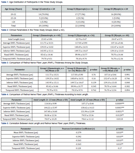
A total of 116 eyes from 62 participants were included in the study, comprising 40 emmetropic eyes from 20 subjects, 36 hyperopic eyes from 22 subjects, and 40 myopic eyes from 20 subjects. Participants ranged in age from 20 to 46 years. Peripapillary retinal nerve fiber layer (RNFL) thickness was assessed using the 3D Spectral-Domain Optical Coherence Tomography (OCT) system. The demographic and clinical characteristics of the study population are summarized in Table 1.
Retinal Nerve Fiber Layer Thickness Results
No statistically significant differences in age were observed among the three study groups. Visual acuity was 20/20 in all participants except for one subject in the hyperopic group, who had a visual acuity of 20/30.
The mean spherical equivalent (SE) refractive error was −0.30 ± 0.33 D in the emmetropic group, +1.27 ± 1.02 D in the hyperopic group, and −3.05 ± 1.25 D in the myopic group. Mean astigmatism was less than 1.00 D in all groups. The mean average retinal nerve fiber layer (RNFL) thickness in the emmetropic (control) group was 111.73 ± 10.31 µm and was used as the reference value for comparison. A trend toward reduced RNFL thickness was observed with increasing myopia, whereas RNFL thickness tended to increase with increasing hyperopia. However, these variations did not reach statistical significance. Detailed RNFL thickness measurements for the three groups are presented in Table 2.
The emmetropic group (Group I) demonstrated a mean RNFL thickness of 111.73 ± 10.31 µm and a mean axial length of 23.43 ± 0.86 mm. The hyperopic group (Group II) exhibited the greatest mean RNFL thickness (117.00 ± 8.90 µm) and the shortest mean axial length (22.74 ± 0.67 mm). In contrast, the myopic group (Group III) showed the lowest mean RNFL thickness (107.16 ± 8.46 µm) and the longest mean axial length (24.47 ± 0.71 mm). These findings suggest a tendency toward thicker RNFL measurements in eyes with shorter axial lengths and thinner RNFL measurements in eyes with longer axial lengths. Compared with the emmetropic control group, the hyperopic group (Group II) demonstrated higher mean RNFL thickness values in the average measurement as well as in all four quadrants (superior, inferior, nasal, and temporal).
However, these differences did not reach statistical significance for the average RNFL thickness (p = 0.760) or for the superior (p = 0.160), inferior (p = 0.139), nasal (p = 0.345), and temporal (p = 0.762) quadrants. Similarly, the myopic group (Group III) exhibited lower mean RNFL thickness values compared with the control group across the average and all quadrant measurements. Nevertheless, the observed reductions were not statistically significant for the average RNFL thickness (p = 0.991) or for the superior (p = 0.746), inferior (p = 0.848), nasal (p = 0.382), and temporal (p = 0.894) quadrants. These findings indicate that refractive status alone was not associated with significant differences in RNFL thickness measurements within the study population (Table 3).
Relationship Between Axial Length and Retinal Nerve Fiber Layer Thickness
Pearson correlation analysis revealed a significant negative correlation between axial length and retinal nerve fiber layer (RNFL) thickness. Greater axial length was associated with reduced RNFL thickness, with statistically significant correlations observed for the average RNFL measurement (p = 0.0009), as well as the superior (p = 0.0003), inferior (p = 0.0031), and nasal (p = 0.0129) quadrants. However, no statistically significant association was identified between axial length and temporal RNFL thickness (p = 0.8118). These results suggest that axial elongation is associated with progressive thinning of the RNFL in most peripapillary sectors, whereas the temporal quadrant remains relatively stable and unaffected by variations in axial length (Table 4).
Correlation Between Axial Length and Retinal Nerve Fiber Layer Thickness
Pearson correlation analysis demonstrated a weak but statistically significant negative correlation between axial length and mean retinal fiber layer (RNFL) thickness (r = −0.379, p = 0.01). This finding indicates that eyes with greater axial lengths tend to exhibit thinner RNFL measurements. Although the strength of the correlation was modest, the association remained statistically significant, suggesting that axial elongation may contribute to a reduction in peripapillary RNFL thickness (Table 5).
RNFL thickness in the superior, inferior, and nasal quadrants demonstrated weak but statistically significant negative correlations with axial length (r = −0.392, p = 0.01; r = −0.363, p = 0.01; and r = −0.365, p = 0.01, respectively), indicating progressive thinning of these peripapillary regions with increasing axial length. In contrast, the temporal quadrant showed a weak positive, non-significant correlation with axial length (r = 0.128, p = 0.170), suggesting relative stability of temporal RNFL measurements despite axial elongation (Table 5).
Spherical equivalent and retinal nerve fiber layer thickness results in Hyperopic group
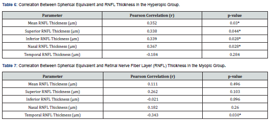
In the hyperopic group, mean RNFL thickness demonstrated a weak positive correlation with spherical equivalent (r = 0.352, p = 0.03). Quadrant-wise analysis showed weak positive correlations between spherical equivalent and RNFL thickness in the superior (r = 0.338, p = 0.044), inferior (r = 0.339, p = 0.043), and nasal (r = 0.367, p = 0.028) quadrants. In contrast, the temporal quadrant did not exhibit a statistically significant correlation with spherical equivalent (r = −0.184, p = 0.284), indicating relative independence of temporal RNFL measurements from refractive status in hyperopic eyes (Table 6).
Spherical Equivalent and Retinal Nerve Fiber Layer Thickness in the Myopic Group
In the myopic group, mean RNFL thickness showed no statistically significant correlation with spherical equivalent (r = 0.111, p = 0.496). Similarly, RNFL thickness in the superior (r = 0.262, p = 0.103), inferior (r = −0.021, p = 0.896), and nasal (r = 0.182, p = 0.260) quadrants did not demonstrate significant correlations with spherical equivalent. In contrast, the temporal quadrant exhibited a weak but statistically significant negative correlation with spherical equivalent (r = −0.343, p = 0.030), indicating a reduction in temporal RNFL thickness with increasing myopic refractive error (Table 7 and Figures 4-7).
Discussion
The present study demonstrated that retinal nerve fiber layer (RNFL) thickness was not significantly associated with refractive error. This finding contrasts with previous reports [11-13]. This discrepancy may be attributed to the relatively limited sample size and restricted refractive range, as the study was conducted over a three-month period at the King Saud University Female Campus, which may have influenced the variability of refractive error representation. In contrast, a significant inverse relationship was observed between axial length and RNFL thickness, indicating progressive RNFL thinning with increasing axial elongation.
This finding is consistent with earlier studies [14,15], which also reported a reduction in RNFL thickness in eyes with longer axial lengths. [11], using spectral-domain OCT, reported a significant reduction in RNFL thickness in the inferior and temporal quadrants in myopic eyes, while no significant differences were observed in the superior and nasal quadrants. In hyperopic eyes, they reported a significant increase in RNFL thickness in the inferior and temporal quadrants, with no significant changes in the superior or nasal quadrants.
Similarly, Veysi Oner et al. [12], using Stratus OCT, demonstrated a significant reduction in RNFL thickness across all quadrants (superior, inferior, temporal, and nasal) in myopic eyes. In hyperopic eyes, they observed a significant increase in RNFL thickness in the nasal quadrant, whereas no significant differences were detected in the superior, inferior, or temporal quadrants. In the present study, RNFL thickness showed a significant negative correlation with axial length in the superior, inferior, and nasal quadrants, while the temporal quadrant did not demonstrate a statistically significant association (r = −0.392, p < 0.01; r = −0.363, p < 0.01; r = −0.365, p < 0.01; r = 0.128, p = 0.170, respectively). These findings agree with the results reported by Christopher et al. [15].
Conclusion
The present study demonstrated that refractive status, including both hyperopia and myopia, does not significantly influence retinal nerve fiber layer (RNFL) thickness. In the hyperopic group, mean RNFL thickness and quadrant-wise measurements in the superior, inferior, and nasal regions showed weak positive correlations with spherical equivalent, while the temporal quadrant did not demonstrate any significant association. In the myopic group, RNFL thickness and RNFL measurements in the superior, inferior, and nasal quadrants showed no significant correlation with spherical equivalent.
However, the temporal quadrant exhibited a weak negative correlation with spherical equivalent. In contrast, axial length showed a significant inverse relationship with RNFL thickness. Increasing axial length was associated with statistically significant thinning of the RNFL in the average, superior, inferior, and nasal quadrants, whereas the temporal quadrant remained unaffected. Overall, mean RNFL thickness and quadrant-specific measurements in the superior, inferior, and nasal regions demonstrated weak negative correlations with axial length, while the temporal quadrant showed a weak, non-significant positive correlation.
Compliance with Ethical Standards
Ethical Approval: All procedures performed in this study were conducted in accordance with the ethical standards of the institutional research committee and with the 1964 Helsinki Declaration and its later amendments or comparable ethical standards.
Informed Consent: Informed consent was obtained from all individual participants included in the study.
References
- Leung CK, Chan WM, Yung WH, et al. (2005) Comparison of macular and peripapillary measurements for the detection of glaucoma: an optical coherence tomography study. Ophthalmology 112: 391-400.
- G Wollstein, JS Schuman, LL Price, A Aydin, SA Beaton, PC Stark, et al. (2004) Optical coherence tomography macular and peripapillary retinal nerve fiber layer measurements and automated visual fields. American Journal of Ophthalmology 138(2): 218-225.
- Giovannini A, Amato G, Mariotti C (2002) The macular thickness and volume in glaucoma: an analysis in normal and glaucomatous eyes using OCT. Acta Ophthalmologica Scandinavica 236: 34-36.
- Greenfield DS, Bagga H, Knighton RW (2003) Macular thickness changes in glaucomatous optic neuropathy detected using optical coherence tomography. Archives of Ophthalmology 121(1): 41-46.
- V Guedes, JS Schuman, E Hertzmark, G Wollstein, A Correnti, et al. (2003) Optical coherence tomography measurement of macular and nerve fiber layer thickness in normal and glaucomatous human eyes. Ophthalmology 110(1): 177-189.
- David E Lederer, Joel S Schuman, Ellen Hertzmark, James Heltzer, Leonardo J Velazques, et al. (2003) Analysis of macular volume in normal and glaucomatous eyes using optical coherence tomography. American Journal of Ophthalmology 135(6): 838-843.
- Leung CK, Chan WM, Yung WH, et al. (2005) Comparison of macular and peripapillary measurements for the detection of glaucoma: an optical coherence tomography study. Ophthalmology 112: 391-400.
- Medeiros FA, Zangwill LM, Bowd C, Vessani RM, Susanna R, (2005) Evaluation of retinal nerve fiber layer, optic nerve head, and macular thickness measurements for glaucoma detection using optical coherence tomography. American Journal of Ophthalmology 139(1): 44-55.
- Hougaard JL, Bengtsson BJ (2007) Glaucoma detection by Stratus OCT. Acta Ophthalmologica 16(3): 302-306.
- KN Mahdavi, K Nikkhou, DC Hoffman, SK Law, J Caprioli (2008) Detection of early glaucoma with optical coherence tomography (Stratus OCT). Journal of Glaucoma 17(3): 183-188.
- Öner V, Aykut V, Taş M, Alakuş M, Işcan Y (2012) Effect of refractive status on peripapillary retinal nerve fibre layer thickness: A study by RTVue spectral domain optical coherence tomography. British Journal of Ophthalmology 96(1): 75-79.
- Öner V, Taş M, Türkcü FM, Alakuş M, Işcan Y, Yazıcı AT (2013) Evaluation of peripapillary retinal nerve fiber layer thickness of myopic and hyperopic patients: A controlled study by Stratus optical coherence tomography. Current Eye Research 38(1): 102-107.
- Sowmya V, Venkataramanan VR, Vishnu P (2015) Effect of refractive status and axial length on peripapillary retinal nerve fiber layer thickness: An analysis using 3D OCT. Journal of Clinical and Diagnostic Research 9(9): 1-4.
- Tariq Y, Li H, Burlutsky G, Mitchell P (2012) Retinal nerve fiber layer and optic disc measurements by spectral domain OCT: Normative values and associations in young adults. Eye 26: 1563-1570.
- Christopher L, Mohamed S, Leung CK, et al. (2006) Retinal nerve fiber layer measurements in myopia: An optical coherence tomography study. Investigative Ophthalmology & Visual Science 47(12): 5171-5176.

























