




 JOJO Home




Abstract
Purpose: To assess zonular anatomy and zonular integrity of subluxated lens by using ultrasound bio microscopy and its relation to the clinical and intra operative finding.
Patients and Methods: 32 Patients with congenital and acquired subluxated lens were included in the study. Patients underwent full personal; medical and ocular history including time of trauma..Patients underwent full Ophthalmic examination including Visual acuity measurement using Landolt’s broken ring chart, Slit lamp Bio microscopy, Fundus examination. Intra-ocular pressure measurement, Ultrasound biomicrchart, examination (byVuMAX HD, Sonomed, Inc.1979 Marcus Avenue, Suite C 105, Lake Success, NY 11042, USA).
Results: The current study included 32 cases with subluxated lenses there were 14 males (43.8%) and 18 females (56.2%) congenital ectopia lentis in 24 cases (75.0%), acquired hyper mature cataractous subluxation in 4 cases (12.5%) and acquired traumatic sublaxated lens in 4 male cases (12.5%). The most common lens displacement was upward position in 10 cases (31.3%) followed by lateral displacement and upward and lateral displacement in 6 cases (18.8%).
Conclusion: Identification of the site and extent of zonular dialysis prior to the surgery helps the surgeon to plan and modify the surgical technique appropriately.
Keywords: Ultrasound Bio Microscopy; Congenital; Hyperlysinemia; Ocular Trauma; Uncorrected Visual Acuity
Abbreviations: UBM: Ultrasound Bio microscopy; UCVA: Uncorrected Visual Acuity; BCVA: Best Corrected Visual Acuity; SPSS: Statistical Package for Social Sciences
Introduction
Human tears are not a uniform secretion; rather, they are divided into three distinct types-basal (for continuous lubrication), reflex (evoked by irritants), and emotional (triggered by strong affective states)-each with unique composition and function [1]. The tear film itself is a complex, multilayered structure containing nearly 1,000 different constituents, and recent multi-omics profiling has revealed over 1,500 unique proteins within this fluid, many of which possess antimicrobial or anti-inflammatory properties [2]. Among these proteins, lysozyme and lactoferrin are particularly abundant, with lysozyme constituting 20-30% of total protein in basal and reflex tears, and lactoferrin serving as an iron-binding antimicrobial agent [3].
Beyond proteins, tears contain electrolytes such as sodium, potassium, and chloride, as well as trace elements including manganese and iron; studies using Particle Induced X-ray Emission (PIXE) analysis have shown that tear iron concentrations vary with gender, physical activity, and dietary supplementation, while manganese in emotional tears can reach levels 30-fold higher than in serum, implying an excretory function [4]. The biochemical signature of emotional tears distinguishes them sharply from basal and reflex types. They contain elevated concentrations of stress-related hormones such as adrenocorticotropic hormone (ACTH) and cortisol-the latter decreasing after a crying episode-as well as prolactin, which supports immune function [5]. Emotional tears are also unique in containing leucine-enkephalin, an endogenous opioid that acts as a natural painkiller [6], and they exhibit higher potassium levels compared to other tear types [7].
The release of these tears is not a random event but is orchestrated by a well-defined neural circuit. The lacrimal gland receives its primary innervation from the parasympathetic nervous system, with fibres arising from the pterygopalatine ganglion and travelling via the greater petrosal nerve and the nerve of the pterygoid canal to reach the gland [8]. This parasympathetic drive, mediated largely by muscarinic M₃ receptors, is the principal stimulus for fluid and protein secretion [9]. Sympathetic fibres, originating from the superior cervical ganglion, also innervate the gland and release norepinephrine, which modulates secretion through both α- and β-adrenergic receptors, although their role is generally considered secondary to parasympathetic control [10]. The physiological dynamics of emotional crying have been systematically investigated through meta-analysis of autonomic correlates. At crying onset, there is a consistent increase
in sympathetic activity, reflected in elevated heart rate, blood pressure, and skin conductance; this is followed by a sympathetic withdrawal and a subsequent parasympathetic rebound, with heart rate variability increasing approximately five minutes post-crying, suggesting a homeostatic recovery function [11]. This recovery process, however, appears to be impaired in depression; non-depressed individuals show a vagal rebound (increased respiratory sinus arrhythmia) upon resolution of tearful crying, whereas depressed individuals do not [12]. Beyond its role in emotional regulation, the tear film has emerged as a promising non-invasive diagnostic window into systemic health. Changes in tear protein, lipid, and metabolite concentrations have been linked to a wide range of diseases, including neurodegenerative disorders, cancers, autoimmune conditions, and endocrine diseases, and a growing body of literature supports the use of tear biomarkers for screening and monitoring [13].
Early work by Frey and colleagues established that the chemical composition of tears varies with the type of stimulus, with emotional tears containing higher levels of protein and hormones than reflex tears [14]. More recent metabolomic studies have further refined this picture, revealing distinct metabolic profiles between positively and negatively valenced emotional tears, indicating that tear chemistry is sensitive to the specific affective state [15]. The autonomic changes during crying are also reflected in heart rate variability patterns, with weeping producing a measurable increase in parasympathetic modulation [16]. In clinical practice, the relationship between tear dysfunction and systemic conditions is perhaps most evident in dry eye disease (DED), which affects hundreds of millions worldwide and is often accompanied by psychiatric comorbidities such as depression, anxiety, and sleep disturbances [17]. Moreover, the antimicrobial proteins lysozyme and lactoferrin, which are critical for ocular surface defence, show altered concentrations in both basal and reflex tears of patients with ocular surface disease [18].
The neurobiology of human crying has been comprehensively reviewed, highlighting the interplay between limbic structures, the lacrimal nucleus, and autonomic outflow, and underscoring the unique position of emotional tears as a window into affective neuroscience [19]. Critically, recent advances in bioinorganic chemistry and neuroimaging have revealed that the human brain contains significant deposits of biogenic magnetite (Fe₃ O₄) and other ferrimagnetic iron species, which are concentrated in the hippocampus, cortex, and basal ganglia-regions intimately involved in memory, emotion, and motor control [20-23]. These iron-oxide nanoparticles are not inert; they participate in electron transfer, contribute to the brain’s endogenous electromagnetic field, and generate reactive oxygen species (ROS) via Fenton chemistry, particularly during periods of high metabolic and neural activity.
We propose that the iron excreted in emotional tears-whose concentrations are modulated by diet, exercise, and gender [4]-is not a passive waste product but a functional component of a homeostatic loop: intense emotional arousal mobilises labile iron pools and magnetite turnover in the brain; the subsequent parasympathetic-mediated lacrimal response clears excess iron-bound complexes, reducing oxidative burden and restoring electromagnetic coherence. This framework-the "Tears of Iron" hypothesis-elevates the lacrimal system to a central role in cerebral redox homeostasis and positions emotional crying as a physiological necessity rather than a mere behavioural epiphenomenon. The bidirectional link between dry eye disease and psychiatric disorders has received increasing attention, with neuroimmune mechanisms and monoaminergic dysregulation being implicated in both conditions [24].
The lacrimal gland’s neural regulation, extensively characterised by Dartt and colleagues, involves complex interactions between parasympathetic, sympathetic, and sensory inputs that together form the lacrimal gland functional unit [25]. Walcott has further elaborated on the detailed innervation patterns of the gland, emphasising the direct parasympathetic contacts with each acinus [26]. In the context of systemic disease, tear biomarkers have been identified for Parkinson’s disease, Alzheimer’s disease, multiple sclerosis, and other neurodegenerative conditions, offering a potentially accessible route for early diagnosis [27]. The field of tear biomarker discovery has matured to the point where comprehensive reviews now outline standardised collection protocols, analytical methods, and clinical validation pathways [28].
Finally, the intracellular signalling cascades that couple receptor activation to secretion-involving Ca²⁺, diacylglycerol, and protein kinases-have been elucidated in detail, providing a molecular basis for tear production [29], while longitudinal studies have confirmed that emotional crying reduces circulating cortisol levels, reinforcing its stress-buffering function [30]. With this comprehensive background, the present review synthesises current knowledge on the biochemical, neurophysiological, and clinical aspects of tear types, with a special emphasis on emotional lacrimation, its autonomic correlates, and its emerging role as a diagnostic biofluid. We propose an integrated model of emotional crying as a homeostatic mechanism that restores autonomic balance and may have therapeutic implications for both ocular and mental health.
Introduction
Various congenital and systemic conditions are associated with ciliary zonular abnormalities. Congenitally subluxated lens may occur as incases of Marfan syndrome, Homocystinuria, Ehlers-Danlos syndrome, hyperlysinemia, Sulfite oxidase deficiency, Simple primary ectopia lentis, and congenital aniridia syndrome [1,2]. Acquired Subluxations of the lens can be traumatic (the most frequent cause) [3], Postsurgical or Spontaneous subluxation occurs secondary to ocular pathologies due to rupture of the zonular fibres because of degenerative and inflammatory factors as in long-standing glaucoma, high myopia, hypermature cataract, retinal detachment and pseudo exfoliation syndrome [4]. Slit lamp signs such as phacodonesis, iridodesis, lenticular dislocation can suggest that zonular abnormalities exist. Although these signs of increased lenticular mobility can imply only that there is zonular defect, however ;the nature and extent of this defect is frequently uncertain because of difficulty in visualizing the zonule through the intact iris [5].
Ultrasound biomicroscopy (UBM) has become main imaging technique for visualizing structures in the angle of anterior chamber and the ciliary body area [6]. The location of the ciliary processes and integrity of zonules can also be assessed, thus enabling preoperative detection of subtle degrees of subluxation. Occult zonular defects detected preopatively, can be managed more effectively [7]. Which have a significant benefit to the surgeon and might reduce the chance of intra operative complications [8]. UBM provides high-definition, reliable, and repeatable images of the anterior segment, as well as quantitative measurements. UBM uses high frequency ultrasound at 50–100 MHz for anterior segment imaging and a computer program then converts these sound waves into a high-resolution image [9]. The aim of the study is to assess zonular anatomy and zonular integrity of subluxated lens by using ultrasound biomicroscopy and its relation to clinical and intra operative finding.
Patients and Methods
This study included 32 cases with subluxated lenses from patients attending. Ophthalmic center, Faculty of Medicine, Mansoura University, Egypt. The study was conducted between September 2022 and November 2026. This study was a cross-sectional analytic case with written informed consent obtained from all participants before inclusion in the study, explaining the value of the study, plus the procedures that will be conducted. The whole study design was approved by the institutional review board, faculty of medicine, Mansoura University. Confidentiality and personal privacy were respected in all levels of the study. Patients felt free to withdraw from the study at any time without any consequences. Collected data were not and will not be used for any other purpose.
1. Inclusion Criteria
• Age: all age groups.
• Gender: both genders.
• Patients: with congenital and acquired subluxated lens.
2. Exclusion Criteria
• Patients with open globe injury.
For all subjects, the following were done:
History Taking: General history; Personal demographic data; Complaint and present history; General medical history; Family history; Ophthalmic history:(History of ocular trauma, History of ocular surgery. History of wearing glasses, History of contact lens wearing.
Full ophthalmic examination was done;
Visual acuity (both distant and near, Uncorrected visual acuity (UCVA), Best corrected visual acuity (BCVA) using Landolt’s VA chart (Snellen’s) and then transformed for statistical analysis to logarithm of minimal angle of resolution units (Log MAR). External ocular examination (For detection of any pathology like ptosis, lid hemangioma, Strabismus or nystagmus. Patient's refractive error using Autorefractometer Used (Canon Auto Ref R-30). Cyclopiegic refraction retinoscopic refraction (through phakic and aphakic areas), intraocular pressure, was measured using Goldmann applanation tonometer.
Slit Lamp Biomicroscopy Must be Performed Under Maximum Mydriasis
By the slit lamp biomicroscopy (Haag Streit BP 900) (Haag-Streit, Koeniz, Switzerland) to assess: Corneal clarity, AC for depth and regularity, Pupil shape, size, regularity and reactivity, Lens: any signs of exfoliation, dehiscence of zonules, subluxation and grading of nuclear hardness using LOCS lll classification system Direction of subluxation.
•Number of clock hours involved. Degree of phacodonesis.
•Presence or absence of any virtues in anterior chamber. Presence of any lens coloboma along with subluxation Lens coloboma
Posterior Segment Examination:
Using indirect ophthalmoscope and slit lamp bio microscopy with non-contact Volk 90 lens (Dilated fundus examination to exclude any posterior segment disorder).
Ultrasound Biomicroscopy Examination:
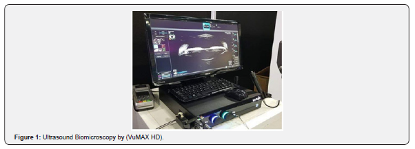
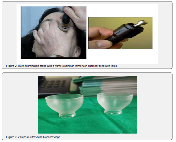
It was conducted with non-dilated pupils and with the patient in a supine position, using UBM model, probe frequency 35 MHz (VUMAX II; Sonomed Inc.) (Figure 1-3). The UBM probe was used to obtain transverse and axial scans at the four quadrants, while kept perpendicular to the ocular surface and central on the pupil, together with transverse immersion chamber filled with liquid. The depth of scans ranged from the anterior corneal surface to the posterior lens capsule. Examination was performed in the supine position; an eye cup was used to separate the eyelids maintaining upward pressure on the eye cup to hold it in place after a drop of benoxinate Hcl 0.4% had been applied. The eye cup was filled with distilled water as a coupling medium. Placing the probe close to the anatomical structure and changing the globe to 360 degrees was done with interest to the cilary body, Zonules, posterior capsule and lens position were done.
Intra Operative Observation
It was done to the anterior segment anomaly, Lens configuration and evaluation of Zonules configuration. Statistical analysis and data interpretation. The collected data was coded, processed and analyzed using the
SPSS (Statistical Package for Social Sciences) version 22 for Windows® (IBM SPSS Inc, Chicago, IL, USA).
Qualitative Data
Were presented with frequency tables (Number and percentages). Quantitative variables: the normality of data was first tested with Shapiro-Wilk test and presented data by central indices and dispersion:
• Mean ± Standard deviation (SD) for normally distributed variables.
• Median and interquartile range (Minimum – Maximum) for non- normally distributed variables
• Data analysis Agreement analysis Kappa agreement coefficient was used to test the agreement between different assessment modaliti from (0: 0.3) …… weak agreement.
• r from (0.3: 0.6) …… moderate agreement
• r from (0.6: 1) ………… strong agreement. Level of significance (P-value)
In all applied tests, the P-values associated with test statistics indicated the significance level at which the null-hypothesis (the hypothesis of no difference) was rejected and it was set at 0.05 so that a P- values ≥0.05 are statistically non-significant, P-values < 0.05 are significant, and P-values < 0.01 are highly significant.
Results
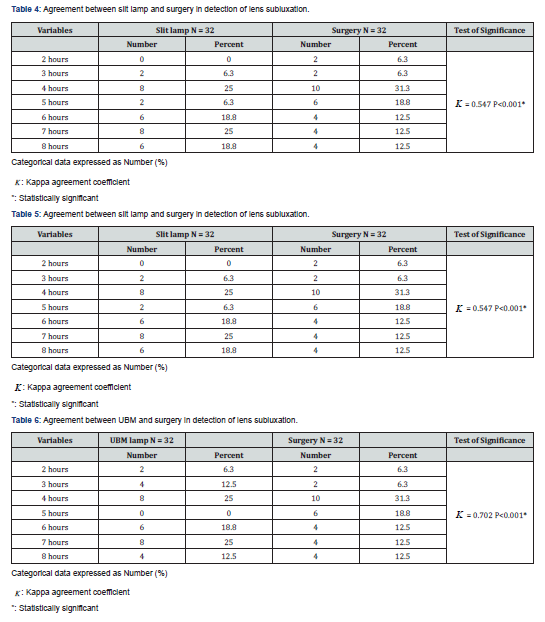
The current study included 32 cases with subluxated lenses who were recruited from the ophthalmic center, Mansoura University of Medicine. The study was conducted between September 2022 and November 2026. The mean age was 13.41 ± 12.85 years, and the median age was 7 years with range between 2 and 42 years. Among the included cases there were 14 males (43.8%) and 18 females (56.2%) The right eyes were affected in 15 cases (46.9%) and the left eyes were affected in 17 cases (53.1%). Positive family history was reported in 13 cases (54.2%). The diagnosis in the cases included congenital ectopia lentis in 24 cases (75.0%), acquired hyper mature cataractous subluxation in 4 cases (12.5%) and acquired traumatic sublaxated lens in 4 male cases (12.5%). The UBM evaluation of posterior capsule integrity was 30 cases (93.7%) have intact posterior capsule, and 2 cases (6.3%) have open posterior capsule. The extension of zonular dialysis by slit lamp was extended 4 and 7 hours in each case (25%), extended 6 and 8 hours in each case (18.8%), extended 3 and 5 hours in each case (6.3%). The most common lens displacement was upward in 10 cases (31.3%) followed by lateral displacement and upward and lateral displacement in 6 cases (18.8%) as shown in Table1.
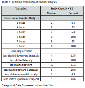
The position of lens subluxation as detected during surgery, Table 2, the most affected positions where 4 hours were detected 31.3%, 5 hours were detected 18.8% and 7 hours and 8 hours each were detected in 12.5% of the cases. The extension of lens subluxation as detected by UBM, the most affected positions were 4 hours, 7 hours each were detected 25.0% and 3 hours and 8 hours each were detected in 12.5% of the cases Lens subluxated as detected by slit-lamp, occupied 3 hours in 2 cases (6.3%), 4 hours in 8 cases (25%), 5 hours in 2 cases (6.3%), 6 hours in 6 cases (18.8%), 7 hours in 8 cases (25%) and 8 hours in 6 cases (18.8%).As detected by UBM, the subluxated lenses occupied 2 hours in 2 cases (6.3%), 3 hours in 4 cases (12.5%), 4 hours in 8 cases (25%), 6 hours in 6 cases (18.8 ), 7 hours in 8 cases (25%) and 8 hours in 4 cases (12.5%). As detected by during surgery, the subluxated lenses occupied 2 hours in 2 cases (6.3%), 3 hours in 2 cases (6.3%), 4 hours in 10 cases (31.3%), 5 hours in 6 cases (18.8%), 6 hours in 4 cases (12.5%), 7 hours in 4 cases (12.5%) and 8 hours in 4 cases (12.5%) as shown in (Table 8).
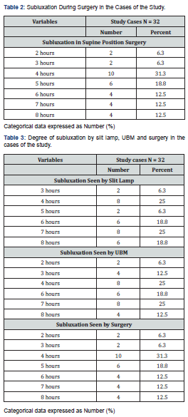
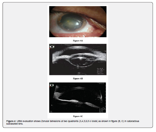
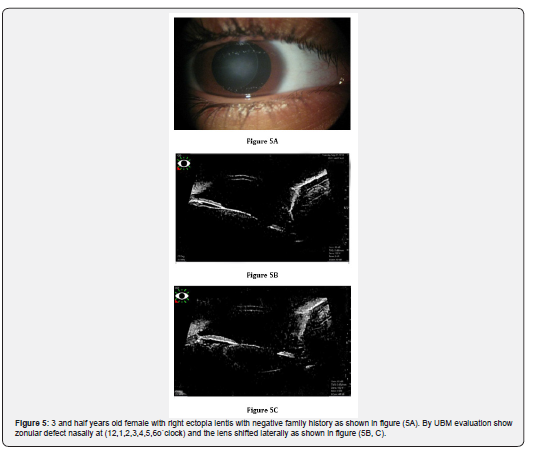
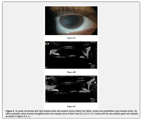
The degree of agreement of subluxation between the slit-lamp and surgery in detection in the extension in lens subluxation was (0.547). The degree of agreement was moderate with high statistically significant value (p0.001) (Table 3). The degree of agreement of subluxation between the UBM and surgery in detection in the extension in lens subluxation was (0.702). The degree of agreement was strong with high statistically significant value (p 0.001) Table 4. The degree of agreement of subluxation between the slit-lamp and UBM in detection in the extension in lens subluxation was (0.536). The degree of agreement was moderate with high statistically significant value (p0.001). The highest degree of agreement was detected between the UBM and the position during surgery (Table 5-8). There was moderate degree of agreement of subluxation between slit lamp, UBM and surgical findings in detection of the extension in lens subluxation (0.595). This degree of agreement showed high statistically significant value (p0.001). The highest degree of agreement was detected between the UBM and the surgical findings followed by the agreement between the slit lamb and surgical findings and finally between the slit lamb and UBM (Figures 4-6).
Discussion
Lens subluxation is a change of lens position caused by a zonal abnormality. The causes of lens subluxation are varied, including ocular trauma, hereditary diseases (such as Marfan syndrome), and spontaneous subluxation [10]. Accurate preoperative localization of the lens is necessary for surgical planning. Complete luxation of the lens can be detected by low frequency B-scan ultrasound, a slit lamp, or an ophthalmoscope. However, subluxation of the lens, especially slight subluxation of the lens, cannot be determined directly by the above means [11]. In the present study, there were 14 males (43.8%) and 18 females (56.2%) while in the study done by Patel and Danayak [12] majority of the cases were males accounting for 68%, whereas females are accounting for 32%. Also, in another study by Bhatt et al. [13] more than two-thirds of the patients were male (67.8%) while around one-third were females (32.2%) respectively.
Özdal et al. [14] found that males are more frequently implicated in ocular trauma than females. When compared to other research, such as Dannenberg et al. (1992) where males made up 97% of the study population, our study showed that all traumatic cases were males represent 12.5 % of the cases. Lens subluxation is often suspected when irregular anterior chamber depth is observed [15]. However, for patients with ocular trauma, the factors are more complex. First, the source and direction of external force could impair the zonule to different degrees; second, the examination of anterior chamber depth is also affected by the examination position [16]. In our study, Congenital lens subluxation was more common and was detected in 24 cases (75%) while acquired subluxation was detected in 8 cases (25%), that disagreed with [13] who stated that the most common cause of the zonular defect was trauma, which was followed by pseudoexfoliation, surgical procedures, congenital spherophakia, and Marfan syndrome Dhar et al. [17] conducted a study of ultrasound biomicroscopy for 50 patients with mature senile cataract, pseudoexfoliation, trauma, or those with clinical suspicion of zonular dehiscence [n=5].
Ten had clinical zonular dialysis with phacodonesis /iridodonesis which was confirmed by UBM. Three patients were noted to have zonular dehiscence on UBM. Areas of zonular dehiscence were mapped on UBM and the UBM findings are described. Surgery could be planned accordingly and complications prevented. Accuracy for preoperative detection of zonular loss is high with UBM [17]. In the current study, the most common lens displacement was upward in 10 cases (31.3%) followed by lateral displacement and upward and lateral displacement in 6 cases each (18.8%). UBM can show the accurate location of the lens and can display the integrity of the posterior lens capsule, which is particularly important for traumatic cataracts. Preoperative determination of the presence of an intact posterior lens capsule through echographic confirmation assists the physician in surgical planning [18,19].
In the present study, the zonular integrity detected by UBM, the most common finding was the zonular defect downward and nasal in 8 cases (25%) followed by zonular defect downward, zonular defect nasally and zonular elongated downward nasal in 4 cases each (12.5%). Bhatt et al. [13] found that zonular tears occurred between 3 and 6 clock hours in 23.8% of cases and zonular stretching occurred between 3 and 6 clock hours in 21% of cases, respectively. In a series of patients with zonular dialysis reported by Gimbel et al. (1997). Only 6 of the 14 cases of ruptured zonules were identified preoperatively. The remaining 8 were noted during cataract surgery. Zonular damage might be clinically visible if the iris is also disrupted or if the lens is dislocated. Other signs include phacodonesis or the presentation of vitreous. However, occult damage to the zonules that is not clinically obvious might also occur [8]. In our study, the degree of agreement of subluxation between the slit-lamp and surgery in detection in the extension in lens subluxation was (0.547).
The degree of agreement was moderate with high statistically significant value (p< 0.001). Clinical observation through slit-lamp examination found iridodonesis, vitreous prolapse, and partial quadrant lens subluxation; the diagnosis of obvious lens subluxation is not difficult. If the above signs are found in the slit-lamp examination, it is believed that the range of subluxation may be very large (Loo et al., 2002). Zonular status is an important issue in deciding for the surgical technique that will be done. Although observing signs of increased lenticular mobility, such as phacodonesis and iridodonesis in slit-lamp examination can imply a zonular defect, the nature and extent of this defect cannot be correctly visualized [14]. In the current study, the degree of agreement of subluxation between the UBM and surgery in detection in the extension in lens subluxation was (0.702). The degree of agreement was strong with high statistically significant value (p< 0.001).
In this study, there was moderate degree of subluxation between slit lamp, UBM and surgical findings in detection of the extension in lens subluxation (0.595). This degree of agreement showed high statistically significant value (p< 0.001). that agreed with Patel and Danayak [12] who found that UBM examination was able to identify zonular dialysis in 59% patients, whereas clinical examination was able to identify zonular dialysis in only 15% patients. UBM was far superior in identifying and localizing areas with zonular defects (in clock hours) than clinical examination. In cases having fewer clock hours of zonular dialysis, clinical examination may be equivocal, resulting in under-diagnosis of zonular dialysis. Studies carried out by Özdal et al. [14] have shown that zonular deficiency was the most frequent finding in blunt ocular injury and is far underestimated by clinical examination alone. Prompt identification of the site and extent of zonular dialysis prior to the surgery helps the surgeon to plan and modify the surgical technique appropriately.
Shi et al. [16] conducted a study to investigate the role of 25 MHz ultrasound biomicroscopy (UBM) in detecting subluxated lenses and comparing it with 50 MHz UBM. 45 patients (49 eyes) with suspected subluxation of the lens and 20 normal volunteers (40 eyes) were included. The position of the lens on axial sections could be clearly shown by using 25 MHz UBM. There was a statistically significant difference between 25 and 50 MHz UBM in showing subluxation of the lens, the testing time was significantly faster (2.0 min versus 7.5 min), and the diagnostic accuracy was much higher (98.0% versus 71.4%) with 25 versus 50 MHz UBM [16]. In our study, 35 MHz UBM is used, it provides high definition and reliable images of the anterior segment. A significant advantage of the 35 MHz frequency over the higher 50 MHz standard is its ability to capture the entire anterior segment in a single image. Higher frequencies (50-100 MHz) often require multiple images to be stitched together to view the same area.
Summary
Subluxation of the lens is a common lens disease, and most cases require surgical treatment. Accurate preoperative localization of the lens is necessary for surgical planning. High frequency UBM is a useful imaging tool of the anterior segment of the eye, providing high-resolution images with both qualitative and quantitative information about anatomical relationships of the anterior segment and zonular structures. During surgery for traumatic cataracts, the presence of zonular damage raises the risk of vitreous loss and posterior dislocation of the lens into the vitreous cavity. Thus, UBM is useful for zonular status analysis before surgery in individuals with a history of ocular damage, that is why the current study is to assess zonular anatomy and zonular integrity of subluxated lens by using ultrasound biomicroscopy and its relation to the clinical and intra operative finding.
Conclusion
· This study showed that zonular abnormalities are commonly seen congenital and after ocular trauma.
· Zonular damage can be missed or underestimated by clinical examination. Such situations are frequently encountered as intraoperative surprises, necessitating a change in surgical care.
· In the absence of proper visualization of zonules due to opaque media in cases of ocular trauma, this is where imaging, in the form of UBM, plays an unrivaled role. It provides excellent real-time images of the anterior segment of the eye with good contrast.
· UBM has a great diagnostic value in verifying the status of the lens location, provides accurate and reliable information about the status of lens subluxation and posterior capsular integrity.
· UBM is especially superior to clinical examinations in the evaluation of the zonular anatomy, zonular integrity of subluxated lens, and extension of the zonular damage which is very helpful in selection of the surgical plane.
Recommendation
· We recommend incorporating UBM analysis as an integral part of the work-up of patients with ocular trauma and cases of cataract, especially with opaque media for optimal patient management and outcomes.
· Further large multicentric prospective studies and randomized clinical trials are needed to improve our understanding of zonular anatomy and zonular integrity of subluxated lens by using ultrasound biomicroscopy and its relation to clinical and intra operative finding.
References
- Dureau P (2008) Pathophysiology of zonular diseases. Curr Opin Ophthalmol 19(1): 27-30.
- Kohnen T (2010) Pseudoexfoliation: impact on cataract surgery and long-term intraocular lens position. J Cataract Refract Surg 36: 1247-1248.
- Roldan Pallares M, Vilar Maseda N (2001) Vitrectomy and perfluorocarbons in the management of luxated lenses. Arch Soc Esp Oftalmol 76: 431-436.
- K, Shentu X, Jiang J, Du X (2002) Phacofragmentation without perfluoro-carbon Yao liquid for dislocated crystalline lenses or lens fragments after phacoemulsification. Eur J Ophthalmol 12(3): 200-204.
- Charles J Pavlin, Yvonne M Buys, T Pathmanathan (1998) Imaging zonular abnormalities using ultrasound biomicroscopy. Arch ophthalmol 116(7): 854-857.
- Meenakshi Dhar MS, Abhijeet S Khake, H Sujithra DO, Niranjan Pehere (2008) Is UBM Useful for Zonular Integrity? Kerala journal of ophthalmology 2008: 144-147.
- Agarwal T, Saxena R, Vajpayee RB (2003) Ultrasound biomicroscopy in lens(coloboma) Eur J Ophthalmol 13(4): 390-391.
- McWhae JA, Crichton AC, Rinke M (2003) Ultrasound biomicroscopy for the assessment of zonules after ocular trauma. Ophthalmology 110(7): 1340-1343.
- Maslin JS, Barkana Y, Dorairaj SK (2015) Anterior segment imaging in glaucoma: An updated review. Indian J Ophthalmol 63(8): 630-640.
- Bhatt V, Bhatt D, Barot R, Sheth J (2021) Ultrasound biomicroscopy for zonular evaluation in eyes with ocular trauma. Clinical Ophthalmology 15: 3285-3291.
- Patel RB, Danayak PM (2014) Ultrasound biomicroscopic evaluation of traumatized eyes. International Journal of Advances in Medicine 1(3): 247-251.
- Lee S, Hayward A, Bellamkonda VR (2015) Traumatic lens dislocation. International journal of emergency medicine 8: 16.
- Dhar M, Khake AS, Sujithra H, Pehere N (2008) Is UBM Useful for Zonular Intergrity? Kerala Journal of Ophthalmology 4(1): 144-147.
- Özdal M, Mansour M, Deschenes J (2003) Ultrasound biomicroscopic evaluation of the traumatized eyes. Eye 17(4): 467-472.
- Shafique M, Muzaffar W, Ishaq M (2016) The eye as a window to a rare disease: ectopia lentis and homocystinuria, a Pakistani perspective. International ophthalmology 36(1): 79-83.
- Shi M, Ma L, Zhang J, Yan Q (2018) Role of 25 MHz ultrasound biomicroscopy in the detection of subluxated lenses. Journal of ophthalmology 2018: 3760280.
- Shi Y, Tu Y, De Maria A, Mecham RP, Bassnett S (2013) Development, composition, and structural arrangements of the ciliary zonule of the mouse. Investigative ophthalmology & visual science 54(4): 2504-2515.
- Stachs O, Martin H, Behrend D, Schmitz K, Guthoff R (2006) Three-dimensional ultrasound biomicroscopy, environmental and conventional scanning electron microscopy investigations of the human zonula ciliaris for numerical modelling of accommodation. Graefe's Archive for Clinical and Experimental Ophthalmology 244(7): 836-844.
- Wang A, Mou D, Wang N and Wang H (2022) The Imaging Characteristics of Lens Subluxation on the Ultrasound Biomicroscopy. Contrast Media & Molecular Imaging 2022: 7030866.