




 JOJO Home



Abstract
Background: Central retinal artery occlusion (CRAO) is an ophthalmic emergency characterized by sudden, painless monocular vision loss and is considered an ocular analogue of ischemic stroke [1]. Although typically seen in elderly patients with vascular risk factors, its occurrence in young adults is uncommon and often indicates underlying systemic or vascular pathology.
Case Presentation: We describe two young male patients who presented with acute painless monocular vision loss due to CRAO associated with ipsilateral internal carotid artery (ICA) occlusion and concurrent cerebral infarction. Both patients had a preceding febrile illness and a recent alcohol binge. Fundoscopic examination revealed retinal pallor with a cherry-red spot and a relative afferent pupillary defect. Neuroimaging revealed ICA occlusion with acute cerebral infarction. Cardiac evaluation was unremarkable and neither patient had any conventional vascular risk factors. Laboratory findings showed elevated haemoglobin and haematocrit levels, suggestive of haemoconcentration. Infectious and autoimmune workup were negative. Both patients presented beyond the therapeutic window and were managed conservatively [2].
Conclusion: CRAO in young individuals should prompt an urgent evaluation for underlying vascular pathology. ICA occlusion may simultaneously compromise both retinal and cerebral circulation. Early recognition is critical for stroke risk assessment and the prevention of further neurological morbidity.
Keywords: CRAO (Central retinal artery occlusion); ICA (Internal Carotid Artery); Retinal stroke; Monocular vision loss; Young stroke
Abbreviations:CRAO: Central Retinal Artery Occlusion; ICA: Internal Carotid Artery; FAZ: Foveal Avascular Zone
Key Messages
CRAO is a type of retinal stroke that requires urgent systemic evaluation. In young patients, CRAO warrants investigate carotid pathology and hypercoagulable states. ICA occlusion simultaneously affects retinal and cerebral perfusion. Haemo concentration due to dehydration and alcohol binge drinking may increase the thrombotic risk. CRAO should be treated as a neurovascular emergency.
Introduction
Central retinal artery occlusion (CRAO) is an acute ischemic event resulting from the obstruction of the central retinal artery, leading to sudden painless monocular vision loss. It is widely regarded as the ophthalmic equivalent of ischemic stroke and necessitates urgent systemic and neurovascular evaluations.
The central retinal artery arises from the ophthalmic artery which is a branch of the internal carotid artery (ICA). Therefore, ICA pathology can directly impair retinal perfusion. Although CRAO commonly occurs in older individuals with established vascular risk factors, its occurrence in young adults is rare and often signals atypical etiologies such as hypercoagulable states, carotid artery disease, inflammatory vasculopathies, or hematologic abnormalities. We report two cases of young male patients presenting with CRAO associated with ipsilateral ICA occlusion and cerebral infarction, highlighting the importance of early neurovascular assessment.
Case Presentation
Case 1:
A 25-year-old man presented with fever for four days followed by right-sided headache. He developed sudden painless blurring of vision in his right eye which progressed to complete vision loss within seconds. There was a history of continuous alcohol consumption for four days before the onset.
Clinical Findings:
Right Eye: only perception of light present
Relative Afferent Pupillary Defect: present
Left Eye: visual acute was 6/6
Left Pupil: normal and reactive
No focal neurological deficits
Fundus Examination:
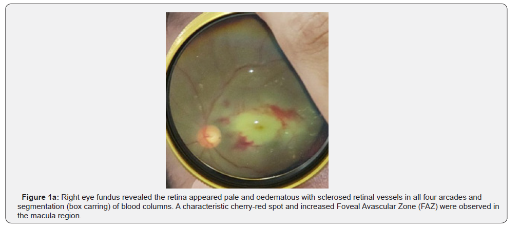
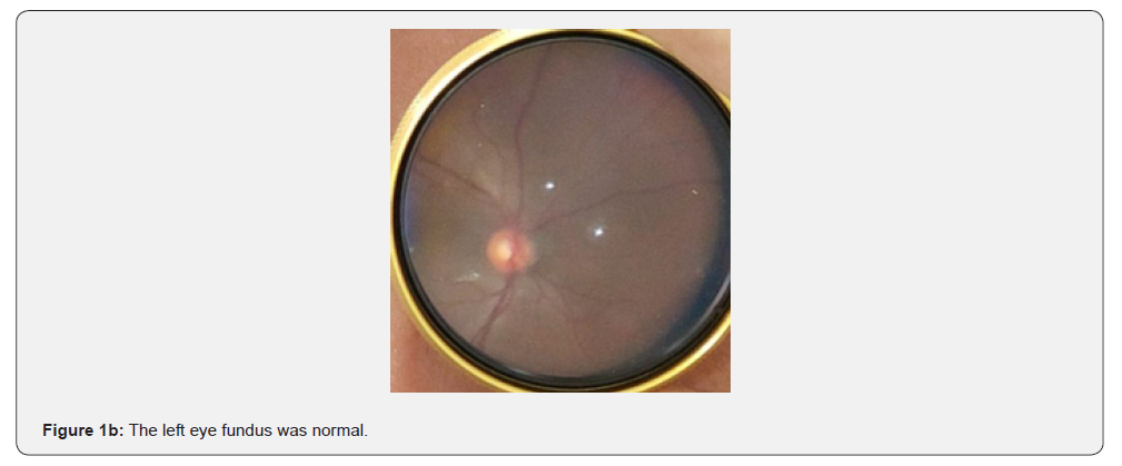
Fundus examination of the right eye revealed clear ocular media with a cup-to-disc ratio of 0.3 and optic disc pallor. The retina appeared pale and oedematous with sclerosed retinal vessels in all four arcades and segmentation (box carring) of blood columns. A characteristic cherry-red spot was observed in the macula (Figure 1a). The fundus of the left eye was normal (Figure 1b).
OCT Examination:
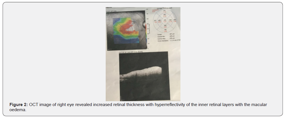
OCT imaging of right eye demonstrated increased retinal thickness with hyperreflectivity of the inner retinal layers, consistent with acute retinal ischemia. The central macular thickness was approximately 497 μm, indicating macular oedema secondary to a vascular compromise. These findings support the diagnosis of acute central retinal artery occlusion (Figure 2).
Investigations:
Haemoglobin: 17.3 g/dL
Haematocrit: 49.5%
Lipid profile: low HDL
Autoimmune and Infectious Workup: negative
Echocardiography: normal
Neuroimaging:
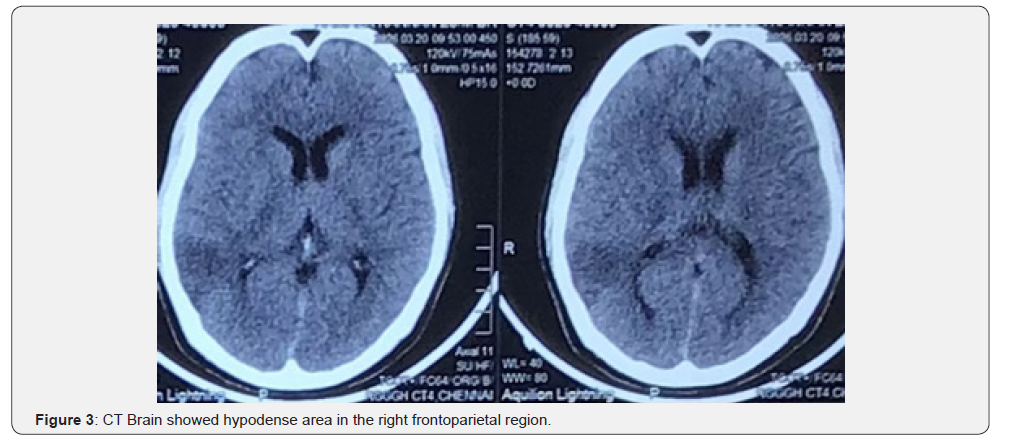
CT Brain: Axial CT imaging demonstrates hypodense areas in the right frontoparietal region, suggestive of acute cerebral infarction (Figure 3).
CT Cerebral Angiography:
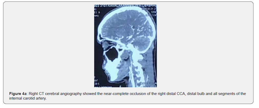
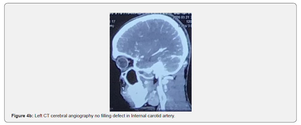
CT cerebral angiography demonstrates near-complete occlusion of the right distal CCA, distal bulb, all segments of the internal carotid artery, reformed flow noted in the right MCA and ACA, right posterior communicating artery through collaterals (Figure 4a, 4b).
Fundus Fluorescein Angiography:
Fluorescein angiography of the right eye demonstrated normal choroidal flush with delayed arterial filling at approximately 20 seconds and delayed venous filling at around 30 seconds. Early hypo fluorescence in the macular region suggested macular ischemia secondary to CRAO. Non filling areas in the capillaries of the macular region with increased Foveal Avascular Zone (FAZ).
Management:
The patient presented beyond the thrombolysis window period and was treated with antiplatelets, statins, ocular hypotensive agents, and hydration.
Case 2:
A 32-year-old man presented with sudden painless vision loss in the right eye following a febrile illness with chills and vomiting. He reported heavy alcohol intake for seven days before the onset.
Clinical Findings:
Right Eye: only perception of light present
Relative Afferent Pupillary Defect: present
Left Eye: visual acute was 6/6
Left Pupil: normal and reactive
Neurological Exam: left-sided ataxic hemiparesis
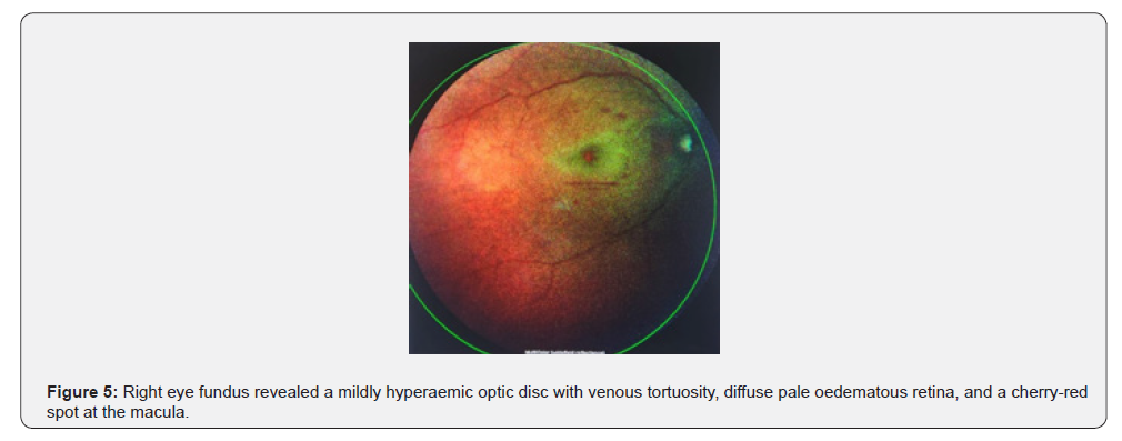
Fundus examination of the right eye revealed a mildly hyperaemic optic disc with venous tortuosity, diffuse pale oedematous retina, and a cherry-red spot at the macula. The fundus of the left eye was normal (Figure 5).
OCT Examination:
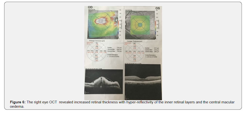
OCT imaging of right eye demonstrated increased retinal thickness with hyperreflectivity of the inner retinal layers, consistent with acute retinal ischemia. The central macular thickness was approximately 497 μm, indicating macular oedema secondary to a vascular compromise (Figure 6).
Investigations:
Haemoglobin: 17.9 g/dL
Haematocrit: 53.7%
Autoimmune And Infectious Profile: negative
Echocardiography: normal
Neuroimaging:
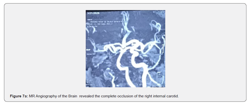
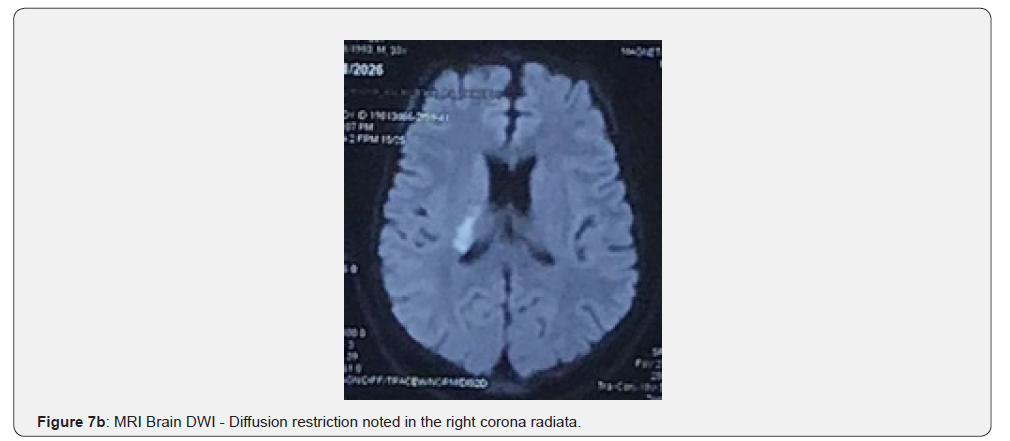
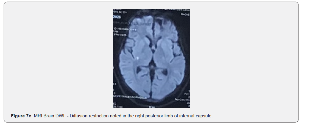
MRI brain: MRI brain demonstrated complete occlusion of the right internal carotid artery from the petrous to supra-clinoid segment. Acute infarctions were noted in the right corona radiata and Posterior limb of internal capsule (Figure 7a 7b 7c).
CT Cerebral Angiography:
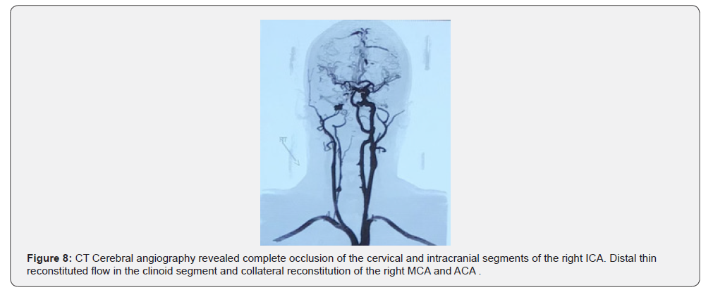
CT Cerebral angiography revealed complete occlusion of the cervical and intracranial segments of the right ICA. Distal thin reconstituted flow in the clinoid segment and collateral reconstitution of the right MCA and ACA (Figure 8).
Management:
Conservative management like Case 1 was initiated.
Discussion
CRAO represents an acute retinal ischemic insult and shares pathophysiological mechanisms with cerebral ischemia [3]. The close anatomical relationship between the ICA and retinal circulation explains the coexistence of retinal and cerebral infarctions in these patients [4].
Both cases highlight ICA occlusion as the underlying etiology,
likely contributing via:
• Embolic mechanisms
• Hemodynamic compromise
Notably, both patients lacked conventional vascular risk factors. Elevated haemoglobin and haematocrit levels suggested haemoconcentration, likely secondary to dehydration from febrile illness and alcohol binge. Increased blood viscosity may predispose to thrombosis [2].
Clinical Implications
CRAO in young individuals should prompt evaluation for:
• Carotid artery disease or dissection
• Hypercoagulable states
• Vasculitis
• Hematologic abnormalities
• Cardioembolic sources
CRAO carries a high risk of concurrent or subsequent ischemic stroke, especially in the early period, necessitating urgent stroke workup [4].
Management Considerations
Treatment options for CRAO remain limited. Conventional
therapies such as ocular massage and intraocular pressure
reduction are often attempted but lack strong evidence. Current
emphasis is on:
• Early stroke evaluation [5]
• Secondary prevention strategies [1]
Both the patients presented with CRAO, ICA occlusion and subsequent major cerebral ischemia, reinforcing that monocular vision loss can be an early manifestation of a larger ischemic event [6].
Both the patients presented with beyond therapeutic window period, so the early reperfusion therapy could not be done. Early reperfusion strategies appear most promising within a narrow time window, including intravenous alteplase within 4.5 hours and selected hyperbaric oxygen pathways, but the evidence is still incomplete and randomized data remain sparse [7]. Intra-arterial thrombolysis remains debated: the EAGLE randomized trial found no visual benefit over conservative therapy and more adverse events, although isolated recent case reports suggest technical feasibility in carefully selected ICA occlusion cases with collateral access and treatment within 4 to 4.5 hours [8].
Conclusion
These two cases highlight the potential association between excessive alcohol consumption and delayed recognition of central retinal artery occlusion (CRAO). In both patients, acute visual loss occurred while they were under the influence of alcohol, resulting in impaired awareness of the visual deficit and significant delay in hospital presentation. By the time they were evaluated, the therapeutic window for retinal reperfusion had likely been exceeded, and vision could not be restored. These cases emphasize that CRAO is an ocular emergency in which time to diagnosis and reperfusion is critical. Clinicians should maintain a high index of suspicion for acute retinal ischemia in patients presenting after heavy alcohol consumption, particularly when the history is unreliable or the patient is unaware of the visual deficit. In these two cases, the combination of retinal ischemia, ipsilateral ICA occlusion, and acute cerebral infarction underscores the need for early recognition, prompt ophthalmological and stroke evaluation, and timely reperfusion assessment may provide the best opportunity to prevent permanent visual disability.
References
- Celia Chen, Gurfarmaan Singh, Reema Madike, Sudha Cugati (2024) Central retinal artery occlusion: a stroke of the eye. Eye (Lond) 38(12): 1324-1332.
- A Dagra, BL‐Wold, K McGrath, I Mehkri, Y Mehkri, et al. (2023) Central Retinal Artery Occlusion: A Review of Pathophysiological Features and Management. Stroke: Vascular and Interventional Neurology 4(1): e000977.
- Kambara A, Yamamoto Y, Sato H, H Kakita, F Shimizu, et al. (2025) Central retinal artery occlusion associated with carotid artery occlusion: case report and literature review. Surg Neurol Int 16: 352.
- Tripathy K, Sharma YR, Venkatesh P (2024) Central retinal artery occlusion. In: StatPearls Internet. Treasure Island (FL): StatPearls Publishing.
- BM Grory, M Schrag, V Biousse, KL Furie, MG Herman, et al. (2021) Management of central retinal artery occlusion: a scientific statement from the American Heart Association. Stroke 52(6): e282-e294.

























