




 JOJO Home



Abstract
Introduction: Phacomorphic glaucoma is a vision-threatening secondary angle-closure glaucoma caused by an intumescent cataract that produces pupillary block, anterior displacement of the iris-lens diaphragm, iridotrabecular contact, and acute intraocular pressure (IOP) elevation. Delayed cataract surgery in low-resource settings across sub-Saharan Africa continues to increase the burden of preventable blindness from this condition. This report describes the diagnosis and management of phacomorphic glaucoma in a Ghanaian presenting with acute ocular pain and severe visual loss. The case emphasizes the importance of rapid pressure reduction, detailed clinical assessment, urgent cataract extraction, and follow-up care in preserving visual function and preventing further irreversible glaucomatous optic neuropathy.
Case Report: A 54-year-old Ghanaian woman presented with a 10-day history of severely painful, red right eye (RE), progressive visual loss, photophobia, colored halos around lights, nausea, and frontal headache. She reported gradual visual decline in the RE for three years without prior ophthalmic care. Uncorrected visual acuity (VA) was light projection in the RE and 6/18 in the LE. Slit-lamp examination of the RE revealed circumcorneal injection, microcystic corneal edema, a shallow anterior chamber, a mid-dilated sluggish pupil, and a large white intumescent cataract displacing the iris-lens diaphragm anteriorly. Goldmann applanation tonometry measured IOP at 49 and 16 mmHg in the RE and LE, respectively. Gonioscopy demonstrated closed angles in the RE and open angles in the LE. The LE showed an immature cataract, Van Herick grade 3, and a healthy optic disc with a CDR of 0.4. B-scan ultrasonography revealed no vitreoretinal pathology. After excluding primary acute angle-closure glaucoma, aqueous misdirection, and phacolytic glaucoma, phacomorphic glaucoma was confirmed. Emergency medical management was initiated with topical timolol 0.5%, brimonidine 0.2%, and dorzolamide 2%, oral acetazolamide 500 mg stat, and prednisolone acetate 1%; IOP decreased from 49 to 22 mmHg within three hours, resulting in improved corneal clarity (topical glycerol being supportive). Urgent manual small-incision cataract surgery was performed without complications. Postoperatively, RE best-corrected visual acuity was 6/9, and IOP normalized without the need for additional glaucoma therapy. Fundus examination revealed moderate glaucomatous optic neuropathy with a CDR of 0.7. IOP remained stable at 15 mmHg bilaterally at four weeks. Gonioscopy confirmed a widely open angle in all quadrants.
Conclusion: Phacomorphic glaucoma remains an important cause of preventable blindness in sub-Saharan Africa due to delayed cataract presentation and limited access to timely cataract surgery. This case demonstrates that early recognition of lens-induced secondary angle closure, prompt intraocular pressure reduction, and urgent cataract extraction can restore useful vision and prevent permanent optic nerve damage.
Keywords: Phacomorphic Glaucoma; Intumescent Cataract; Lens-Induced Glaucoma; Secondary Angle-Closure; Sub-Saharan Africa
Abbreviations: IOP: Intraocular Pressure; VA: Visual Acuity; RE: Right Eye
1. Abstract
Introduction
Lens-induced glaucoma encompasses a group of secondary glaucoma’s triggered by pathological changes in the crystalline lens. Phacomorphic glaucoma is the angle-closure variant and results from an intumescent, rapidly enlarging lens that displaces the iris-lens diaphragm anteriorly. This forward displacement reduces the anterior chamber depth and narrows the iridocorneal angle, ultimately producing pupillary block, iris bombe, and iridotrabecular contact that obstructs aqueous outflow, precipitating an acute rise in intraocular pressure [1,2]. Phacomorphic glaucoma is well documented as an important cause of preventable blindness in developing regions of the world, particularly in sub-Saharan Africa and South Asia, where barriers to routine cataract care, including cost, geographic isolation, and limited specialist infrastructure, allow cataracts to progress to the intumescent or hypermature stage [3,4].
In Ghana, cataract remains the leading cause of blindness, accounting for approximately 43% of all blindness cases nationally [5]. Under these circumstances, phacomorphic glaucoma is an anticipated, albeit avoidable, complication. This case report describes the clinical course of a Ghanaian patient who developed phacomorphic glaucoma secondary to a neglected intumescent cataract. The presentation, differential diagnosis, and management are described in detail. Prompt identification of this condition and institution of the appropriate treatment pathway are essential to minimising the risk of permanent optic nerve damage and maximising the potential for visual recovery. No identifiable patient health information is included in this report.
Case Report
A 54-year-old woman of Akan ethnicity was referred by a community health nurse to the Eye Clinic with a 10-day history of acute pain and blurred vision in the right eye. She described a throbbing periorbital headache, photophobia, rainbow-colored halos around lights, and nausea accompanying the ocular symptoms. The patient reported a longstanding history of gradual visual decline in the right eye over the preceding three years, which she had attributed to old age and for which she had not previously sought ophthalmic care. She denied any prior history of ocular trauma or use of ophthalmic medications. She reported no medical or surgical histories. Past ocular, family ocular, and family medical histories were unremarkable. She had not been on any medications for several months, except for paracetamol, which she had taken for the past 10 days of severe eye pain. She had no known drug allergies, did not use corrective lenses, and reported no chemical exposures that could exacerbate her ocular condition.
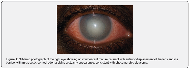
On presentation, uncorrected distance visual acuity was light projection in the right eye and 6/18 in the left eye, with pinhole improving to 6/12. The right pupil was irregular, mid-dilated, and sluggish. Assessment for a relative afferent pupillary defect was limited by the dense cataract and poor fixation, although an afferent response asymmetry was suspected. Confrontation visual fields were grossly full in the left eye but could not be reliably assessed in the right eye because of severely reduced acuity. Anterior segment evaluation of the right eye by slit-lamp bio microscopy revealed conjunctival and episcleral vessel injection, 3+ microcystic corneal oedema, and a markedly shallow anterior chamber, consistent with angle closure (Figure 1). The iris appeared bowed forward (iris bombe configuration). Examination of the right lens disclosed a large, white intumescent cataract with an anterior cortex that appeared milky and swollen, filling the posterior chamber and displacing the iris-lens diaphragm anteriorly (Figure 1). No phacodonesis or zonular laxity was observed. Anterior segment findings in the left eye were within normal limits, apart from an immature cataract with a Van Herick angle grade of 3.
Intraocular pressure measured by Goldmann applanation tonometry was 49 mmHg in the right eye and 16 mmHg in the left eye. Because microcystic corneal oedema limited visualization of the anterior chamber angle, topical anhydrous glycerol was applied briefly to the corneal surface as a temporary diagnostic aid. Its local hyperosmotic effect transiently dehydrated the corneal epithelium, reducing epithelial oedema sufficiently to improve clarity for gonioscopic assessment. Following this transient improvement in corneal clarity, gonioscopy of the right eye with a Goldmann three-mirror contact lens revealed complete appositional iridotrabecular contact in all four quadrants, with no identifiable angle structures, consistent with Shaffer grade 0 angle closure. The left eye demonstrated an open angle to the posterior trabecular meshwork in all quadrants (Shaffer grade 3). Posterior segment evaluation was not possible in the right eye because of the dense cataract. B-scan ocular ultrasonography confirmed a shallow anterior chamber, an acoustically solid lens mass consistent with an intumescent cataract, and an otherwise intact vitreous and retina with no evidence of suprachoroidal haemorrhage or choroidal effusion.
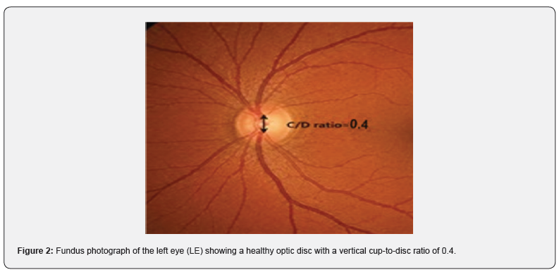
Undilated fundus examination of the left eye showed a healthy optic disc with a cup-to-disc ratio of 0.4 (Figure 2), normal rim tissue, and unremarkable macula and vessel. The differential diagnoses considered in this patient included primary acute angle-closure glaucoma, aqueous misdirection (malignant glaucoma), phacomorphic glaucoma, and phacolytic glaucoma. Primary acute angle-closure glaucoma was considered given the acute presentation with a shallow anterior chamber and elevated intraocular pressure; however, the demonstrably intumescent lens as the anatomical driver of the anterior displacement, and the absence of a prior history of symptomatic episodes in the fellow eye, argued against an idiopathic primary mechanism [6,7]. Aqueous misdirection was considered in view of the extreme shallowing of the anterior chamber but was regarded as less likely in the absence of a recent surgical history. Phacolytic glaucoma, which presents as open-angle glaucoma in the setting of a hypermature cataract with leaking lens proteins, was excluded on the basis of closed angles on gonioscopy, the absence of large translucent macrophages or white flocculent material in the anterior chamber, and the appearance of the lens consistent with an intumescent rather than a hyper mature morphology [8-10].
The clinical constellation of acute secondary angle closure, a markedly shallow anterior chamber, microcystic cornea oedema, iris bombe, and a demonstrably intumescent cataract led to the diagnosis of phacomorphic glaucoma. Medical management was initiated immediately with the aim of lowering intraocular pressure and reducing corneal oedema in preparation for definitive surgical treatment [1,2]. One drop each of timolol maleate 0.5%, brimonidine tartrate 0.2% and dorzolamide 2% was instilled in the right eye at 15-minute intervals. Oral acetazolamide 500 mg was also administered to further reduce aqueous production. Topical prednisolone acetate 1% was prescribed four times daily to control secondary anterior segment inflammation associated with the acute angle-closure episode and the anticipated postoperative inflammatory response. Within three hours of initiating emergency pressure-lowering treatment, intraocular pressure decreased to 22 mmHg, indicating an adequate initial response to topical aqueous suppression and oral acetazolamide.
Corneal oedema partially resolved; however, slit-lamp examination still demonstrated a shallow anterior chamber with persistent angle closure, consistent with an ongoing phacomorphic mechanism. Given the lens-induced nature of the attack and the need for definitive management, the patient was urgently referred for specialist ophthalmologic care. Following ophthalmologic assessment, urgent cataract extraction was performed. Manual small-incision cataract surgery was selected as the operative approach because of the intumescent nature of the cataract and the resource setting [11,12]. The procedure was completed without intraoperative complications. At the one-week postoperative review, the patient reported significant subjective improvement in vision and complete resolution of ocular pain (Figure 3).
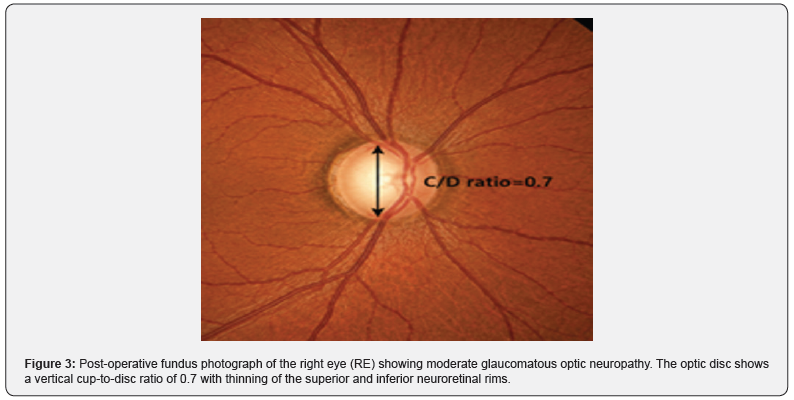
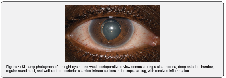
Best-corrected visual acuity in the right eye was 6/9. Intraocular pressure was 14 mmHg in the right eye and 15 mmHg in the left eye, without any pressure-lowering medications. The anterior chamber was deep and quiet, the cornea was clear, and the implanted intraocular lens was well-centred in the capsular bag (Figure 4). Postoperative fundus examination of the right eye demonstrated moderate glaucomatous optic neuropathy with a vertical cup-to-disc ratio of 0.7 (Figure 3), with thinning of the superior and inferior neuroretinal rims consistent with glaucomatous optic nerve damage secondary to prolonged intraocular pressure elevation. Gonioscopy of the right eye demonstrated a widely open angle in all quadrants, confirming complete resolution of the angle-closure mechanism following removal of the intumescent lens. The postoperative reopening of the angle supported a predominantly appositional, rather than synechial, angle-closure mechanism secondary to the intumescent lens. At the four-week postoperative visit, intraocular pressures remained stable at 15 mmHg bilaterally without medication.
Discussion
Phacomorphic glaucoma is a secondary acute angle-closure glaucoma that results directly from the mechanical effects of an enlarging intumescent lens. As the lens absorbs water and increases in volume during the intumescent phase of cataractogenesis, it pushes the iris anteriorly and reduces the depth of the anterior chamber. This reduction in anterior chamber depth narrows the iridocorneal angle. When the swollen lens produces complete pupillary block, preventing aqueous from flowing from the posterior to the anterior chamber, the iris bows anteriorly in the configuration known as iris bomb, opposing the trabecular meshwork and obstructing outflow, with a consequent precipitous rise in intraocular pressure. The pathophysiology of phacomorphic glaucoma differs fundamentally from that of phacolytic glaucoma. In phacolytic glaucoma, the angle remains open, and obstruction of the trabecular meshwork is caused by high-molecular-weight soluble lens proteins and macrophages that have leaked through microperforations in the intact capsule of a hypermature cataract.
In phacomorphic glaucoma, the angle is closed by iridotrabecular apposition secondary to forward displacement of the iris-lens diaphragm. This distinction is diagnostically critical because the treatment and prognosis differ substantially between these two conditions [8-10]. In the Ghanaian and broader West African context, phacomorphic glaucoma carries particular public health significance. Structural barriers to eye care, including the cost of surgery, travel distances to tertiary facilities, limited availability of trained ophthalmic surgeons, and cultural health-seeking behaviours, contribute to delayed presentation. A multicentre study from tertiary hospitals in West Africa found that patients with lens-induced glaucoma presented on average more than two weeks after symptom onset, substantially increasing the risk of permanent optic nerve damage at the time of presentation [13].
In the present case, the patient had experienced three years of progressive visual decline in the affected eye before seeking care, a pattern entirely consistent with the epidemiology of delayed cataract care in this setting [14]. The initial medical management of phacomorphic glaucoma is directed toward rapidly lowering intraocular pressure to reduce the risk of irreversible ischaemic damage to the optic nerve and to improve corneal clarity before surgery. Aqueous suppressants, including topical beta-blockers, alpha-2 agonists, and carbonic anhydrase inhibitors, form the cornerstone of medical therapy [1,2]. Topical steroids help suppress the secondary iridocyclitis that accompanies acute angle closure. Importantly, miotics such as pilocarpine should be used with caution or avoided entirely in phacomorphic glaucoma; in a pupillary-block mechanism, a miotic may paradoxically worsen the block by increasing lens-iris contact [2,7].
Unlike the situation in primary acute angle closure, laser peripheral iridotomy-while theoretically capable of bypassing the pupillary block - typically provides only transient pressure reduction in phacomorphic glaucoma because the primary anatomical cause, the intumescent lens, remains in situ. Definitive treatment is therefore prompt lens extraction [2]. Manual small-incision cataract surgery is the preferred surgical technique for phacomorphic glaucoma in resource-limited settings, including most Ghanaian tertiary hospitals [11,12]. MSICS offers several advantages over phacoemulsification in this context: it requires less sophisticated equipment, has a shorter learning curve, and is less vulnerable to intraoperative complications that can arise when attempting to phacoemulsify a dense, potentially pressurised intumescent lens.
Published series from sub-Saharan Africa and South Asia have consistently demonstrated that MSICS for phacomorphic glaucoma achieves excellent intraocular pressure control and favourable visual outcomes comparable to those achieved in planned cataract surgery, provided the procedure is performed before irreversible optic nerve atrophy supervenes [3,11]. From a systems perspective, this case highlights the importance of community-level eye health education and accessible, affordable outreach cataract screening programs in Ghana. The Ghanaian National Eye Health Policy and the Vision 2030 framework for Universal Eye Health identify the elimination of cataract blindness as a priority objective [5]. Strengthening primary eye care infrastructure to ensure timely identification of visually significant cataracts, before they progress to the intumescent stage, is the most effective strategy to eliminate phacomorphic glaucoma as a clinical entity in this setting [5,14].
Conclusion
Phacomorphic glaucoma is a secondary acute angle-closure glaucoma precipitated by a mechanically obstructing intumescent cataract. In Ghana and other sub-Saharan African countries, it continues to present as a cause of acute, potentially irreversible visual loss, largely because of delayed access to routine cataract care. The diagnosis rests on clinical recognition of the characteristic triad of a shallow anterior chamber, an intumescent cataract, and closed angles on gonioscopy, in the context of an acute, painful red eye with elevated intraocular pressure. Initial medical management to lower pressure and suppress inflammation, followed by urgent lens extraction, typically results in normalisation of intraocular pressure and meaningful visual recovery. Eyecare providers in resource-limited settings play an indispensable role in the timely identification of intumescent cataracts at risk of phacomorphic complications, and in ensuring appropriate and prompt surgical referral.
References
- Epstein DL (1982) Diagnosis and management of lens-induced glaucoma. Ophthalmology 89(3): 227-230.
- Johnson SM (2025) Cataract-Induced Glaucoma: Phacolytic/Phacomorphic. In Cataract Surgery in the Glaucoma Patient. Cham: Springer Nature Switzerland pp: 257-265.
- Pradhan D, Hennig A, Kumar J, Foster A (2001) A prospective study of 413 cases of lens-induced glaucoma in Nepal. Indian Journal of Ophthalmology 49(2): 103-107.
- Lee JW, Lai JS, Lam RF, Wong BK, Yick DW, et al. (2011) Retrospective analysis of the risk factors for developing phacomorphic glaucoma. Indian Journal of Ophthalmology 59(6): 471-474.
- Wiafe B, Quainoo A, Antwi P (2015) Ghana Blindness and Visual Impairment Study. Accra: Ghana Health Service and Operation Eyesight Universal.
- Stein JD, Khawaja AP, Weizer JS (2021) Glaucoma in adults - screening, diagnosis, and management: a review’, JAMA 325(2): 164-174.
- Kanski JJ, Bowling B (2015) Kanski’s Clinical Ophthalmology: A Systematic Approach. 8th edn. Edinburgh: Elsevier Health Sciences.
- Flocks M, Littwin CS, Zimmerman LE (1955) Phacolytic glaucoma: a clinicopathologic study of one hundred thirty-eight cases of glaucoma associated with hypermature cataract. AMA Archives of Ophthalmology 54(1): 37-45.
- Braganza A, Thomas R, George T, Mermoud A (1998) Management of phacolytic glaucoma: experience of 135 cases. Indian Journal of Ophthalmology 46(3): 139-143.
- Mandal AK, Gothwal VK (1998) Intraocular pressure control and visual outcome in patients with phacolytic glaucoma managed by extracapsular cataract extraction with or without posterior chamber intraocular lens implantation. Ophthalmic Surgery and Lasers 29(11): 880-889.
- Chandrashekharan S, Chakrabarty S, Tanwar M, Madhuvarasu B, Uduman MS, et al. (2022) Outcomes and favourable prognostic factors in patients of phacomorphic and phacolytic glaucoma managed by manual small-incision cataract surgery. Indian Journal of Ophthalmology 70(4): 1216-1221.
- Antwi-Adjei EK, Owusu E, Kobia-Acquah E, Dadzie EE, Anarfi E, et al. (2021) Evaluation of postoperative refractive error correction after cataract surgery. PLoS ONE 16(6): e0252787.
- Ayub R, Tom LM, Venkatesh R, Srinivasan K (2021) Outcomes and reasons for late presentation of lens induced glaucoma: a prospective study’, Ophthalmology Glaucoma 4(5): 504-511.
- Ocansey S, Kumi-Kyereme A, Awusabo-Asare K, Ilechie AA, Boadi-Kusi SB, et al. (2014) Utilization of eye care services among Ghanaian elderly population: evidence from a peri-urban community', Ophthalmology Research: An International Journal 1(2): 89-101.

























