




 JOJO Home



Abstract
The major goal of this case report is to describe the consequences of acute corneal hydrops and their resolution following intervention. This case
report’s etiology is unclear, making it extremely relevant for eye care specialists who can also grasp these treatments modalities and their results.
A case of severe acute corneal hydrops with uveal prolapse described in a 27-year-old female patient presented with a sudden unilateral (Right
eye) painful visual loss with watering of the eye (Aqueous humour coming out- siedel positive) and having no history of trauma, ocular surgery,
or any systemic illness. The anterior segment of her right eye showed a severe perforated corneal ulcer with iris prolapse, superior infiltrates,
superior vascularization & hypotonic (digitally).
Corneal hydrops is an uncommon complication, and, in this case, the preferred and suggestive intervention was to perform Tectonic penetrating
keratoplasty to gain a clear corneal interface with improved vision, and further follow-ups for better management was planned, followed by
cataract surgery as well as RGP contact lens trial. In addition to keratoconus, other ecstatic conditions, like the one in the present case, can
also cause corneal hydrops. However, even a slight increase in vision might be detrimental to the prognosis, necessitating a later penetrating
keratoplasty usually. Due to high cylinder prescription or any irregularity following suture removal, special contact lenses (RGP & Scleral) may
be required after penetrating keratoplasty.
Keywords: Corneal Hydrops; Tectonic Penetrating Keratoplasty; Trauma; Uveal Prolapse; Extraocular Muscles
Abbreviations:OD: Oculus Dexter (Right eye); OS: Oculus Sinister (Left eye); HM: Hand Movements; R/E: Right Eye; EOM: Extraocular Muscles; PVD: Posterior Vitreous Detachment; ONH: Optic Nerve Head; RCS: Retinal, Choroidal, Scleral; WNL: Within Normal Limit; CFCF: Count Finger- Close To Face; NI: No Improvement; AC: Anterior Chamber; BD: Twice Daily; Mg: Milligram; IOP: Intraocular Pressure; CF: Count Finger; TDS: Three Times Daily; QD: Quarter Times Daily; PC: Posterior Chamber; IOL: Intraocular Lens; PK: Penetrating Keratoplasty; RGP: Rigid Gas Permeable; DM: Descemet Membrane; UBM: Ultrasound Biomicroscopy
Introduction
Acute corneal hydrops is an uncommon disorder that is characterised by stromal edema brought on by aqueous fluid leakage through a Descemet membrane tear [1-3]. It frequently occurs in conditions connected to the ectatic cornea, such as advanced keratoconus. The patient had eye pain, photophobia, and a dramatic loss in vision. It typically shows up as an abrupt loss of eyesight. Nearly every patient in hospital-based practice needs a keratoplasty to enhance their vision, with the percentage rising with longer follow-up. After a hydrops has resolved, the presence of a corneal scar advises keratoplasty as soon as possible to restore eyesight [4-7].
Case Presentation
Initial visit
Patient description
A 27-year-old female presented for examination in tertiary eye hospital with complaints of sudden unilateral painful visual loss with watering of eyes (Aqueous coming out- siedel positive) in her right eye. Referred from outside to Cornea department at tertiary eye care hospital for further management. The history of the patient included eye pain, sudden loss of vision, but the patient denied any history or occurrence of ocular trauma, past ocular surgery, or any systemic illness.
Her past medical history was unremarkable with no use of systemic medication. Her family history was nil, her social history was negative for tobacco, alcohol, or recreational drug use. The patient reported that the aqueous coming out during sneezing started approximately 15 days before her visit. She had been medicating with Moxicip eye drops 3 times a day in the right eye while denied any similar ocular incidences in the past.
Examination findings
At the time of the initial presentation (Table 1), her visual acuity was found to be OD- HM+ (Pinhole- Not improving) & OS- 6/6. During retinoscopy, the glow was not visible in the R/E, and on subjective refraction, no improvement was seen. Pupil examination revealed, the R/E pupil was slightly reactive to light (with no afferent pupillary defects), confrontation visual field was unremarkable while EOM was full and free in all gazes. On Slit-lamp examination, slight redness in bulbar conjunctiva, abnormal tears flow, perforated corneal ulcer, Iris Prolapse, Anterior capsular cataract (central), disturbed anterior chamber (siedel positive), and digital hypotonicity were observed in the right eye, the fundus was not visible, so recommended B-scan. B-Scan depicting incomplete PVD and little vitreous opacity, with Axial, ONH, and RCS Complex was WNL, and no evidence of Retinal and Choroidal detachment or any intraocular foreign body (Figure 1).

Prognosis explained to the patient and their relatives about penetrating keratoplasty may need optical correction (glasses or contact lenses), regular follow-ups for 6 months. The patient was further investigated for her blood sugar level was normal, after that she had been admitted for tectonic penetrating keratoplasty under Ophthalmologist/Optometrist care. The patient was started on the medicine of Brimonidine + Timolol 3 times a day, Sodium Hyaluronate (0.1%) 4 times a day, Cyclosporine (2%) 2 times a day, 4 Quinpfs 0.5 ml (Moxiflox), 12 times a day, Tacrolimus (0.03%) once a day & Acetazolamide (250mg) 3 times a day for 2 days. Follow-up was scheduled after 2 days in the cornea specialty unit of the eye department. Prognosis explained to the patient and their relatives.
Follow-up visit-1
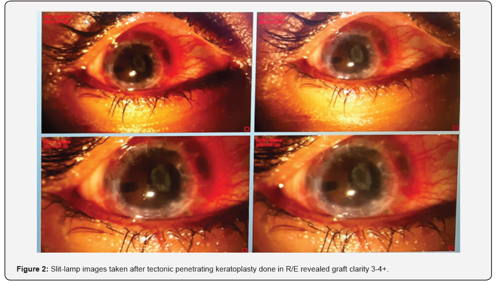
The patient reported good compliance with the prescribed treatment regimen, but no more changes in her symptoms. The patient’s visual acuity was CFCF in OD (pinhole- NI) & 6/6 in OS. Slit-lamp biomicroscopy revealed (Table 2) graft clarity 3-4+, blood clot in AC though it was well-formed, advised continuing with same medicines as previously prescribed (adding oral Ultracet- 325+37.5mg, BD) (Figure 2). IOP was non-recordable in R/E. The patient was instructed to take rest for 5 days and continue to use the prescribed drugs. A follow-up appointment was scheduled after 5 days to assess the changes.


Follow-up visit- 2
The patient reported no significant change in her vision. Her visual acuity was CF at 50 cm in OD (pinhole- NI) & 6/6 in OS. Slit-lamp biomicroscopy evaluation of right eye showed graft clarity 3+, AC formed, lens- complicated cataract with graft diffuse edema. Her corneal scrapping culture report was negative. IOP measured 01:00 PM was 8mm of Hg OD & 12 mm of Hg OS. Advised Acetazolamide (250mg) TDS, Brimonidine+Timolol TDS, Cyclosporine (2%) BD, Sodium Hyaluronate (0.1%) QD, Tacrolimus (0.03%) OD, Loteprednol QD. Follow-up was scheduled after 1 week.
Follow-up visit- 3
The patient returned as scheduled for her follow-up. She reported good compliance with the prescribed treatment regimen. Her visual acuity was found to be CF at 1m in OD (pinhole- NI) & 6/6 in OS. Slit-lamp biomicroscopy revealed graft clarity 3+, AC quiet, lens-complicated cataract, no evidence of infections, loose sutures removal done. Advised topical same as previously prescribed (add Prednisolone 6 times a day). IOP measured 02:05 PM was 9 mm of Hg in OD & 14 mm of Hg in OS. The patient was instructed to continue her prescribed topical & rest for 5 days, follow-up appointment was scheduled after 5 days and explained about the proposed cataract surgery after 6 months and explained the prognosis after cataract surgery and the importance of cataract surgery..
Results
The patient’s small incision cataract surgery with PC IOL was done on 31/12/2021. Examination findings on follow-up next day, IOL in situ, AC quiet, inferior PAS+, Advised topical (Brimonidine+ Timolol-BD, Cyclosporine-BD, Gatifloxacin+ Prednisolone- QD, Sodium hyaluronate- QD, Tacrolimus ointment-OD), Follow-up scheduled after 1 week. Examination findings on follow- up (08/01/2022), IOL in situ, AC quiet, inferior PAS+, Advised topical in standard dosages (Brimonidine+ Timolol-BD, Cyclosporine- BD, Gatifloxacin+ Prednisolone-TD, Sodium hyaluronate- TD, Tacrolimus ointment- OD), Follow-up scheduled after 1 month for refraction.
The patient returned as scheduled for her follow-up (05/02/2022). She reported good compliance with the prescribed treatment regimen. Her visual acuity was found to be 6/60 in OD (pinhole- 6/15) & 6/6 in OS.
Refraction: R/E +4.00/-7.75@10 (6/15), +2.50 Additions (N8 at 33cm), and L/E Plano (6/6), Patient was facing diplopia with given correction binocularly. Advised post-PK RGP trial.
RGP trial was done (12/02/2022): R/E Rose K2 post-PK 6.60/-3.75/8.70 (6/9, N12). No diplopia, over-refraction +2.00D.
RGP trial was done (12/02/2022): R/E Rose K2 post-PK 6.60/-3.75/8.70 (6/9, N12). No diplopia, over-refraction +2.00D.
Discussion
Plaut first described the condition as a sudden opacification of the corneal apex caused by Descemet membrane (DM) rupture [3]. Keratoconus is discovered in cases of impaired corneal biochemical activity, leading to secondary thinning and ectasia. The acute corneal hydrops happens once the intraocular aqueous passes through a rupture of DM, and it is characterized by stromal edema [2,3]. Commonly, the onset is in the second - third decade of life. The condition progresses until the fourth decade, once it stabilizes [2]. on examination, a significantly reduced visual acuity, photophobia, and eye pain is found. Rubbing the eyes is usually one of the commonest reported symptoms.
On slit-lamp evaluation, a well-defined micro-cystic corneal stromal and epithelial edema, stromal cysts, and injection are proved. However, the diagnosis is based on the patient’s history, and bio-microscopy, ultrasound biomicroscopy (UBM), or anterior optical coherent tomography were suggested [8]. The treatment could be conservative or surgical. The conservative therapy includes topical lubricants, antibiotics, cycloplegic agents, hyperosmotic solutions, anti-glaucoma drugs, topical corticosteroids, and non-steroid anti-inflammatory drugs. Surgical intervention includes intra-cameral injection of air or gas (pneumatic descemetopexy), but the definitive treatment is penetrating keratoplasty [1,9]./p>
Though local and systemic factors have been identified may predispose an individual with keratoconus to develop acute corneal hydrops. But in our patient, no local and systemic factors and even no history of rubbing eyes or trauma were observed. We used conservative and surgical treatment immediately because it is a rare event. Follow-up was recommended and a future about the proposed cataract surgery after 6 months and the prognosis was explained (keratoplasty to improve their vision the proportion increasing with further follow-up for cataract surgery and prevent from other complications or chance of failed graft after keratoplasty).
Conclusion
In addition to keratoconus, other ectatic conditions, like the one in the present case, can also cause corneal hydrops. However, even a slight increase in vision might be detrimental to the prognosis, necessitating a later penetrating keratoplasty usually. The intra-cameral gas injection decreases the length of morbidity and the danger of problems such corneal nervousness that could compromise the future graft, even if it has no effect on the final visual results. Due to high cylinder prescription or any irregularity following suture removal, special contact lenses (RGP & Scleral) may be required after penetrating keratoplasty, for an optometrist to monitor or provide pre- and post-operative treatment for this situation.
Acknowledgement:
Authors are also grateful to patients, authors/editors/publishers of all those articles, journals and books from where the literature for this article has been reviewed and discussed.
Patient consent: Written informed consent was obtained for identifiable health information included in this case report.
Funding: The authors declare that no funds, grants, or other support were received during the preparation of this manuscript.
References
- Maharana PK, Sharma N, Vajpayee RB (2013) Acute Corneal Hydrops in Keratoconus. Indian J Ophthalmol 61(8): 461-464.
- A Barsam, H Petrushkin, N Brennan, C Bunce, W Xing, et al. (2015) Acute Corneal Hydrops in Keratoconus: A National Prospective Study of Incidence and Management. Eye 29(4): 469-474.
- Sharma N, Mannan R, Jhanji V, et al. (2011) Ultrasound Biomicroscopy-Guided Assessment of Acute Corneal Hydrops. Ophthalmology 118: 2166-21671.
- SJ Tuft, LC Moodaley, WM Gregory, CR Davison, RJ Buckley (1994) Prognostic factors for the progression of keratoconus. Ophthalmology 101(3): 439-447.
- Weed KH, McGhee CN (1998) Referral Patterns, Treatment Management, and Visual Outcome in Keratoconus. Eye (Lond) 12(Pt 4): 663-668.
- Tuft SJ, Gregory WM, Buckley RJ (1994) Acute Corneal Hydrops in Keratoconus. Ophthalmology 101(10): 1738-1744.
- Basu S, Reddy JC, Vaddavalli PK, et al. (2012) Long-term Outcomes of Penetrating Keratoplasty for Keratoconus with Resolved Corneal Hydrops. Cornea 31: 615-620.
- Greenwald MF, Vislisel JM GK (2016) Acute Corneal Hydrops.; EyeRounds.org 8: 123-125.
- S Grewal 1, P R Laibson, E J Cohen, C J Rapuano (1999) Acute Hydrops in the Corneal Ectasias: Associated Factors and Outcomes. Trans Am Ophthalmol Soc 97: 187-203.

























