




 JOJO Home



Abstract
Keywords: Cerebral Venous Sinus Thrombosis; CVST; Idiopathic Thrombocytopenic Purpura; Papilledema; Eltrombopag
Abbreviations:CVST: Cerebral Venous Sinus Thrombosis, ITP: Idiopathic Thrombocytopenic Purpura, CE-MRV: Contrast-Enhanced Magnetic Resonance Venography, BMI: Body Mass Index, OCT: Optical Coherence Tomography, CSF: Cerebral Spinal Fluid, INR: International Normalized Ratio
Introduction
Cerebral venous sinus thrombosis (CVST) is a relatively rare form of stroke associated with increased mortality in young adults, with an incidence rate of 3-4 cases per 2 million people per year [1]. The causes of CVST include various factors such as genetics, pregnancy, infections, and tumors. CVST can manifest with several signs and symptoms, making it challenging to differentiate from other neurological disorders [2]. Thus, radiological investigation is important in the diagnosis of CVST. Managing CVST focuses on early detection and prevention of thrombus growth and related complications. Prognosis is usually good, with up to 80% of patients making a complete recovery [3]. However, conflicting treatment approaches often make clinical management challenging.
This case highlights the uncommon scenario of a 19-year-old female patient diagnosed with CVST in the context of ITP. The intricate management of her condition involved careful adjustments in her treatment regimen, underscoring the importance of a multidisciplinary approach. The combination of increased dosages of methylprednisolone alongside fondaparinux demonstrated ef fective management, providing valuable insights into therapeutic strategies for similar cases. Furthermore, the favorable outcomes observed at follow-up emphasize the potential for positive intervention in patients suffering from these overlapping conditions. We obtained written informed consent from the patient and then analyzed the collected data.
Case Description
Our study subject is a 19-year-old female. She experienced blurred vision and headaches lasting for one month. She has been taking methylprednisolone and platelet-raising drugs for 1 year because of idiopathic thrombocytopenic purpura. Before the onset of illness, she took eltrombopag olamine tablets 75 mg for ITP for 4 weeks. There was no family history suggestive of cranial mass, infection, or demyelinating disease. Physical examination: The patient’s body mass index (BMI) was 26 kg/m² (normal range: 18.5–23.9 kg/m²). Her visual acuity was 20/20 and 20/40 in the right and left eye, respectively, and the intraocular pressure was R/L 24.1/21.6mmHg.
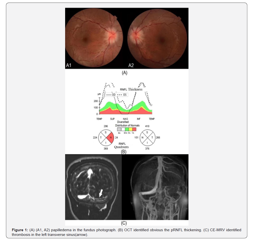
Fundus examination showed optic disc edema in both eyes (Figure 1(A1, A2)), and optical coherence tomography (OCT) indicated increased pRNFL thickness (Figure 1(B)). No obvious abnormalities were found in the nervous system. The blood platelet count was 99*10^9/L (100~300) 10^9/L), the thrombocytocrit was measured at 0.095% (0.11~0.28%). An examination of the cerebral spinal fluid (CSF) yielded the following results: pressure >400mm H2O; protein 14.4mg/dl; glucose 3.31mmol/L; protein C 138%; protein S 52.9% and antithrombin III 112.2%. No obvious abnormality was revealed in genetic testing for thrombophilia. The nucleic acid of SARS-CoV-2 was negative.
An examination of intracranial venous contrast-enhanced magnetic resonance venography (CE-MRV) and black blood imaging revealed thrombosis in the left transverse sinus, and mural thrombus in the superior sagittal sinus (Figure 1(C)). Since there is a contradiction between the treatment of CVST and immune thrombocytopenia, anticoagulation causes a progressive decrease in platelets. Based on multidisciplinary consultation, the dosage of methylprednisolone tablets was increased, while selective factor Xa inhibitor fondaparinux sodium replaced unfractionated heparin to reduce bleeding risk. Oral warfarin was subsequently introduced, with the international normalized ratio (INR) closely monitored and targeted to a range of 1.8–2.0.
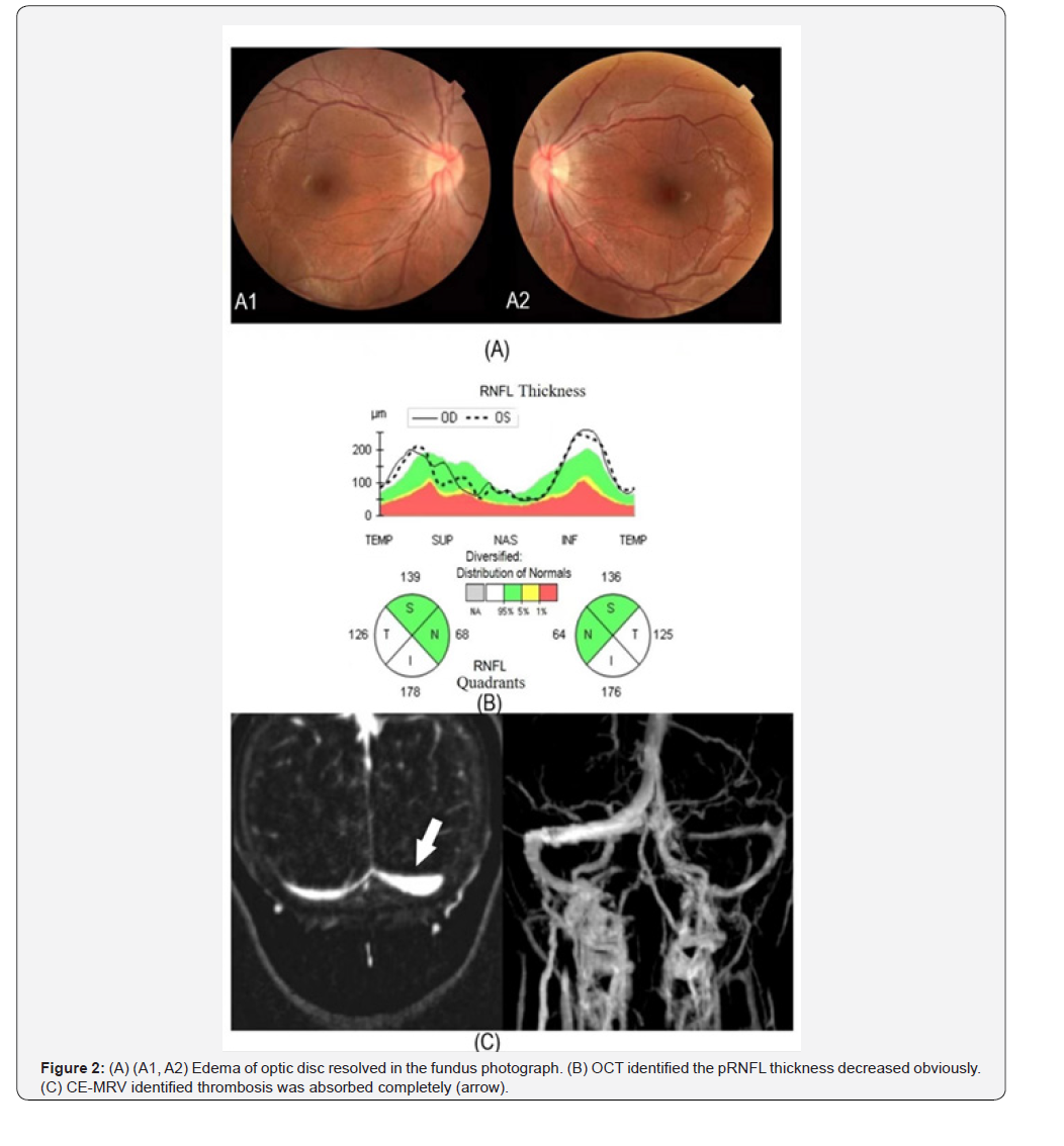
At the one-month follow-up, the patient’s best corrected visual acuity improved to 20/20 in both eyes, with a gradual reduction in optic disc edema, OCT results indicated a decrease in pRNFL thickness, and CE-MRV imaging showed a significant reduction in thrombosis within the left transverse sinus. At the six-month follow-up, her condition remained stable, with complete resolution of optic disc edema (Figure2(A (A1, A2)), and OCT findings indicated a notable decrease in pRNFL thickness (Figure2(B)). CE-MRV imaging confirmed complete absorption of the thrombus (Figure2(C)). At the one-year follow-up, her visual acuity was maintained at 1.0, and OCT revealed no signs of thinning or atrophy in the pRNFL.
Discussion and Conclusions
The case of the 19-year-old female patient suffering from visual disturbances and headaches highlights the complex interplay between idiopathic thrombocytopenic purpura (ITP) and cerebral venous sinus thrombosis (CVST). This patient’s condition is particularly significant as it underscores the importance of recognizing potential thromboembolic complications in individuals with underlying hematological disorders, which may not always present with overt symptoms. Because such cases are rare, researchers need to explore diagnostic and therapeutic approaches specifically for patients diagnosed with both ITP and CVST.
In this case, the multidisciplinary approach to management was crucial, as the treatment of CVST in the context of ITP presents a unique challenge. While anticoagulation is essential in managing CVST, it poses a risk of exacerbating thrombocytopenia, thus complicating the clinical decision-making process [4]. The decision to increase the dose of methylprednisolone while introducing fondaparinux as an anticoagulant reflects a strategic balance between managing thrombotic risk and preserving platelet function. The presence of optic disc edema, as observed in this patient, is a clinical manifestation that warrants careful evaluation, especially when coupled with the findings of elevated intracranial pressure from cerebrospinal fluid analysis and imaging studies revealing venous thrombosis.
Papilledema due to idiopathic intracranial hypertension was not considered in this patient, despite her high BMI. Genetic testing for thrombophilia was also normal. After taking the eltrombopag tablets 75mg for 4 weeks, the patient developed headaches and decreased vision. Moreover, thrombosis caused by drug-induced improvements in platelet technology cannot be excluded. In addition, if there is a risk of thrombotic formation of PLT, it should be stopped immediately to prevent thrombosis. Thrombosis has been suggested as a side effect of eltrombopag tablets.
Furthermore, the patient’s favorable recovery, with her visual acuity and optic disc edema showing significant improvement at follow-up, reinforces the potential for successful outcomes in managing patients with concurrent ITP and CVST when appropriate treatment strategies are employed. This case serves as a reminder of the necessity for ongoing vigilance and the adaptation of treatment protocols in complex clinical scenarios involving thrombosis and hematological conditions [5].
Summary
In conclusion, this case contributes to the scarce literature on managing CVST in patients with ITP and highlights the importance of a tailored, it is essential to acknowledge its limitations. Additionally, the relatively short follow-up period restricts the ability to draw definitive conclusions regarding long-term outcomes and the efficacy of the treatment regimen employed. Future research should aim to address these gaps by exploring larger cohorts and extended follow-up durations to better elucidate the interplay between ITP and CVST. Furthermore, the insights gained from this case serve as a reminder of the need for heightened awareness among clinicians regarding the diagnostic complexities and therapeutic challenges posed by these coexisting conditions, ultimately improving patient management and outcomes.
Funding
The study was supported by granting the Project of Zhejiang Provincial TCM Science and Technology (No.2023ZL388)
The Research Project of Zhejiang Chinese Medical University (No.2025JKZKTS50)
Foundation of Zhejiang Medical Science and Technology Project (No.2024KY1211)
National Natural Science Foundation of China for Young Scientists (No. 82505676)
Author Contributions
XZ and FL designed and conducted the study; XZ and KZ collected the data; XZ prepared the manuscript. All the authors reviewed and approved the final manuscript.
Acknowledgments
I would like to thank the patient and her family for supplying the data.
Ethics Approval and consent to participate
This study was approved by the First Affiliated Hospital of Zhejiang University of Traditional Chinese Medicine Ethics Committee (2023-KLS-172-01) and was conducted following the Declaration of Helsinki in its currently applicable version and applicable Chinese laws.
Consent for Publication
Written informed consent was obtained from the patient for publication of this case report and any accompanying images. A copy of the written consent is available for review by the Editor- in-Chief of this journal.
Availability of data and material
The data involved in this study are owned by the Department of Ophthalmology of the First Affiliated Hospital of Zhejiang Chinese Medical University.
Data Availability Statement Missing on Editorial System
All data generated or analysed during this study are included in this published article.
References
- Saposnik G, Barinagarrementeria F, Brown RD, Bushnell CD, Cucchiara B, et al. (2011) Diagnosis and management of cerebral venous thrombosis: a statement for healthcare professionals from the American Heart Association/American Stroke Association. Stroke 42(4): 1158-1192.
- Idiculla PS, Gurala D, Palanisamy M, Vijayakumar R, Dhandapani S, et al. (2020) Cerebral Venous Thrombosis: A Comprehensive Review. European neurology 83(4): 369-379.
- José M Ferro (2005) Prognosis and treatment of cerebral vein and dural sinus thrombosis. Clinical advances in hematology & oncology: H&O 3(9): 680-681.
- Cui J, Li X, Xia D, Gu X, Zhao Y, et al (2024) Peritumoral vessel characteristics on magnetic resonance venography reflect the sinus invasion status of para-sinus meningioma. Quantitative imaging in medicine and surgery 14(12): 8183-8195.
- Toohey TP, Shortt J, John N, Al-Qureshi S, Wickremasinghe SS (2024) Intravitreal anti-vascular endothelial growth factor therapy in the treatment of vision loss associated with hematologic malignancy. American journal of ophthalmology case reports 36: 102095.

























