




 JOJO Home



Abstract
A healthy 10-year-old boy presented with a conjunctival lesion following eye trauma. The patient was injured while playing in the pool. He was accidentally hit in the left eye while wearing googles, and in the act of removing the dislodged goggles, a brief negative suction pressure against the conjunctiva was created. A mobile conjunctival lesion in the inferior fornix was appreciated on initial silt lamp exam. The eye exam was otherwise normal including visual acuity. The patient was prescribed erythromycin ophthalmic ointment and was closely monitored due to the lack of literature on this mechanism of injury. Despite notable improvement following erythromycin use, the lesion began to protrude out of the inferior fornix and keratinize on repeat examination three weeks after the injury. Conjunctival excision was performed in the operating room without the use of an amniotic membrane graft due to the small, narrow base of the lesion. Complete recovery was achieved without symblepharon formation or forniceal shortening. This case demonstrates that mild negative suction pressure injuries to the conjunctiva can be successfully managed with erythromycin ointment and surgical excision of tissue outgrowth.
Keywords: Negative Pressure Suction Injury; Suction Injury; Conjunctival Trauma; Pediatric Conjunctival Trauma; Case Report
Introduction
Ocular trauma to the anterior segment, including the conjunctiva, cornea, sclera, iris, and lens, is typically classified as mechanical, chemical, or thermal [1]. Anterior segment trauma is commonly described in the context of foreign bodies, abrasions, and chemical burns [1], yet ocular suction injuries are rarely reported. To our knowledge, this is the first report in the English literation of a negative pressure suction injury to the human conjunctiva; a PubMed query of MeSH derivations of conjunctival suction injury yielded no relevant publications.
Case Description
A healthy 10-year-old boy presented to the emergency room with a left conjunctival lesion following eye trauma. The patient’s left eye was accidentally hit while he was wearing googles in the pool, causing the goggles to shift in position from the eyelid skin to the conjunctiva. When the patient removed the goggles, a negative suction pressure was created, and he reported discomfort in the left eye immediately. Later that day, he noticed a red lesion protruding from the left inferior fornix and mild left lower eyelid edema. He denied any ophthalmic or relevant past medical history. On initial examination, visual acuity was 20/15 in each eye and intraocular pressures were normal. Pupils were round and reactive with no relative afferent pupillary defect. Extraocular movements and confrontation visual fields were full. The patient was orthotropic in primary position at distance and near. Color perception by Ishihara was normal (10/10 in each eye). A mobile erythematous palpebral conjunctival lesion in the left inferior fornix was appreciated. The anterior segment examination was normal including no corneal abrasion or cell/flare in the anterior chamber. Dilated fundoscopic examination was unremarkable. Cycloplegic refraction demonstrated age-appropriate hyperopia. The patient was prescribed erythromycin ophthalmic ointment (5 mg/gm) into the left eye three times per day.
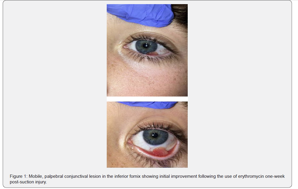
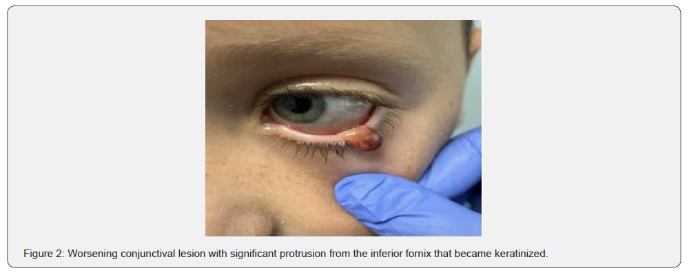
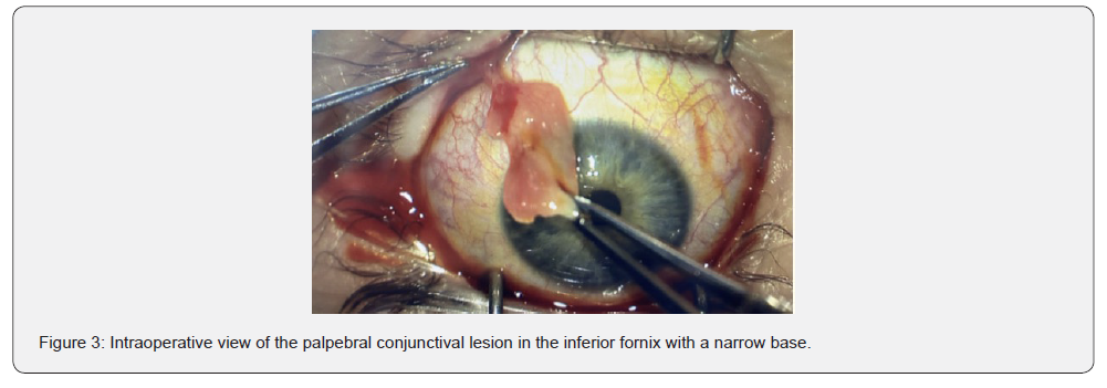
Eight days following the injury, the patient presented to the pediatric ophthalmology clinic for further evaluation. The patient’s mother reported adherence to the erythromycin ointment and noted significant improvement. Visual acuity remained 20/15 in each eye. A mobile palpebral conjunctival lesion in the left inferior fornix was again appreciated on slit lamp exam, which extended two millimeters on to the bulbar conjunctiva (Figure 1). The exam otherwise remained normal. The patient was instructed to continue the erythromycin ointment for two additional weeks. Three weeks after injury, the mobile conjunctival lesion continued to protrude out of the inferior fornix and had begun to keratinize (Figure 2), so surgical excision was recommended. The patient and his family elected to undergo conjunctival excision one week later. Intraoperatively, the conjunctival lesion was noted to be eight millimeters in length, originating from the left lower inferior palpebral conjunctiva, with a narrow base (Figure 3). The lesion was excised with Westcott scissors and gentle cautery was utilized for hemostasis. An amniotic membrane graft was not utilized given the less than one-millimeter, narrow base of the lesion and corresponding small conjunctival defect. The pathology report noted granulation tissue with acute and chronic inflammation, necrosis, and reactive vascular changes. Maxitrol ointment was prescribed postoperatively three times per day for one week. One week postoperatively, the inferior palpebral conjunctiva was white and quiet and there was no symblepharon formation. Three months postoperatively, the conjunctiva remained white and quiet with no symblepharon formation or forniceal shortening. Visual acuity, intraocular pressures, extraocular pressures, and color perception were unchanged from baseline.
Conclusions
While conjunctival lesions can be isolated, they may be the presenting sign of intraocular trauma such as traumatic iritis, hyphema, iridodialysis, vitreous hemorrhage, commotio retinae, and ruptured globe, to name a few. As a result, it is imperative to rule out intraocular trauma on initial conjunctival trauma presentation. Due an unremarkable anterior segment, dilated fundoscopic exam, and sensory exam on initial presentation, our patient was started on medical management with erythromycin ointment and followed closely. Upon repeat examination, the lesion had become significantly displaced and keratinized. Keratinization occurs at the cellular level as a reaction to irritation in an attempt to shed off old cells and replace with a mature population [2]. This process created a displaced lesion that was uncomfortable for the patient and was cosmetically unacceptable, thereby warranting surgical excision. The pathology report indicated granulation tissue with acute and chronic inflammation, necrosis, and reactive vascular changes. These changes are consistent with an ischemic injury to the conjunctival tissue that was pulled off from the ocular surface [3]. This injury occurred via a negative suction mechanism to the conjunctiva, which was created from the force applied to the surface of the patient’s dislodged googles.
Suction injuries occur when a part of the body is exposed to negative pressure. Small suction injuries to the skin cause mild bruising due to rupture of small superficial blood vessels [4]. Suction injuries can range from mild to severe depending on the length of exposure to the negative pressure, the magnitude of the suction pressure, and the area of injury. A severe complication of suction injuries is compartment syndrome [4]. Although the conjunctiva is a thin, delicate structure that is highly vascularized in its deepest fibrous layer, the negative pressure insult experienced by this patient was fortunately not damaging enough to cause severe disruption in blood flow. Suction injuries to children commonly occur from the drains of swimming pools, and several case reports have been published about extremity injuries or bowel eviscerations from these drains [5-6]. Although this is the first case report of a suction injury to the conjunctiva, Fernandes et al. reported a case of a teenager whose eyes were briefly exposed to the negative suction pressure from a pool drain after getting his face stuck inside [7].
The suction pressure caused a brief expansion and traction of the vitreous base onto the retinal surface which resulted in vascular leakage with macular edema, mild elevation of retinal layers, and acute vision loss. The patient had a visual acuity of 20/20 bilaterally and a complete resolution of macular edema one week after the trauma without any treatment [7]. After surgical excision, our patient had complete resolution of symptoms and of the conjunctival lesion. He was followed postoperatively to monitor for symblepharon formation and forniceal shortening, which would indicate chronic inflammation of the conjunctival layers, but there was no symblepharon or forniceal shortening after three months. Our case suggests that mild negative pressure suction injuries to the palpebral conjunctiva can be successfully managed with erythromycin ointment and surgical excision of tissue outgrowth, without the use of an amniotic membrane graft if the base of the lesion is very small. Injuries to the ocular surface, in particular from suction injuries that can potentially damage vascular supply to delicate tissues, warrant prompt evaluation by an ophthalmologist.
Conflicts of Interest
The authors declared no potential conflicts of interest with respect to the research authorship, and/or publication of this article.
Ethical Approval and Informed Consent
The present study did not require IRB approval. Written informed consent was obtained from the patient and family to publish this case report.
References
- Comez AT, Ozbas M (2022) Evaluation and Management of Ocular Traumas. In: Vision Correction and Eye Surgery.
- Deo PN, Deshmukh R (2018) Pathophysiology of keratinization. J Oral Maxillofac Pathol 22(1): 86-91.
- Eagle RC Jr (2016) Conjunctiva In: Eye Pathology: An Atlas and Text, 3rd pp: 90-133.
- Ihama Y, Ninomiya K, Noguchi M, Fuke C, Niki H, et al. (2010) An autopsy case of suction injury. Leg Med (Tokyo) 12(4): 188-191.
- Škach J, Kašák P, Šrám J (2015) Úraz podtlakem v bazénu: etiologie, prevence a management [Swimming pool suction injury: etiology, profylaxis and management]. Rozhl Chir 94(1): 34-38.
- Saborido Fiaño R, Illade Quinteiro L, Fariña Nogueira S, Baña-Souto A, Regueiro-García A, et al. (2015) Suction entrapment of an upper limb in a swimming pool. Pediatr Int 57(5): 1049-1050.
- Fernandes T, Freitas C, Mendonça L, Sousa K (2018) Suction-induced retinopathy: optical coherence tomography correlations. BMJ Case Rep 2018: bcr2018224531.

























