Abstract
Retinal tears occurring more than one year after acute posterior vitreous detachment (PVD) are rare. However, patients with notable risk factors, such as myopia and lattice degeneration, and those aged <60 years require prolonged monitoring to prevent vision-threatening complications [1,2]. This case report presents a patient who developed a retinal tear 21 months after initial PVD, underscoring the importance of extended follow-up protocols.
Keywords: Floaters; Posterior Vitreous Detachment; Retinal Tear; Laser Retinopexy
Abbreviations: PVD: Posterior vitreous detachment
Introduction
The retinal tears are known to occur few weeks after PVD, in the present publication we report a case of delayed retinal tear formation following acute posterior vitreous detachment (PVD).
Case Report
Initial Presentation
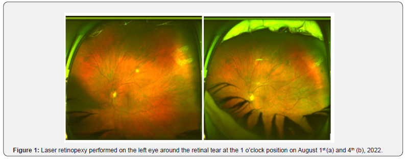
On August 1, 2022, a myopic male patient (-1.75 diopters) reported a 10-day history of floaters and flashes in the left eye. Examination following pupillary dilation revealed a posterior vitreous detachment with moderate vitreous hemorrhage. Retinal assessment showed a tear at the 1 o’clock position. Urgent argon laser retinopexy was performed without complications (Figure 1a & b) and a structured follow-up plan involving hemorrhage monitoring and three-monthly exams were initiated. complications, and a structured follow-up plan involving hemorrhage.

Disease Progression and Subsequent Events
• June 2023: While in France, the patient experienced
posterior vitreous detachment in the right eye, complicated
by retinal detachment.
• Pars-plana vitrectomy and gas injection were successfully
performed.
• Follow-up in our department resumed after his return
from France and continued until October 29, 2023.
• Consultation Gap: No ophthalmologic assessments occurred
between October 2023 and April 2025, as the patient
did not return during this period.
April 2025
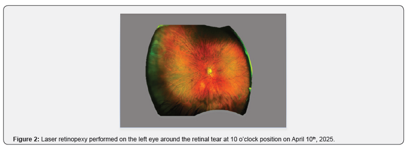
On April 10, 2025, the patient presented with floaters in the left eye. Examination revealed a retinal tear at the 10 o’clock position. Immediate laser retinopexy was performed in our department to stabilize the condition (Figure 2).

Discussion
Although retinal tears following acute PVD usually appear
within weeks, delayed cases remain uncommon. However, such
occurrences highlight the need for extended monitoring in highrisk
patients [1-3].
• Risk Assessment: Patients with myopia, lattice degeneration,
or those under 60 years require follow-up beyond standard
guidelines.
• Long-Term Follow-up Protocols: Educating patients on
symptom recognition and ensuring routine ophthalmic evaluations
is essential.
Disclosure Statement:
There are no conflicts of interest to report for author.
Acknowledgement
The author wishes to thank Mr. Joseph Bennett for editing the manuscript.
References
- Jindachomthong KK, Cabral H, Subramanian ML, Ness S, Siegel NH, et al. (2023) Incidence and risk factors for delayed retinal tears after an acute, symptomatic posterior vitreous detachment. Ophthalmol Retina 7(4): 318-324.
- Patricia Bouweraerts K (2022) Delayed retinal tears may occur long after acute vitreous detachment. Ophthalmology Advisor.
- Uhr JH, Obeid A, Wibbelsman TD, Wu CM, Levin HJ, et al. (2020) Delayed retinal breaks and detachments after acute posterior vitreous detachment. Ophthalmology 127(4): 516-522.


