IS/OS-Ellipsoid Zone Complex Anomaly of Benign Concentric Annular Macular Dystrophy
Fubin Wang* and Aijun Wang
Shanghai Bright Eye Hospital, 899 Maotai Road, Changning District, Shanghai 200336, Shanghai, China
Submission: May 8, 2023;Published: May 12, 2023
*Corresponding author: Fubin Wang, Shanghai Bright Eye Hospital, 899 Maotai Road, Changning District, Shanghai 200336, Shanghai, China
Fubin W, Aijun W. IS/OS-Ellipsoid Zone Complex Anomaly of Benign Concentric Annular Macular Dystrophy. JOJ Ophthalmol. 2023; 10(1): 555776. DOI: 10.19080/JOJO.2023.10.555776
Abstract
Aim: To report IS/OS-Ellipsoid zone complex anomaly of benign concentric annular macular dystrophy.
Methods: A case of adult-onset macular dystrophy was observed with BCAMD associated with the IS/OS-Ellipsoid zone complex anomaly by CFP, OCT, en-face analysis, FA and FAF, etc.
Results: CFP revealed concentric rings in the parafoveal area prominent and drusen in the macula of right eye and not concentric rings in the left eye. No concentric ring changes were observed in FA and FAF. En-face analysis found that the IS/OS-Ellipsoid complex was obviously abnormal. Although the left eye did not show the concentric ring-like changes in the CFP, but IS/OS-Ellipsoid complex was abnormal with semi-annular blotch-like dark areas.
Conclusion: BCAMD is a rare macular dystrophy. En-face analysis found that the IS/OS-Ellipsoid complex was obviously abnormal.
Keywords: Benign Concentric Annular Macular Dystrophy; OCT; En-Face Analysis; IS/OS-Ellipsoid Complex; Drusen
Introduction
Benign concentric annular macular dystrophy (BCAMD) is a rare macular dystrophy, first described by Duetman in 1974 [1]. This is caused by mutation in the interphotoreceptor matrix proteoglycan 1 gene on chromosome 6 [2]. This report describes a case of adult-onset macular dystrophy with BCAMD associated with the IS/OS-Ellipsoid zone complex anomaly.
Subjects and Methods
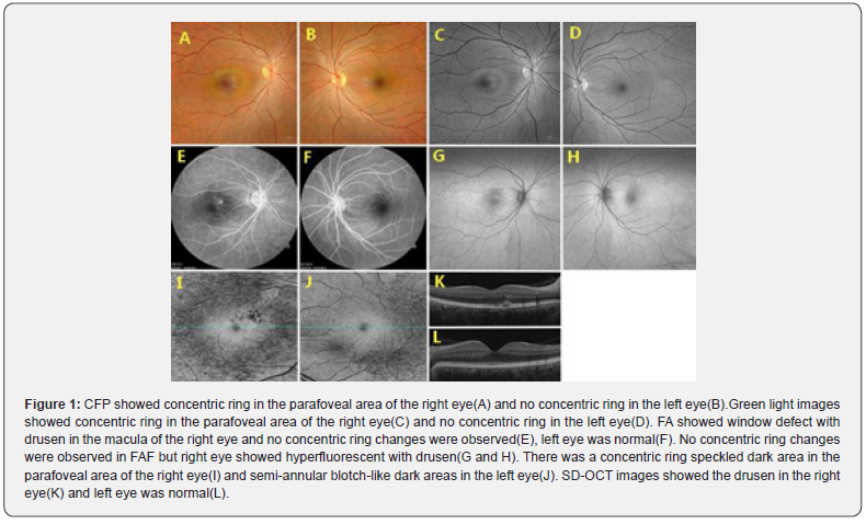
Benign concentric annular macular dystrophy is associated with gene mutation and is clinically rare. In the multi - mode images, different image characteristics are presented. A 42-year-old woman was referred to the retina unit of our hospital. She complained of photophobia. Best corrected visual acuity(BCVA) in the right eye and left eye was 1.0, respectively. The anterior segment examination was normal in both eyes. Fundus bio microscopy and color fundus photograph(CFP, Clarus 500, Carl Zeiss Meditec, Inc) revealed concentric ring in the parafoveal area prominent and drusen in the macula of right eye and not concentric rings in the left eye. The green light image revealed a concentric ring in the parafoveal area of right eye. Fluorescein angiography (FA, Visucam 524, Carl Zeiss Meditec AG) showed window defect with drusen in the macula of the right eye. No concentric ring changes were observed in FA and fundus autofluorescence(FAF, Daytona P200T). FAF showed hyperfluorescent with drusen in the right eye.
Except for drusen, SD-OCT in both eyes showed no significant abnormalities. However, en-face analysis found that the IS/OS-Ellipsoid complex was obviously abnormal by OCT (Cirrus HD-OCT 5000, Germany). There was a concentric ring speckled dark area in the parafoveal area of the right eye. Although the left eye did not show the concentric ring-like changes in the CFP, but IS/OS-Ellipsoid complex was abnormal with semi-annular blotch-like dark areas observed. (Figure 1) The perimetry (Humphrey field analyzer 860) and the flash electroretinogram (GT-2008V-1) in both eyes were within normal range (Figure 2).
Discussion
Benign concentric annular macular dystrophy (BCAMD) is a rare macular dystrophy, Good visual acuity is retained till late, which explains the term “benign” [3]. It is characterized by the presence of bull’s-eye maculopathy with annular atrophy of the pigment epithelium in the perifoveal retina with central respect. Several reports have described abnormal changes such as CFP, FA, FAF, OCT, etc. But not all of these abnormalities are present, and there may be variations in the severity of the condition [4,5].


En-face analysis is a useful examination technique by OCT, especially in the early stages of the disease. This preset is designed to highlight disruptions to the IS/OS-Ellipsoid zone due to a variety of causes. It follows the RPE contour and is elevated slightly to put it at the level of the IS/OS-Ellipsoid zone (Upper: 39um above the RPE layer; Lower: 19um above the RPE layer). Disturbances to the IS/OS-Ellipsoid zone show as dark areas. It facilitates early detection of IS/OS-Ellipsoid zone exceptions.
References
- Deutman AF (1974) Benign concentric annular macular dystrophy. Am J Ophthalmol 78(3): 384-396.
- Van Lith-Verhoeven JJ, Hoyng CB, van den Helm B, Deutman AF, Brink HM, et al. (2004) The benign concentric annular macular dystrophy locus maps to 6p12.3-q16. Invest Ophthalmol Vis Sci 45(1): 30-35.
- Gomez-Faiña P, Alarcón-Valero I, Buil Calvo JA, Calsina-Prat M, Martín-Moral D, et al. (2007) Distrofia macular anular benigna concéntrica. Arch Soc Esp Oftalmol 82(6): 373-376.
- Aarti Jain, Giridhar Anantharaman, Anubhav Goyal, Mahesh Gopalakrishnan (2019) Multimodal imaging of benign concentric annular macular dystrophy. Indian J Ophthalmol 67(10): 1719-1720.
- Karina Fernández-Berdasco, José Galvez-Olortegui, Sussan's Pamela Guillén-Lozada, Noelia García González, Joaquín Castro-Navarro (2023) PRPH2 mutation c.582-1G>A causing adult-onset macular dystrophy with a benign concentric annular macular dystrophy phenotype in a family. Arq Bras Oftalmol S0004-27492023005001202.


