Irido-Chorio-Retinal Colobomas: About 3 Cases in Eye Center of Abass Ndao Hospital
Diagne Jean Pierre1*, Gueye Abou1, Aw Aissatou1, Ka Aly Mbara1, Sy El Hadji Malick1, Mbaye Soda1, Ousmane Ndiaga sengho, Marguerite Edith Quenum De Meideros1, Hawo Madina Diallo1, Joseph Matar Mass Ndiaye2 and Ndiaye Papa Amadou1
1Abass Ndao Hospital Eye Center, USA
2Ophtalmology Department of Aristide Le Dantec Hospital, USA
Submission: February 02, 2022;Published: February 16, 2022
*Corresponding author: Abass Ndao, Ophtalmology department of Aristide Le Dantec hospital, USA
How to cite this article: Diagne J P, Gueye A, Aw Aissatou, Ka Aly M, Sy El Hadji M,. Irido-Chorio-Retinal Colobomas: About 3 Cases in Eye Center of Abass Ndao Hospital. JOJ Ophthalmol. 2022; 9(1): 555755. DOI: 10.19080/JOJO.2022.09.555755
Abstract
Aim: To describe clinical and prognostic characteristics of irido-chorio-retinal colobomas through 3 cases.
Observations: The average age of the three patients was 12,66 years. The sex-ratio was 0,5 with female predominance (66,7%). The three patients were admitted for a severe visual impairment and automated refraction test did not work for the two eyes. They all presented a nuclear cataract and an inferior iris coloboma for the two eyes. All the patients had bilateral chorio-retinal colobomas including papilla. The pediatric examination found one case of cryptorchidism but was normal for the two other patients. B-scan ocular ultrasound found a chorio-retinal defect facing the optical nerve for one patient. We managed the low vision and prescribed sunglasses to avoid photophobia. A cataract surgery was realized, and we also gave a genetic counselling. No patient had a retinal detachment.
Discussion & Conclusion: Chorio-retinal coloboma is a rare and serious disease, because of its evolution and complications. It requires early, appropriate, and multidisciplinary treatment.
Keywords: Coloboma; Iris; Choroid; Retina; Amblyopia
Introduction
Eye colobomas are congenital malformations of the eye secondary to an abnormality of closure of the embryonic fissure. All eye structures may be affected [1]. Chorio-retinal coloboma is an abnormality of choroid and retinal development. The purpose of our work was to describe the clinical and prognosis aspect of irido-chorio-retinal colobomas through three cases.
Observations
Case 1
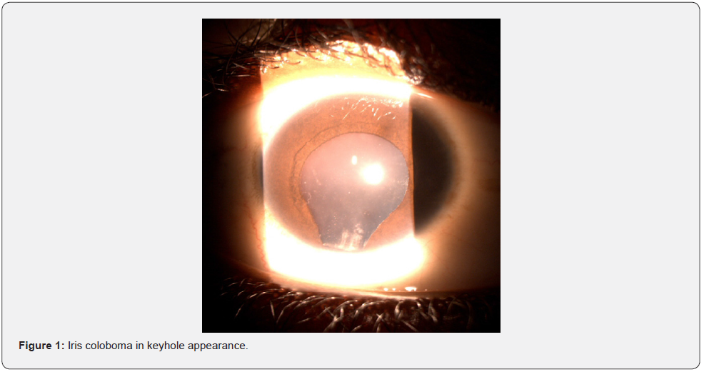
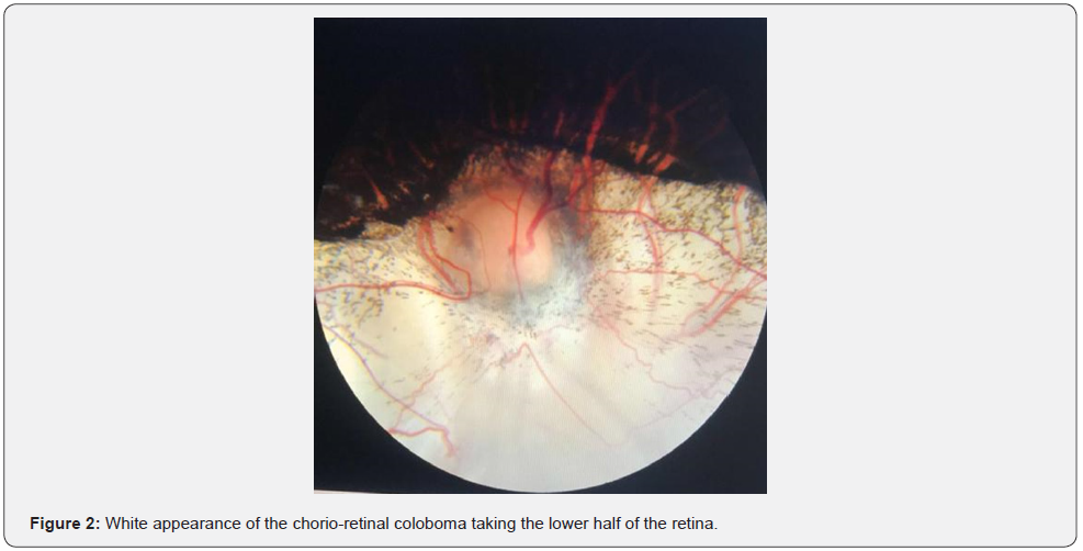
It was a male subject, 12 years old, taken in consultation by his parents for school difficulties. Personal history was not unique. Her older sister had an iris coloboma and a chorio-retinal coloboma in both eyes. The ophthalmological examination showed: a reduced visual acuity in the fingers at one meter in both eyes, a bilateral horizontal nystagmus, an iris coloboma at the bottom taking the appearance of a keyhole in both eyes (Figure 1), a nuclear cataract in both eyes, and a chorio-retinal coloboma taking the lower half encompassing the papilla in both eyes (Figure 2). A cryptorchidism was diagnosed and managed in a pediatric surgery department. We did low vision rehabilitation, prescription sunglasses to avoid photophobia, genetic counseling. Cataract surgery was performed without improving visual acuity.
Case 2
She was a 14-year-old female subject who was taken to a consultation by her parents for school difficulties. Personal history was not unique. Her younger brother (previous observation) had an iris coloboma and a chorio-retinal coloboma in both eyes. No parental inbreeding was found. The ophthalmological examination showed: a reduced visual acuity to account the fingers at one meter in both eyes, a horizontal bilateral nystagmus, an iris coloboma at the bottom taking the appearance of a keyhole in both eyes, a nuclear cataract in both eyes, and a chorio-retinal coloboma taking the lower half encompassing the papilla in both eyes. The pediatric examination revealed no particularities. Genetic testing was requested but was not available in our laboratories. The treatment consisted of low vision management, prescription sunglasses to avoid photophobia, genetic counseling, and monitoring. Cataract surgery was performed without improving visual acuity.
Case 3
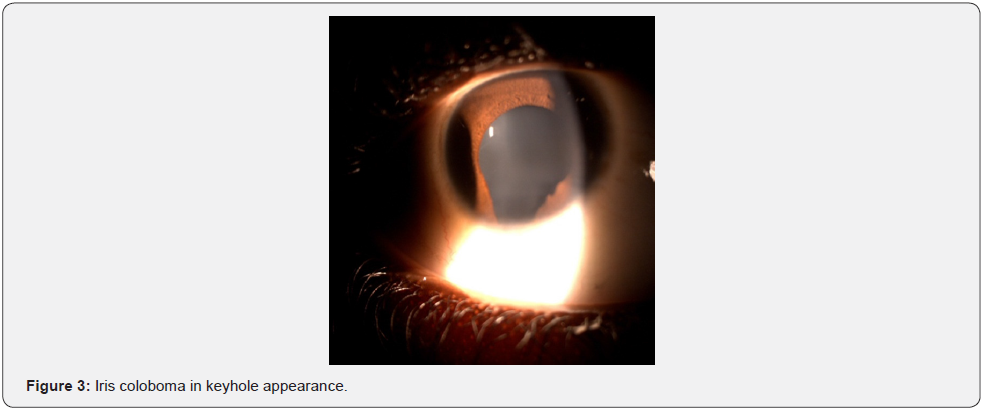
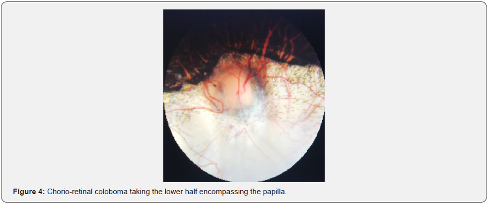
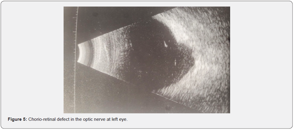
She was a 12-year-old female subject who had been receiving visual blur in both eyes since birth. Personal history was not unique. No parental inbreeding was found. The ophthalmological examination showed: a visual acuity of 0.5/10 in the right eye and reduced to a light perception in the left eye, a bilateral inferior iris coloboma (Figure 3), a dense nuclear cataract in the left eye, limiting the examination of the fundus; and right eye: a chorio retinal coloboma taking the lower half encompassing the papilla (Figure 4). The B-Mode ocular ultrasound showed a chorio-retinal defect in the optic nerve (Figure 5). The pediatric exam was normal. The treatment consisted of low vision management, prescription of sunglasses to avoid photophobia, genetic counseling, and monitoring. Cataract surgery was performed without improving visual acuity.
Discussion
Chorio-retinal colobomas are rare congenital pathologies, with a prevalence of 0.4% of the general population [2]. Its incidence is 1/100,000 births [3]. They represent the most common types of all coloboma eye conditions [4]. Over 5 years (between June 2014 and June 2019), we found 3 cases of chorioretinal coloboma out of a total of 23,270 visits or a frequency of 0.00013 or 0.013%. Colobomas are congenital malformations of the eye that are present from birth. The average age of our patients was 12.66. This delay in diagnosis is linked to the socio-economic level, the lack of awareness about the signs of calls, unknown by pediatricians, midwives, and parents, who are the first to be in contact with children. All patients had low visual acuity that could not be improved. Visual acuity is very variable depending on the location of the deficit. It is lower if the macula and optic nerve are affected [5]. All our patients had nuclear cataracts. This association is also described in the literature [6], as well as microphthalmia [7]. The iris coloboma objectified in our patients is also described by several authors. Hafidi [8] in 2014, found in a 7-year-old girl, a full-thickness iris coloboma in both eyes. Denis [3] in 2013 had found in a series of 35 cases that coloboma was bilateral in 54.2% of cases and unilateral in 45.8% of cases. The inferior nasal localization of chorio-retinal colobomas is also reported in the literature [5].
This inferior nasal localization is explained by the fact that during embryonic development, between the 5th and 7th week of life, there is the invagination of the optical vesicle and the closure of the fetal fissure. The embryo has the shape of a bud, then a kind of cupule that will gradually close at the level of the fetal fissure [9]. DIALLO [10] found in a 6-year-old child, on the examination of the fundus at the level of the 2 eyes a very extensive chorio-retinal coloboma ranging from 3 hours to 10 hours encompassing the entire papilla. ESSALIME [11] in his study, had a 9-year-old boy who had a total retinal detachment on a chorio-retinal coloboma in the right eye and a chorio-retinal coloboma in the left eye.
Chorio-retinal colobomas may be isolated or associated with other malformations such as CHARGE syndrome [12]. We had found an association with a surgical cryptorchidism in a pediatric surgery department. KAMOUN [13], in a 15-month-old infant describes an association with a malformative syndrome of KLIPPEL FEIL, characterized by a very short neck, low implantation of the hair, extreme limitation of head motion and welding of cervical vertebrae to spine radiography. Hafidi [8], shows in his study the interest of additional tests in the search for pathologies associated with colobomas and in the search for possible complications.
There is no significant link between the severity of the coloboma and the abnormalities found (p =1.0) nor between the abnormalities of gyration and the severity of the coloboma attack (p =1.0) [3]. Our management procedure is in line with what is done in the literature which also proposes the treatment of associated complications such as retinal detachment or nuclear cataract [5]. Cataract surgery was performed in all our patients but without improvement.
During retinal detachment on chorio-retinal coloboma, the dehiscence is often at the level of the hypoplastic retina in the coloboma area. They are difficult to identify and treat, often requiring prolonged internal tamponing. In case of peripheral dehiscence, conventional surgery often provides good results [11]. Retinal detachment associated with ocular coloboma accounts for 0.5% of retinal detachments in children [13]. These detachments are more often acquired than congenital. In our study, none of our patients had developed retinal detachment. The prevention of these retinal detachments consists in performing a complete and frequent eye examination, as well as Argon laser photocoagulation.
Conclusion
Chorio-retinal colobomas are rare congenital abnormalities of the eye, secondary to an abnormality of closure of the embryonic fissure. These are serious abnormalities because they involve the functional or anatomical prognosis of the eye. They therefore require a comprehensive ophthalmological and general examination for other associated malformations.
References
- Gunderson CA, Stone R, Peiffer R, Freedman S (1996) Corneal coloboma, aphakia and retinal neo vascularization with anterior segment dysgenesis (Peters anomaly). Ophthalmologica 210: 361-366.
- Exana J, Gordon M (2008) Coloboma bilateral de iris, cristalino, coriorretina y nervio optico, asociado a desprendimiento retiniano. Revista Mexicana de Oftalmologia Julio Agosto 82(4): 267-268.
- Denis D, Girard N, Levy-Mozziconacci A, Berbis J, Matonti F (2013) Colobome oculaire et résultats de l’IRM cérébrale: résultats pré Journal Français d’Ophtalmologie Elsevier Masson édition 36(3): 210-220.
- Ammann F, Klein D, Johr P (1964) Une famille atteinte de colobomes irido-choroidiens d’hétérochromie irienne et syndrome de Marian abortif. Ophthalmologica 147: 118-124.
- Elkhoyaali A, El Asri F, Chatoui S, Iferkhass S, Reda K, Oubaaz A (2015) Colobome chorio-retinien bilatéral Journal Français d’ Elsevier Masson édition 38(7): 674-675.
- Giuffre G (1989) Colobomas of the optic area 12(4): 100-102.
- Warburg M (1993) Classification of microphthalmos and coloboma. Journal of Medical Genetics 30: 664-669.
- Hafidi Z (2014) Formes cliniques inhabituelles des colobomes du segment postérieur: à propos de deux cas. Journal Français d’Ophtalmologie. Elsevier Masson édition 37(5): 75-77.
- Magli A, Greco A, Alfieri MC, Pignalosa B (1986) Hereditary colobomatous anomalies of the optic nerve head. Journal of Ophthalmic Pediatric Genetics 7(2): 127-130.
- Diallo S, Bakayoko S, Coulibaly B, Sidibe MK, Guirou N (2018) Colobome chorioretinien bilateral: à propos d’un cas. The Pan African Medical Journal 30: 261.
- Essalime K, Mazzouz H, Lahbil D, El Kettani A, Lamari H, et al. (2008) Décollement de rétine sur colobome choriorétinien: à propos d’un cas. Journal Français d’Ophtalmologie. Elsevier Masson édition 32: 1-226.
- Sanlaville D, Lyonnet S, Amiel J (2002) Le syndrome CHARGE. Journal de Pédiatrie et de Pué Elsevier Masson édition 15(4): 209-214.
- Kamoun R, Kria L, Erraies K, Anane R, Ouertani AM (2006) Colobome oculaire associé au syndrome cervico-oculo-acoustique. Journal Français d' Elsevier Masson édition 29(7): 836.


