Two Cases of Idiopathic Acquired Dacryocystocele
Ceyhun Arici* and Rengin Yildirim
Department of Ophthalmology, Cerrahpasa Medical Faculty, Istanbul University, Istanbul, Turkey
Submission: September 12, 2017; Published: September 27, 2017
*Corresponding author: Ceyhun Arici, Istanbul Universitesi, Cerrahpasa Tip Fakultesi, Goz Hastakiklari AD, Istanbul, Turkey Tel: +90 532 340 93 51; Fax: +90 212 570 70 36; Email: ceyhundr@gmail.com
How to cite this article: Ceyhun A, Rengin Y. Two Cases of Idiopathic Acquired Dacryocystocele. JOJ Ophthal. 2017; 5(2): 555658. DOI: 10.19080/JOJO.2017.05.555658
Abstract
Idiopathic acquired dacryocystocele accompanied only by epiphora without complicating dacryocystitis is very rarely seen. We report two such cases (one of them was the youngest case among previous published cases (using Pubmed database”)” in which epiphora and non-tender, firm, bluish cystic swelling at and below the medial canthal area. Orbital MRI revealed a high-intensity, cystic lesion in T1 and T2 weighted sequences in the inferomedial aspect of the left orbit. Both patient underwent external dacryocystorhinostomy. Pathologic examination revealed columnar epithelium without any neoplastic tissue. Epiphora disappeared postoperatively. No recurrence of either the epiphora or the orbital mass were detected during follow-up period.
Keywords: Dacryocystitis; Dacryocystocele; Dacryocystorhinostomy; Epiphora; Nasolacrimal duct obstruction
Introduction
Distention of the nasolacrimal sac manifests as a bluish cystic swelling at and below the medial canthal area by captured or entrapped mucoid material results in the formation of a dacryocystocele. The retention of mucus results from an obstruction in the distal nasolacrimal duct together with a proximal obstruction at the junction of the common canaliculus and the sac (valve of Rosenmuller) [1,2].
It is typically congenital in origin and often presents in the first few weeks of life. Dacryocystocele also may occur rarely as an acquired disorder in adults [3,4]. Although the nature of the disorder is rarely in question, one still should consider the possibility of a primary tumor of the nasolacrimal sac whenever decompression of the sac is not possible with manipulation or whenever the mass extends above the medial canthal tendon [5]. Additionally, idiopathic, inflammation, complication of dacryocystitis, facial trauma, nasal surgery or punctal agenesis may be the other reasons [4,6]. In this report, two different age generation cases with idiopathic acquired dacryocystocele were described.
Case Report
Case 1
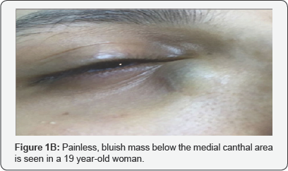
A 60 year-old woman with right medial canthal mass presented to our clinic. The patient had right epiphora for 10 years and right medial canthal mass had developed for one year. 0n examination, visual acuity was 20/20. Anterior segment and fundus were normal. Intraocular pressure (I0P) was 17mmHg. The mass was firm, non-tender and bluish in colour (Figure 1A). Orbital Magnetic resonance imaging (MRI) showed a high intensity cystic expansion in T1 and T2 weighted sequences and 20 x16mm in the right lacrimal sac topography, suggesting an idiopathic acquired dacryocystocele. A soft stop was detected during canalicular probing and lacrimal irrigation leaded to reflux from punctum. The patient underwent external dacryocystorhinostomy (DCR). Pathologic sample of dacryocystocele examination revealed that the lining epithelium was columnar without any neoplastic tissue. The patient was asymptomatic after surgery. The silicone stent was removed 4 months later (Figure 1B). The mass and epiphora did not recur during 2 years follow-up.

Case 2
A 19 year-old woman admitted to our hospital with a medial canthal painless mass in her right eye, which was present for 5 months and accompanied by epiphora. On initial examination, visual acuity was 20/20. Anterior segment and fundus were found to be normal. IOP was 15mmHg. The mass was firm, nontender and non-compressible (Figure 2). Orbital MRI revealed a high-intensity, cystic lesion in T1 and T2 weighted sequences and 16x14mm in the inferomedial aspect of the right orbit. Lacrimal irrigation showed blockage at the nasolacrimal duct. Idiopathic dacryocystocele was diagnosed. External DCR was performed. Pathologic examination revealed columnar epithelium with goblet cells without any neoplastic tissue. Epiphora disappeared postoperatively. The silicone stent was removed 3 months later No recurrence of either the epiphora or the orbital mass were detected during follow-up of 15 months duration.

Discussion
Dacryocystocele is mainly congenital, rare cases for the acquired type have been reported [1,3,6-10]. In conjenital type, swelling of lacrimal sac just below the medial canthal tendon results from distal nasolacrimal duct obstruction and functional proximal obstruction at the junction of the common canaliculus and the sac. Mostly, the proximal obstruction is functional which is supported by the absence of anatomic blockage during probing and by mucus reflux following lacrimal sac region massage. A similar mechanism may ocur in adults. Mucosal distention sendary to chronic inflammation in the localization of the valve of Rosenmuller may prevent mucus reflux from the sac [9].
Differentiaton of the possibility of nasolacrimal sac tumor should be necessary. Early symptoms of nasolacrimal sac malignancy are often nonspecific and can be mistaken for symptoms of benign and more common conditions such as idiopathic nasolacrimal duct obstruction or dacryocystitis. Progressive firm masses in the area of the lacrimal sac/ nasolacrimal duct and displayed overlying skin telangiectasis are more specific clinical signs whereas benign lesions initially presented with epiphora and a bloody discharge without pain or significant mass. Radiographic imaging is recommended in such cases [5,11].
In current study, biopsy was taken from both cases due to suspected mild soft tissue growth in the sac as a precaution. So, any unusual findings other than chronic inflammation during a DCR, the tissue should be sampled at the time of DCR [11,12]. Idiopathic acquired dacryocystocele results from chronic nasolacrimal duct obstruction and same as the congenital type, secondary functional proximal obstruction at the junction of the common canaliculus and sac. In current study, two different generation cases (to our knowledge, one of them was the youngest case among previous published cases (using Pubmed database)) were reported.
Dacryocystorhinostomy is generally curative. Acquired dacryocystocele can be treated by external or endoscopic approaches with similar success rates [13]. Due to the fact that direct visualization of the anatomy that provides the definite removal of bone in the lacrimal fossa and facilitates the precise anastomosis of the nasal mucosa and lacrimal sac. Whereas, preservation of the pumping mechanism of the orbicularis oculi muscle, avoidance of facial scarring, less bleeding and faster rehabilitation are the advantages of endoscopic DCR [6,13].
In conclusion, idiopathic acquired dacryocystocele is a very rare occurrence subsequently to chronic epiphora. In the presence of non-tender cystic swelling at and below the medial canthal area by captured or entrapped mucoid material, dacryocystocele should be considered. DCR is thought as the definitive treatment, and external access is still commonly used approach by ophthalmologists.
References
- Perry LJ, Jakobiec FA, Zakka FR, Rubin PA (2012) Giant dacryocystomucopyocele in an adult: a review of lacrimal sac enlargements with clinical and histopathologic differential diagnoses. Surv Ophthalmol 57(5): 474-485.
- Mansour AM, Cheng KP, Mumma JV, Stager DR, Harris GJ, et al. (1991) Congenital dacryocele: a collaborative review. Ophthalmology 98(11): 1744.
- Mirza AA, Alsharif AF, Elmays OA, Marglani OA (2017) Foreign body mimicking malignancy in acquired dacryocystocele. Clin Case Rep 5(3): 296-299.
- Eloy PH, Martinez A, Leruth E, Levecq L, Bertrand B (2009) Endonasal endoscopic dacryocystorhinostomy for a primary dacryocystocele in an adult. B-ENT 5(3): 179-182.
- Hornblass A, Jakobiec FA, Bosniak S, Flanagan J (1980) The diagnosis and management of epithelial tumors of the lacrimal sac. Ophthalmology 87(6): 476-490.
- Koltsidopoulos P, Papageorgiou E, Konidaris VE, Skoulakis C (2013) Idiopathic acquired dacryocystocele treated with endonasal endoscopic Dacryocystorhinostomy. BMJ Case Rep pii: bcr2013200540.
- Debnam JM, Esmaeli B, Ginsberg LE (2007) Imaging characteristics of dacryocystocele diagnosed after surgery for sinonasal cancer. AJNR Am J Neuroradiol 28(10): 1872-1875.
- Lee JH, Moon SW, Shin YW, Lee YJ (2010) Dacryocystocele in adult: a report of five cases. J. Korean Ophthalmol Soc 51(5): 751-757.
- Woo KI, Kim YD (1997) Four cases of dacryocystocele. Korean J Ophthalmol 11(1): 65-69.
- Britto FC, Rosier VV, Luz TV, Verde RC, Lima CM, et al. (2015) Nasolacrimal duct mucocele: case report and literature review. Int Arch Otorhinolaryngol 19(1): 96-98.
- El-Sawy T, Frank SJ, Hanna E, Sniegowski M, Lai SY, et al. (2013) Multidisciplinary Management of Lacrimal Sac/Nasolacrimal Duct Carcinomas. Ophthal Plast Reconstr Surg 29(6): 454-457.
- Merkonidis C, Brewis C, Yung M, Nussbaumer M (2005) Is routine biopsy of the lacrimal sac wall indicated at dacryocystorhinostomy? A prospective study and literature review. Br J Ophthalmo 89(12): 15891591.
- Ben SGJ, Joseph J, Lee S, Schwarcz RM, McCann JD, et al. (2005) External versus endoscopic dacryocystorhinostomy for acquired nasolacrimal duct obstruction in a tertiary referral center. Ophthalmology 112(8): 1463-1468.


