Evaluation of Aging Disorders by 54 000 Consecutive Ophthalmic Cases
Fouad N Sayegh*
Department of Ophthalmology and Experimental Ophthalmology, University of Jordan, Jordan
Submission: April 27, 2016; Published: May 18, 2016
*Corresponding author: Fouad N Sayegh, Past Dean, Faculty of Medicine, University of Jordan, 11941 Amman, P. O. Box 1885, Jordan, Tel: 0096279 5524711; Email: prof_fuadsayegh@hotmail.com
How to cite this article: F N Sayegh. Evaluation of Aging Disorders by 54 000 Consecutive Ophthalmic Cases. JOJ Ophthal. 2016; 1(3): 555564. DOI:10.19080/JOJO.2016.01.555564
Abstract
The Ciliary body is an unusual location of uveal melanomas and usually these kinds of tumors appear with a reduction of vision due to the formation of sectorial cataract or retinal detachment when the tumor spread to a posterior position. We present a 57 years old woman with sudden and severe ocular pain. Ophthalmologic examination showed a hyper mature cataract and superior displacement of the lens with sectorial angular block. The intraocular pressure was 22 mmHg. After papillary dilation aciliary body tumor was observed. The ultrasound study and magnetic resonance imaging confirmed the diagnosis of uveal melanoma.
Background Information:during the years 1991-2015, about 54,000 consecutive cases were examined at the ophthalmic out patient’s clinic. The aim of this study was to find out if any relationship exists between aging disorders, or each is having its own entity, or one disorder could be the driving factor to produce aging and related complications.
Methods:Findings were entered into computerized documentation program that does simultaneous analysis of clinical data. The correlation between aging disorders was analyzed.
Results:Seven disorders were found to be related to aging; hypertension, glaucoma, cataract, diabetes, age related macular degeneration and ischemic neuropathy, in addition to presbyopia. Hypertension was found to be the leading cause for the development of ischemia and therefore may be the stimulating factor for the development of oxidative stress and oxidation. It shows that basic trends of aging are important while investigating the metabolic disorders at molecular level.
Conclusion:Taking both factors; ischemia and oxidative stress into consideration, it is recommended a strategy be adopted, in addition to antioxidant therapy, prophylaxis and prevention of aging includes medication that guarantees healthy relationship between hypertension and cerebral blood flow.
Keywords: Aging disorders; Diabetes; Hypertension; Cataract; Glaucoma; Age related macular degeneration
Introduction
During the years 1991 and 2015, about 54 000 consecutive cases were examined at the Ophthalmic Clinic in Amman - Jordan. Data were entered directly into a computerized documentation program that performs simultaneous analysis of clinical data. Figure 1 shows the age distribution of examined cases. One notices that Jordan has a relatively young population and a significant decrease of incidence starting at the age group 61-70 years.

It has been noticed that seven diseases increase with age; Hypertension, Diabetes, Glaucoma, Cataract, Age related Macular Degeneration “AMD” and Ischemic Neuropathy, in addition to Presbyopia. The question that arises, is there any relationship between these six disorders? Or each is having its own entity independent of each other? Or one disorder could be the driving factor to produce aging related complications within the other components?
Materials
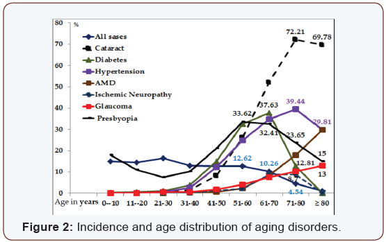
The age distribution of examined cases is shown in Table 1. A comparison between aging subjects to all examined cases (Figure 2) gives us the following information;
- The incidence of aging for all disorders starts to increase at the age of 40 years.
- Diabetic cases start to decline at the age 61-70 years.
- Cataract and hypertension starts to decrease at the age group of 71-80 years.
- Open-angle glaucoma and AMD continue to increase until the age of 80 years and above.
- The accommodative reservoir decreases from birth up to the age of 30 years and Presbyopia starts to develop in average at age 40 and continues to increase up to the age 61- 70 years.

As the incidence of all disorders start to increase at age 40 years, means that the age of 40 is the starting point of aging. This fact could be confirmed as presbyopia starts in average at the age of 40 years. It is possible that the decline of aging incidence (Figure 2) after the age of 61- 70 years by diabetics and at the age of 71-80 years by cataract and hypertension means a relative early death for these two diseases as compared with the normal distribution of all cases with in the same age group.

Cataract
Cataract increases with age, reaching its maximum 72.21% at the age of 71- ≥ 80 years. Its incidence with age is much higher than all other disorders. “Implications of oxidative stress have been examined in the pathogenesis of cataract in vivo treatment with vitamin E, of the Emory mouse led to a decrease in the rate of cataract progression suggesting that in at least in some entrances an oxidative stress could participate in the formation of cataract [1]. It is known that in addition to heredity, environmental factors play a major role in the development of cataract. In addition, 35.5% of all cataract cases are diabetics, 34% hypertensive, 10.6% have AMD and 8.6% Glaucoma. So cataract is involved in a mixture of additional components that might affect its development and patient’s quality of life. It is therefore expected that cataract patients are also affected from ischemic disorders in the same manner like other aging diseases.
Hypertension
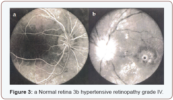
Hypertension increases with age reaching its maximum 39.44% at the age 71-80 years, and then it decreases again. It is known that uncontrolled hypertension causes hypertensive retinopathy. According to its severity, hypertension is classified into four grades (Figure 3) a represents a normal retina.
The ratio of diameter between venules and arterioles is 3:2. (Figure 3) represents hypertensive retinopathy grade IV, showing narrowing of the arterioles, and swelling of the optic nerve head, macular edema, micro hemorrhages and cotton wool spots indicating the presence of severe ischemia. Wayne [2] says “There is increasing evidence that atherosclerosis should be viewed fundamentally as an inflammatory disease. There is evidence that hypertension may also exert oxidative stress on the arterial wall”.
Diabetes
Diabetes increases gradually with age. It reaches the maximum incidence “37.63%” at the age 61-70 years. Its distribution goes parallel to hypertension with the difference that diabetes starts to decline earlier as seen in (Figure 2). There is convincing experimental and clinical evidence that the generation of reactive oxygen species increases in both types of diabetes and the onset of diabetes is closely associated with oxidative stress Some [3] diabetic patient care severely affected with uncontrolled hypertension and the development of hypertensive retinopathy and other vascular disorders in comparison to non-diabetics (Figure 3).

It is expected that diabetic patients with severe hypertension most likely suffer from decreased cerebral blood flow and ischemia of sensitive organs, with the result of getting ischemic neuropathy and renal failure or different vascular obstructions. Similar to hypertension, the prevalence of glaucoma by diabetic patients is 7.7%, much higher than by all examined patients “2.28%”.
Glaucoma
Open angle glaucoma increases with age, starting at the age of 40 years and reaching its maximum “13%” at the age of 80 years. The blood pressure seems to be responsible for aqueous humor formation [4]. The incidence of glaucoma among all patients is 2.28%, and 6.6% among all hypertensive patients. Matt [5] reports of having evidence that antioxidant treatment could help defeat glaucoma. It was found that antioxidant treatment reduces oxidative stress in pressure-treated retinal ganglion cells. This confirms a study done by Weinreb [6] that oxidative damage occurs within hours of elevated hydrostatic pressure or elevated intra ocular pressure.
Age related macular degeneration
Age related Macular Degeneration “AMD” increases with age reaching its maximum 29.81% at the age of ≥ 80 years. The incidence of glaucoma among AMD patients is 6.32% much higher than by all patients included in the study “2.28%” and with hypertension 5.3 %. We experience today exactly what Donders [7] has described Khan HA & Moorhead HB [8] has published, Frank [9] 25 years later that among the four aging blinding diseases, Cataract, Glaucoma, Diabetes and AMD, Agerelated Macular Degeneration is the only one for which there are no effective means of prevention or treatment.
Although it is less common loss of vision in AMD, choroid Neovascularization leading to disc form degeneration produces a more rapid and dramatic and more severe decrease in vision than the atrophic or dry form of AMD. Similar changes were found also among patients in cases of diabetes, AMD, disc form macular degeneration and high myopia, where the clinical appearance of an often round macular scar, that has been recognized to comprise Neovascularization, arising from the chorio-capillaris [10] and proliferative changes at optic nerve head in cases of carotid thrombosis (Figure 4).

Discussion
Aging is a complex of degenerative changes starts at the age of 40 years. Presbyopia is the first measurable sign of aging. The age of 40 years is therefore considered as the starting point for prophylaxis, prevention and rehabilitation in order prolong life expectation and to secure better quality of life for elderly. The presented data show clearly that oxidative stress and oxidative damage initiated much interest by researcher. Oxidative stress is most likely responsible for the ongoing development of oxidative damage and its complications by all mentioned aging diseases. The pathogeneses of these diseases at molecular level gives better insight into a major problem related to aging; the oxidative stress causes a continuous oxidative damage. By the presence of aging diseases and absence of cure, it is expected that prevention with antioxidants might improve or maintain these disorders at acceptable level, but keep fighting to prevent the ongoing oxidative stress is mandatory. Hypertension on the other hand involves all aging diseases as seen in (Table 2).

Ophthalmo-dynamometry is a good method “at least for serial investigations” to measure the blood pressure in both brachialand ophthalmic artery, estimate the vascular resistance and calculate the amount of cerebral blood flow [11] Values that are higher than twice the standard deviation (2xD) of the regression curve are considered as having high cerebrovascular resistance (CVR). The Cerebral Blood Flow (CBF) will be calculated as follows;

In addition to the genetic and environmental factors, uncontrolled hypertension seems to play a leading role in the development of complications related to aging diseases. 47.1% of all hypertensive patients suffer from Glaucoma, and 92.5% of all diabetics are hypertensive. The incidence of glaucoma in hypertension is relatively high “6.6%” in comparison to 2.28% by all examined cases; the incidence of hypertension by AMD patients is 26.7%. As expected hypertension might cause embolism of the central retinal artery CRA the increased vascular resistance measured by ODM on the (Figure 5).Other side not affected, show increased vascular resistance by 59% of all CRA embolism cases, is an alarming sign that such ischemic changes might occur on the other side or elsewhere in the body. Treatment with antihypertensive drugs is important. The positive expected effect of some anti-hypertensive drugs “decrease blood and intra– ocular pressure” could unlikely elevate the cerebral-vascular resistance and decrease the cerebral blood flow as experienced with clonidine [12,13]. It is therefore expected that patients involved within aging diseases suffer from complications related to hypertension and/or a non-wished side-effect of some antihypertensive drugs causing ischemia. Hypertension seems to be the leading and stimulating factor that causes arteriosclerosis, increased vascular resistance and inability to maintain normal blood flow to the whole body, causing severe damage especially to the very sensitive organs, e.g. stroke, myocardial infarction, gangrene, thrombosis and emboli of the central retina or carotid arteries, ischemic neuropathy and renal failure. It might affect the chorio carcinoma and RPE causing ischemia. It is therefore possible that oxidative stress which plays a major role at the molecular level is initiated by ischemia. One comes in agreement with Newell [14] that these macular changes are related to the decreased blood flow of the chorio carcinoma below the fovea. If this theory is correct, increased blood pressure and decreased Blood flow will produce adequate systemic complications within aging diseases. This could be confirmed by the presence of associated disorders as shown in [Table 3] and (Figure 6 a and b).


The majority of cases suffering from vascular obstruction are found frequently in hypertension followed by diabetes, AMD, glaucoma and cataract. Ischemic Neuropathy; “ischemic papillitis, retro bulbar neuritis, and other cranial nerves” were found most frequently by diabetics, followed by AMD, in association with hypertension. By calculation the prevalence related to all cases involved in the study, hypertension was found to be the leading disease causing ischemic disorders; Diabetes increases the prevalence of ischemic neuropathy and renal failure. By observing the distribution curve (Figure 2 & 7) one comes to the following life expectation and survival rate of aging diseases. By considering all cases involved in the study independent of age, the survival rate at age 51-60 years was found to be 12.7% and for the age group ≥ 80 years 1.1%. As aging diseases starts to increase by the age of 40 years where the younger population is excluded, it is expected that the survival rate for aging disorders will be different. The prevalence of Cataract, AMD, glaucoma and hypertension continue to increase till the age of 61-70 years. Then the survival rate becomes less. The best survival rate for the age group 71-80 years is for AMD 34.5%, followed by cataract 23.6%, then glaucoma 20.6%, hypertension 16.8% and diabetes 12.3%. The worst survival rate at the age of ≥ 80 years is for diabetics 1.8% and the best was found to be for AMD patients 13.4%. The survival rate of the same aging group for glaucoma 6.1%, cataract 4.1% and hypertension 3% only.

Conclusion
The central regulation between systemic blood pressure and cerebral blood flow is very important. Deregulation of this mechanism through hypertension seems to be the leading factor that initiates oxidant stress, oxidative damage and aging. Antioxidants are therefore very important to prevent further deterioration of aging disorders. For the prophylaxis of aging diseases it is recommended that in addition to antioxidants, special attention be paid to control hypertension and maintain normal cerebral blood flow.
Highlights
Programmed computerized documentation and analysis of aging disorders among 54000 ophthalmic cases.
Correlates between the most common seven aging disorders in ophthalmology.
Demonstrates the importance of paying attention to ischemic systemic disorders associated with ocular complaints.
Modern eye Journals are not paying enough attention to the basic trends of aging.
Suggests new strategy for the prevention of aging disorders.
References
- Varma SD, Chand D, Sharma YR, Kuck JF, Richards RD (1984) Oxidative
- Alexander RW (1995) Hypertension and the pathogenesis of atherosclerosis, Oxidative stress and the mediation of arterial inflammatory response: a new perspective. Hypertension 25(2): 155- 161.
- Rosen P, Nawroth PP, King G, Moller W, Tritschler HJ, et al. (2001) The role of oxidative stress in the onset and progression of diabetes and its complications. Diabetic Netab. Res Rev 17(3): 189-912.
- WeigelinE, SayeghF, Von Klitzing W, BaurmannH (1975) Aspects of Venous Drainage and TissueFluid Dynamics in the Eye. The Eye Ear Nose Throat Mon 54(2): 47-50.
- Matt Y (2008) Fighting glaucoma with antioxidant treatment. Eye world contributing editor.
- Weinreb RN (2007) Oxidative damage contributes early to glaucomatous optic neuropathy. Hamilton Glaucoma Center and Department of Ophthalmology, University of California San Diego, La Jolla, Calif Cit Matt M Y 48(10): 4580-4590.
- Donders FC (1855) Beitragezur pathologischen anatomie des Auges. GraefesArch KlinOphthalmol. 1:106-112.
- Kahn HA, Moorhead HB (1973) Statistics on Blindness in the Model Reporting Area1969-1970. Washington DC: Dept of Health, Education, and Welfare. Public Health Servic 72-73, 134-141.
- Frank RN (1998) Oxidative protector enzymes in the macular retinal pigment epithelium of aging eyes and eyes with age related macular degeneration. Trans Am Ophthalmol Soc 96: 635-689.
- Verhoef FH, Grossman (1937) Pathogenesis of disc form degeneration of the macula. Arch Ophthalmol 18(1): 561-585.
- Sayegh F N. Anhängigkeit des intraokularenDruckesvomBlutdruck, Durchströmungsvolumen und Strömungs-wiederstande.Adv. Ophthal; Karger, Basel. 1975: 29; 185-189
- Sayegh FN and Weigelin E. Die intraokularedrucksenkungdurchClonidin. IhreBeziehungzucerebralenStrömungs – und Blutdruckänderungen. Klin. Mbl. Augebhk; 1974:63,33 – 47.
- Sayegh FN, Hartmann J, Weigelin E. Das verhalten des Blutduckes, des intrakraniellenKreislaufs und des intraokularenDruckesbeigesundenPersonen, beiHypertonikern und beiPatientenmitGlaucomnachAnwendung von Clonodin (Isoglaucon) alsAugentropfen. KlinMblAugenhk; 1973: 163, 23-29.
- Frank WN (1982) Ophthalmology principles and Concepts (5th Edn.), pp. 301-302.


