




 JOJMS Home




Abstract
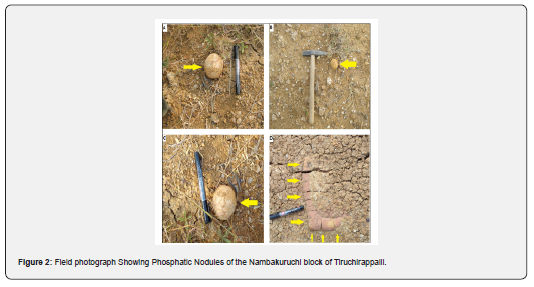
This study focuses on the physical, optical, and X-ray diffraction characterization of phosphate zones from field to fertilizer applications, particularly examining the Cretaceous phosphatic deposits in Tiruchirappalli district, Tamil Nadu. These deposits are significant globally and occur primarily in nodular forms exposed on the surface, often associated with Maruvattur Clay or Karai Shale, exhibiting a badland topography. The nodules, varying in size from 2 to 5cm in width and 6 to 20cm in length, display various shapes, including circular and conical, and feature surfaces that range from yellow to reddish hues with distinct crystal veins. Research aims to identify whether these nodules contain phosphatic, carbonate, or manganese components, and their potential uses in the fertilizer industry. Physical characteristics such as medium to high specific gravity, a hardness of 5 on Mohs scale, and vitreous luster were observed. Optical studies highlighted the fine-grained matrix of carbonate fluorapatite (CFA), essential for microscopic analysis. X-ray diffraction analysis of ten nodule samples revealed that CFA is the primary phosphate mineral, alongside calcite, quartz, feldspar, kaolinite, hematite, and chlorite as gangue minerals, aiding in the differentiation of carbonatite and phosphatic nodules.
Keywords: XRD; Natural; Phosphatic nodules; Minerals; CFA; Physical properties; Tamil Nadu
Abbreviations: ICDD: International Centre for Diffraction Data; PXRD: Powder X-ray diffraction; CFA: Carbonate Fluorapatite
Introduction
The samples were measured by groups based on the transparency and the complexity of molecular structure. The physical appearance of the nodules is very important for the economy as well industry purpose to differentiate to the others one. Colors, luster, fracture, cleavage, specific gravity, and hardness are key parameters for any rocks/mineral’s characteristics. These observations direct affect to the origin and deposition of the nodule’s samples. The optical behavior of the nodules also effects the minerals, texture/structures, flora and fauna remain. Each sample's XRD measurements were carried out concurrently. In order to quickly acquire XRD patterns suitable for phase identification, the major experimental parameters were tuned. Each sample's phase analysis process was thoroughly documented, and the most likely phases were identified. The outcomes were confirmed by standard XRD experiments. The system's benefits, drawbacks, and applicability range were examined. The findings suggest that while specific nodules can be successfully detected using the XRD technique, single-crystal nodules may not be so successfully identified. More widespread acceptance and use of the XRD system in mineralogy and related fields necessitate hardware advancement, software upgrades, and more practical application scenarios.
The present study in physio-chemistry of materials concerns fertilizers. The nodules materials studied are fertilizers as per our latest technology to use in agriculture. X-ray diffraction permits us to characterize nodules from a different location. It can allow us to study the crystalline phases and follow a possible cell parameter evolution between nodules, the transformed nodules verities. Color, gloss, transparency, hardness, cleavage, density, thermal conductivity, refractivity, refractivity, reflectivity, spectral selective absorption, chromatic dispersion, pleochroism, polarized light, fluorescence, phosphorescence, and special optical effects are a few of the physical traits or phenomena that can be used to distinguish nodules. Some of these techniques are based on characteristics of bulk materials in general, such color and hardness, whereas others are exclusively useful for specific types of nodules. Modern analytical techniques have taken a more active role in the field of rock identification along with advancements in science and technology. X-ray diffraction (XRD) is a technique for identifying material phases.
Cretaceous phosphorite deposit is one of the significant deposits in the world. The Uttar group of the Cretaceous period in this district is one of the vital sources of rock phosphate in India. The onshore Cretaceous marine sedimentary sequence of the phosphatic nodules exposed in the Tiruchirappalli district of Tamil Nadu. The mineralogy and composition depend on the types of origin, whether sedimentary or igneous. The origin of the sedimentary deposits is many sources. The phosphatic nodules in the Nambakuruchi are part of the Cauvery basin of South India, which lies in the Uttar group of the Karai formation. The phosphatic buds reported by [1]. Numbers of authors worked on lithostratigraphy, biostratigraphy, paleoceanography, phosphatic nodules, flora, and fauna [2-7]. The purpose of this work is to describe the physical, optical, and mineral components and their application of phosphatic nodules in the Nambakuruchi block of Tiruchirappalli district Tamil Nadu India with the help of physical parameters, a thin section for optical observation and X-ray diffraction analysis.
Samples Collection from Study Area
The Nambakuruchi Varagupadi block of Tiruchirappalli district, Tamil Nadu India, is a part of the Cauvery basin that lies in the Southern part of India. The geographic location of the Nambakuruchi (Lattitude 110 5′ 30″; Longitude 780 52′ 15″ and Varagupadi (Lattitude 110 8′ 50″, Longitude 780 54′ 00″). The study area of phosphatic nodules covers an area of about 27.52 Sq. kms. The most extensive exposure of Cretaceous rocks in Tamil Nadu has been found in Tiruchirappalli district, extending over 400sqkm. The basement rock of the Cretaceous succession of the Cauvery basin is the Precambrian basement overlain by Uttar, Trichinopoly, Ariyalur, and Niniyur groups. The phosphatic nodules have been marked in the Uttar group of Nambakuruchi Varagupadi block of Tiruchirappalli district Tamil Nadu. The buds are generally white with calcareous encrustation.
Material and Methods
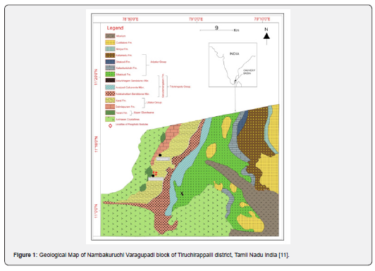
The samples were collected from the Nambakuruchi block of Karai Formation, Uttar Group Tiruchirappalli Tamil Nadu Figure 1. The sampling was carried out in different locations with different shapes and sizes. The representative samples were used for Physical, optical, and XRD analysis. The physical properties of the samples carried out in the laboratory on the physical parameters. For optical properties of the phosphatic nodules sample prepared in thin section cutting lab in the department of geology, AMU Aligarh. First, we cut the chips in different sections/orientations to make a thin section slide for microscopic observation. In the lab, thin sections of representative samples of various lithological units were prepared. Under plane polarized light and in between crossed Nicols, the mineralogical characteristics of the area phosphatic nodules were methodically and meticulously studied. The mineralogical studies were done under transmitted light with the help of an advanced petrological microscope in a petrological lab in the Department of Geology, A.M.U., Aligarh, U.P (India).
The representative samples were used for X-Ray diffraction studies. The phosphatic nodules were cut in two halves, one part was used for X-ray diffraction. The powder was scanned from 3° to 60° 2θ at 0.50 1/min, using nickel filtered Cu Kα radiation at the department of Mechanical Engineering AMU Aligarh. The X-ray diffraction data have been identified by 2θ to d values tables of the National Bureau of Standards, ten series of US Department of Commerce, and also the uses JCPDS cards and Match3 for the identification of minerals Figure 2.
Results and Discussion
Physical properties
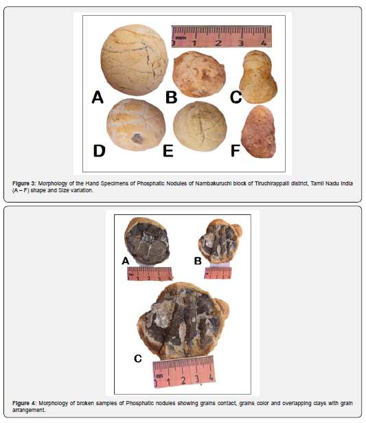
The phosphatic nodule is a sedimentary rock that occurs in the nodular form of various shapes and sizes. Mostly in size, the buds vary in length from 6cms to 20cms and are 2 to 5cms. larger and smaller sizes are occasionally present. They are generally encased in a chalky shell rich in calcium carbonate, or they bear encrustation of ferruginous and calcareous matter. The nodules are hard, and when broken reveal a deep brown to a dark amorphous interior with cracks traversed by veinlets usually of gypsum, calcium carbonate and rarely celestite Figure 3 [8].
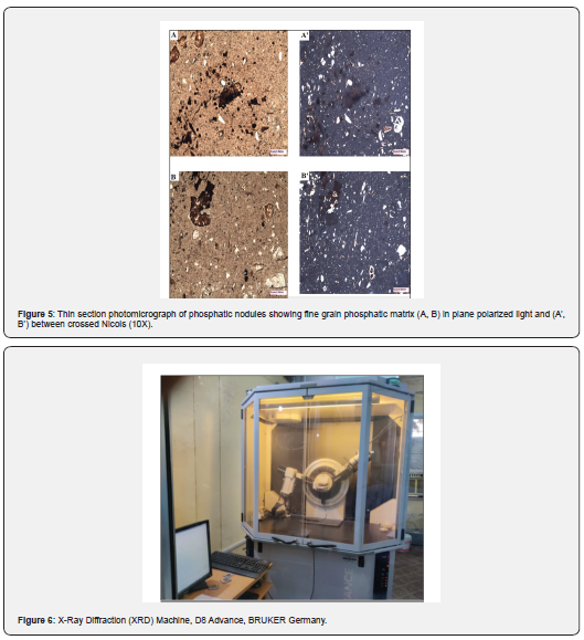
The Uttar group of the Cretaceous period in this district is one of the essential sources of rock phosphate in India. The Cretaceous System here has been divided into four groups viz., Uttattur, Tiruchirappalli, Ariyalur, and Niniyur, based on the lithological, faunal, and stratigraphic characteristics [9]. The Cretaceous belt of the Tiruchirappalli district has revealed that the occurrences of phosphatic nodules are mostly confined to the lower and the middle sub-stages of the Uttattur stage. The smaller and middle sub-stages of the Uttattur Stage predominantly consist of gypseous, fossiliferous clays, faint greenish blue to brown in color, with some intercalations of thin sandy, fossiliferous limestones and a few tiny bands of sandstones. The clays assume buff to brown color on weathering. The clays show weak signs of bedding, and the sandstones in places are red in color. The clays generally contain thin veins of gypsum besides some veinlets of calcite and barite, and Phosphatic nodules are sporadically distributed in clay. The occurrences of phosphatic nodules have been found mostly confined to an area of about 27.52sqkms (10.6 sq. miles) extending between Nambakkurichchi village on the south and Varagupadi village on the north, covering parts of Nambakkurichchi, Uttattur, Varagupadi area Figure 4 [2,8,10].
Optical properties
Briefly relevant are the normal petrographic thin section preparations (cutting, grinding, and polishing). [11-13] all provide in-depth analyses and discussions of this topic [13]. A method for defining the minerals phases in petrographic thin sections of sedimentary rock that contain fossilized plant, animal, and plant remnants. In thin slice, randomly oriented fossils and biogenic fragments are frequently seen. Carbonate and argillaceous sediments are especially good at preserving these artifacts. The preparation of individual fossils that have been preserved in three dimensions and are matrix-free is also possible. The optical behavior is also important for the mineral’s identification parameters. The thin section studies show the fine-grained matrix of the carbonate fluorapatite, which is a significant constituent in microscopic studies Figure 5. The phosphatic nodules of the Nambakuruchi are of different shapes and sizes, and the surface is yellow or white to reddish colors with different veins consisting of crystal contact Figure 5. The crystal of the phosphate is brown to grey color with bryozoan fragments. Thin sections of the nodules indicate the fine-grain phosphate grain with silt-size particles with the fine matrix. Pieces of the foraminifera and other small carbonate particles consist of light to dark lamination: coated grain, poloids, and fine detrital grains with cavities Figure 5. The shape of the poloids is rounded, and structureless may be high-energy environments Figure 5B. Microcracks with microbial mats are also familiar with the algal structure.
Analysis of XRD
X-ray The scattering pattern that is created when an X-ray beam interacts with crystalline materials is used by a diffractometer to analyze their structure. One characteristic that sets a mineral apart is its crystal structure. The most accurate way of identification is based on the regular groupings of cations and anions in a crystal lattice. Every crystalline substance has a distinctive diffraction pattern that acts as the crystal structure's "fingerprint." An understanding of the interior atom array can be gained by an X-ray diffraction investigation. A monochromatic X-ray beam is diffracted when it strikes a crystal surface in a way that is somewhat similar to how light is reflected from a surface. The incident X-rays are scattered by atoms in all directions, and some of these scattered beams will be perfectly in phase, reinforcing one another to generate diffracted beams.
Bragg's law states that when the angle 0 between the incident X-ray beam and a planar array of atoms in a crystal satisfies the relation nʎ, — 2d sin ɵ (n is an integer, X is the wavelength. 'd' is the interplanar spacing and ɵ is the diffraction angle), X-rays are diffracted by the array. For each (hid) plane in the crystal, there is a discrete value of ɵ at which diffraction occurs. Only the interplanar distance and the distinctive X-ray wave length affect this angle. As a result, this method is regarded as a crucial instrument in the analysis of a rock's mineral composition. The preparation of the sample is crucial because the state and grain size of the sample have a significant impact on the intensity of diffracted light. In order to obtain a good diffraction pattern of a mineral, the sample must be ground to approximately <200 mesh size. The finer fraction of -200 mesh powder of ten samples was taken and put into the glass slide holder and loaded on the sample of the machine. The powder was scanned from 30 to 600 2θ at 0.50 1/min, using nickel-filtered Cu Kα radiation at the department of Mechanical Engineering AMU Aligarh Figure 6.
The minerals were recognized by converting 20 values using the table of the National Bureau of Standards, series 10 of the U.S. Department of Commerce, and the X-Ray peaks were meticulously measured. The intensity of each reflection as compared to its corresponding d value to perform the mineralogical identification. Then, with respect to this maximum reflection of 100 percentile, the maximum reflection was taken as 100 and each reflection was translated to the nearest whole integer. This was carried out in order to determine the intensity ratio. The identification of the minerals was carried out using the X-ray identification charts provided by Match Software and the Joint Committee on Powder Diffraction File (JCPDF) or International Centre for Diffraction Data (ICDD) cards. Depending on how organized their small structures or crystalline modes are, solid materials can be categorized as single-crystal, poly-crystal, or amorphous solids according to the principles of crystallography [14]. The most precise method for identifying crystalline minerals, according to [15], is XRD, whereas amorphous solids often result in wider dispersive peaks in XRD patterns. The working mode of XRD instruments is divided into two types, as the single-crystal XRD and the powder XRD, which are respectively suitable for the analyses of single-crystals and poly-crystals in principle. The powder XRD is commoner and has a wider application range. In fact, single-crystal materials can also be identified by powder XRD provided that they are ground to powders as well as that the condition of the Bragg’s Law is met while the systematic absence does not occur [16,17]. On the other hand, non-destructive identification of single-crystal materials by powder XRD is also realizable, but some specific conditions must be met [16]. Sampling is not an essential process in powder XRD experiments for poly-crystal materials as well, but it is the guarantee of getting high-quality standard diffraction patterns [16].
Although the pattern quality might not be very excellent, the non-destructive mode is beneficial for obtaining diffraction data from bulk materials that are not suitable for breaking or sampling. As a result, performing powder XRD through sampling is typically the best option, with the non-destructive mode acting as a helpful supplement. Since it is an established technique, XRD has access to a sizable database provided by the International Centre for Diffraction Data (ICDD). With bulk materials, an X-ray diffraction analysis has been completed. Powder X-ray diffraction (PXRD) is one of the key methods for identifying minerals and has been used in conjunction with the BRUKER Germany D8 Advance X-ray Powder Diffractometer to evaluate the physio-chemical composition of unidentified materials. Ten samples of phosphatic nodules were examined using an X-ray diffraction equipment in powder samples with a mesh size of (-200). At a temperature of 300 K, the proportional counter has been used to measure the diffraction spectrum in the 2 to 60 range. This investigation demonstrates that the stone belongs to a subcategory of phosphatic nodules rather than a subcategory of other nodules. This enables us to examine the samples of phosphoric nodules. Due to the size of the X-ray beam, the mineralogical composition of the bulk natural nodules sample is not uniformly investigated, and there are crystallite orientation problems. We are now able to distinguish between carbonatite and phosphatic nodules to this investigation.
XRD pattern reviled that the minerals CFA, Quartz, Hematite and Calcite (NK-07), CFA, Quartz, Feldspar, Hematite, Calcite (NK-08) in Figure 7 and CFA, Kaolinite, Quartz, Calcite and Hematite (NK-09), CFA, Chlorite, Quartz, Calcite and Hematite (NK-10) in Figure 8. CFA, Quartz, Chlorite, Calcite, Feldspar and Hematite (NK-11) CFA, Quartz and Hematite (NK-12) in Figure 9. CFA, Calcite and Quartz (NK-13) and CFA, Calcite, Hematite and Quartz (NK-14) in Figure 10. CFA, Calcite, Quartz and Hematite (NK-15) and Quartz, CFA, Hematite and Calcite (NK-16) in Figure 11.
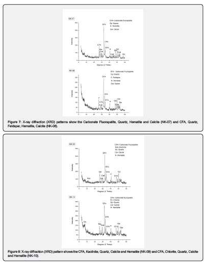
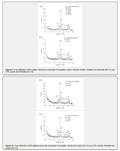
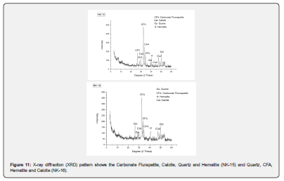
The X-ray diffraction studies revealed that Carbonate Fluorapatite (CFA) is the major phosphate mineral in the nodules of Nambakuruchi block of Trichinopoly district Tamil Nadu India, calcite, quartz, feldspar, kaolinite, chlorite, hematite, is the minor constituents (Figures 7-10), NK-07, NK-08, NK-09, NK-10, NK-11, NK-12, NK-13, NK-14, NK-15, and NK-16). Similar work has been reported by [3].
Conclusion
The Cretaceous phosphatic deposits Nambakuruchi block of Karai formation, Tiruchirappalli Tamil Nadu shows in the nodular form on badland topography. The physical observations of these nodules are associated with Karai Shale having different shapes and sizes. The size of nodules varies from sizes 2 to 5cm in width and 6 – 20cm in length with different shapes like circular, cylindrical, conical, rectangular, and elliptical. And the surface is yellow or white to reddish colors with different veins consisting of crystal contact. The crystal of the phosphate is brown to grey color with bryozoan fragments. In addition to the physical parameters measured such as medium to high specific gravity, hardness 5 on Moh’s scale, vitreous luster, transparent to translucent, fracture and indistinct cleavage, brown color streak and other associated minerals for identification of unknown nodules. The optical behavior is also important for the mineral’s identification parameters. The thin section studies show the fine-grained matrix of the carbonate fluorapatite, which is a significant constituent in microscopic studies. Thin sections of the nodules indicate the fine-grain phosphate grain with silt-size particles with the fine matrix. Pieces of the foraminifera and other small carbonate particles consist of light to dark lamination: coated grain, poloids, and fine detrital grains with cavities. The XRD studies refer to the mineralogical constituents of phosphate is carbonate fluorapatite as a significant and calcite, quartz, feldspar, kaolinite, chlorite, and hematite as gangue constituents.
Acknowledgment
We are thankful to the In-charge of XRD, department of Mechanical Engineering, AMU Aligarh. We also express our gratitude to the chairperson of, the Department of Geology, for providing the necessary facilities for this work.
Conflicts of Interest
The authors declare no conflicts of interest regarding the publication of this paper.
References
- Worth H (1893) Report on the phosphate deposits of Trichinopoly. G.O No: 821, revenue dept, Govt of Madras, India.
- Rao VP, Michard A, Naqvi SWA, Bottcher ME, Krishnaswamy R, et al. (2002) Quaternary phosphorites off the southeast coast of India. Chem Geol 182(2-4): 483-502.
- Rao VP, Dessarkar PM, Nagendra R, Babu EVSSK (2007) Origin of Cretaceous phosphorites from the onshore of Tamil Nadu, India. Journal of Earth System Science 116(6): 525-536.
- Banerjee DM (1986) Proterozoic and Cambrian phosphorites regional review: India subcontinent; In: Cook PJ, Shergold JH (Eds.), Phosphate deposits of the world, Proterozoic and Cambrian phosphorites, Cambridge University Press, USA, pp. 1: 70-90.
- Sundaram R, Rao PS (1979) Lithostratigraphic classification of Ullatur and Trichinopoly groups of Upper Cretaceous rocks of Tiruchirapalli District, South India. Records of the Geological Survey of India 45: 111-119.
- Rao LR (1958) Fossil Algae in India. Nature 181: 544-545.
- Blanford HF (1862) Cretaceous and other rocks of South Arcot and Trichinopoly districts, Madras. Geol Surv India Memoir, India 4: 217.
- Govindan A, Yadagiri K, Ravindran CN, Kalyanasundar R (1998) A field guide on Cretaceous sequences of Tiruchirapalli Area, Cauvery Basin, India; Oil and Natural Gas Corporation Limited, p. 53.
- Nagendra R, Reddy NA (2017) Major geologic events of the Cauvery Basin, India and their correlation with global signatures e A review. Journal of Paleogeography 6(1): 69-83.
- Allman M, Lawrence DF (1972) Geological Laboratory Techniques. Blandford, UK, pp. 1-335.
- Hutchison CS (1974) Laboratory handbook of Petrographic Techniques. Wiley-Interscience New York, USA 12: 527.
- Miller J (1988) Microscopical techniques: 1. Slices, slides, stains and peels. In: Tucker M (Ed.), Techniques in Sedimentology. Blackwell Scientific Publication, pp. 86-107.
- Fang Q, Yu W (2002) Principles of crystallography. National Defense Industry Press, Beijing, China.
- Nakai I, Abe Y (2012) Portable x-ray powder diffractometer for the analysis of art and archaeological materials. Appl Phys A106: 279-293.
- Zhang N, Lin C (2016) Review on the application of x-ray diffraction in gem identification, synthesis and crystal structure research. Rock Miner Anal 35(3): 217-228.
- Yu JS, Lei XR, Zhang JH (2011) X-ray powder identification manual of minerals. Huazhong University of Science and Technology Press, China.
- Venkateshwarlu M, Nagendra R (2016) Drift of the Indian Plate during Upper Cretaceous: An Appraisal from Magnetic and Biostratigraphic Perspectives of Sediment Outcrop Successions from Ariyalur-cauvery Basin, Southern India.