Can Muscle Asymmetry of the Biceps Femoris, Vastus Lateralis and Vastus Intermedius of an Elite Soccer Player be Identified by Ultrasound?
Haniel Fernandes*
Departament of Nutrition, Estácio de Sá College, Brazil
Submission: October 27, 2022; Published: November 14, 2022
*Corresponding author: Haniel Fernandes, Departament of Nutrition, Estácio de Sá College, Brazil
How to cite this article: Haniel F. Can Muscle Asymmetry of the Biceps Femoris, Vastus Lateralis and Vastus Intermedius of an Elite Soccer Player be Identified by Ultrasound?. JOJ Int Med. 2022; 1(3): 555564. DOI:10.19080/JOJIM.2022.01.555564
Abstract
Objectives: How a right-handed elite soccer player can have a greater hypertrophied on vastus lateralis, vastus intermedius and biceps femoris of the left leg than the dominant leg due to the physical functions required by soccer, this work presents an observational narrative of the images of these muscles performed in an elite soccer player through an ultrasound device to know if this method would be important to find asymmetries that can harm this athlete’s performance.
Methods: A 31-year-old right-handed professional soccer player, 179cm, 82.5kg and 6.6% body fat percentage, active in a soccer team of the Campeonato Brasileiro da Série A, with at least 15 years of experience as an athlete, agreed in participate and provide their ultrasound images of the vastus lateralis, vastus intermedius and biceps femoris muscles of both legs for a comparison.
Results: The results on ultrasound image analysis of the right-handed soccer player show a visual difference in favor of the right leg for the biceps femoris muscles and in favor of the left leg for the vastus lateralis and vastus intermedius muscles.
Conclusions: Muscle ultrasound images can serve as a parameter to visualize muscle asymmetry in elite soccer players.
Keywords: Elite soccer player; Muscle ultrasound images; Muscle asymmetry
Introduction
Knowing that muscle asymmetry magnitude is a physical assessment item dependent on the task most performed by the athlete [1] and lower muscles considered symmetrical (difference between 10 and 15%) can be a parameter to define power performance in sports athletes single-legged, such as soccer [2,3]. In this sport, a right-handed professional player who is obviously going to kick with his right leg will have to balance every time he kicks, or makes a pass, on his left leg, can making it more hypertrophied on vastus lateralis than the dominant leg [4,5]. In addition to the muscles used for balance during kicks or passes, hamstring muscles influence the ability to accelerate sprint speed in elite soccer players [6,7]. In this way, this work presents an observational narrative of the image’s comparison of vastus lateralis, vastus intermedius and biceps femoris muscles performed through an ultrasound device in a professional soccer athlete to clarify the following question: the evaluation by ultrasound image would be important to find asymmetries that can harm the elite soccer athlete’s performance?
Methods
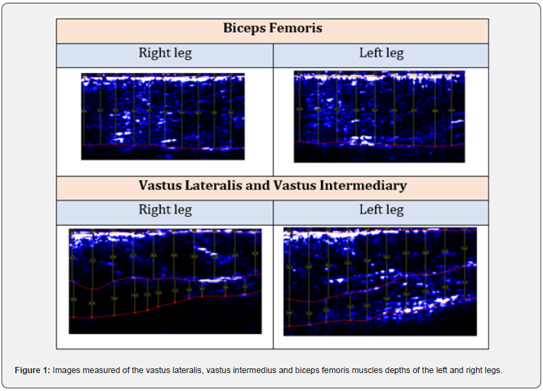
A 31-year-old right-handed professional soccer player, 179cm, 82.5kg and 6.6% body fat percentage, active in a soccer team of the Campeonato Brasileiro da Série A, with at least 15 years of experience as an athlete, agreed to participate and give their ultrasound images to the author, who is their nutritionist. The system used to measure the images was Body Metrix (Intel Metrix, CA), an affordable computer-based ultrasound system that provides rapid assessment of muscle depths in millimeters [8]. The author chose to evaluate the biceps femoris and vastus lateralis muscles of both legs, as they are muscle groups that influence the elite soccer players’ performance and may be in asymmetry for these athletes [4,2,5]. Measurements were performed with the athlete standing, starting from the proximal to distal position of each muscle group evaluated as instructed by the product manufacturer. Due to the anatomical positions, the ultrasound device was capable of measuring, in addition to the vastus lateralis depth, the depth of the vastus intermedius, as shown in figure 1 with the images taken. The gel used in the measurements was provided by the manufacturer and any deviations in the measurement practice, such as measurement speed and selected area, were explained to the athlete during the analysis.
Results
The results of the present study are presented in figure 1, which shows images of the biceps femoris, vastus lateralis and vastus intermediary muscles of the right and left legs of the athlete mentioned in the methodology. Can be noticed a visual difference between the left and right leg portions. For the biceps femoris, a muscle involved in the athlete’s acceleration on the field [6], the difference in detriment to the greater depth of this muscle in the right leg may be linked to the fact that the athlete’s dominant leg is the right leg, on which it exerts greater force during running’s. For the vastus lateralis and vastus intermedius, muscles involved in supporting when he kicks or makes passes [4], the left leg, in this case non-dominant, has an apparent greater depth difference compared to the right leg. In summary, the results on ultrasound image analysis of the right-handed soccer player show a visual difference in favor of the right leg for the biceps femoris muscles and in favor of the left leg for the vastus lateralis and vastus intermedius muscles.

Discussion
Differently from the analyzes brought by other researchers, even used to reference this study, who used assessment practices in groups of athletes [2,3], and clinical analyzes different from an observational model [6,7], this work only deduces from images of ultrasound what could be characterized as asymmetry and only speculates that it may have any impact on the physical performance of the athlete in question. The present work does not have the power to impact a clinical decision nor the statistical strength of a randomized clinical trial with a significant number of participants. However, the author demonstrates that ultrasound can be a method used in clinical practice to identify some form of asymmetry of the vastus lateralis, vastus intermedius and biceps femoris elite soccer athlete muscles to try to explain him about its importance in an attempt optimize his performance in the pitch.
Conclusion
Muscle ultrasound images can serve as a parameter to visualize muscle asymmetry in elite soccer players. However, randomized clinical studies are needed to know whether from these images’ asymmetries can be characterized that may negatively impact athletes’ performance (difference > 15%).
References
- Hewit JK, Cronin JB, Hume PA (2012) Asymmetry in multi-directional jumping tasks. Phys Ther Sport 13(4): 238-242.
- Meyers RW, Oliver JL, Hughes MG, Cronin JB, Lloyd RS (2015) Maximal sprint speed in boys of increasing maturity. Pediatr Exerc Sci 27(1): 85-94.
- Vaisman A, Guiloff R, Rojas J, Delgado I, Figueroa D, et al. (2017) Lower Limb Symmetry: Comparison of Muscular Power Between Dominant and Nondominant Legs in Healthy Young Adults Associated with Single-Leg-Dominant Sports. Orthop J Sports Med 5(12): 1-6.
- Giraldo GJC, Meneses OAN, Hernández EH (2020) Study on the differences in quantitative ultrasound of the quadriceps between schoolchildren who practice different sports 37(6): 379-386.
- Rojas QG, Venegas JP, Valencia O, Guzmán V, Araneda OF, et al. (2021) Hip and thigh muscular activity in professional soccer players during an isometric squat with and without controlled hip contraction Retos 2041(39): 697-704.
- Hammami R, Duncan MJ, Nebigh A, Werfelli H, Rebai H (2022) The Effects of 6 Weeks Eccentric Training on Speed, Dynamic Balance, Muscle Strength, Power, and Lower Limb Asymmetry in Prepubescent Weightlifters. J Strength Cond Res 36(4): 955-962.
- Ishøi L, Aagaard P, Nielsen, MF, Thornton KB, Krommes KK, et al. (2019) The Influence of Hamstring Muscle Peak Torque and Rate of Torque Development for Sprinting Performance in Football Players: A Cross-Sectional Study. Int J Sports Physiol Perform 14(5): 665-673.
- Boucher JP, Chamberland G, Aubertin LM, Jones DH, Rehel R, et al. (2011) Validation of the Body Metrix Ultrasound System for Percent Body Fat.


