




 JOJDC Home



Abstract
Background/Objectives: Pityriasis rosea (PR) is a self-limited papulosquamous eruption with excellent prognosis, often recognized in lighter skin by a herald patch which transforms into the classic Christmas tree distribution. In skin color (SOC), PR presents a diagnostic challenge due to atypical lesion color, morphology, and distribution. This review highlights underrecognized variants, post-inflammatory sequelae, and disparities in diagnosis.
Methods: A narrative review of literature from 2015-2025 was performed, including trials, cohort studies, case reports, and observational studies on PR in darker phototypes. Articles published in PubMed/MEDLINE, Scopus, and Google Scholar were reviewed for thematic relevance. Data on epidemiology, clinical features, diagnostic pitfalls, and outcomes were synthesized with prior foundational studies.
Results: PR prevalence appears consistent across racial groups, but clinical presentations differ in skin of color. Lesions are papular or hyperpigmented with decreased visible scale and less identifiable herald patches in SOC. Black patients may present with atypical distributions involving the face and scalp, as well as oral lesions or absent/multiple herald patches. Misdiagnoses—such as tinea, eczema, or syphilis-are common and can lead to delays in care. Post-inflammatory hyperpigmentation is frequent, while hypopigmentation is also reported. Some studies describe greater pruritus and shorter disease courses in affected children.
Conclusion: PR in SOC frequently deviates from classic teaching, necessitating heightened clinical suspicion. Broader inclusion of SOC in dermatologic training, culturally competent education, and targeted research is essential to improve diagnostic accuracy, management, and equity.
Keywords:Pityriasis rosea; Skin of color; Pigmented skin; Hyperpigmentation; Diagnostic delay; Dermatology education; Health disparities
Abbreviations:PR: Pityriasis Rosea; SOC: Skin Color; PIH: Post-Inflammatory Hyperpigmentation
Introduction
Pityriasis rosea (PR) is an acute, self-limiting skin condition characterized by a distinctive scaly eruption. The most common dermatosis among children and young adults affects patients worldwide at a rate of 0.5%-2% of dermatology outpatients [1]. The disease often presents with a single herald patch in the trunk area and, after 1-4 days, smaller multiple oval erythematous lesions along the lines of skin tension, resulting in the classic “Christmas tree” distribution [2]. The eruption typically lasts 6-8 weeks and then resolves spontaneously. Although epidemiologic data demonstrates. PR is observed with equal frequency across racial groups, with a U.S. prevalence of 0.2%, literature suggests the clinical presentation in skin of color (SOC) diverges from the classical description, which has been based on lighter skin types.
In patients with Fitzpatrick I-III, PR presents as salmon-pink, annular patches with collarette scaling. In those with Fitzpatrick IV-VI, however, these features may be less distinct or absent. Erythema can be masked by melanin, collarette scale can be subtle, and herald patches can be atypically located on the face, scalp or distal extremities [3,4]. These differences are shown in Table 1. In addition to pigmentary differences, PR in SOC can also exhibit atypical morphologies (papular, vesicular, or inverse) or occur at uncommon sites, such as the face, acral areas, or oral mucosa [5]. Figure 1 describes some of these varying morphologies..
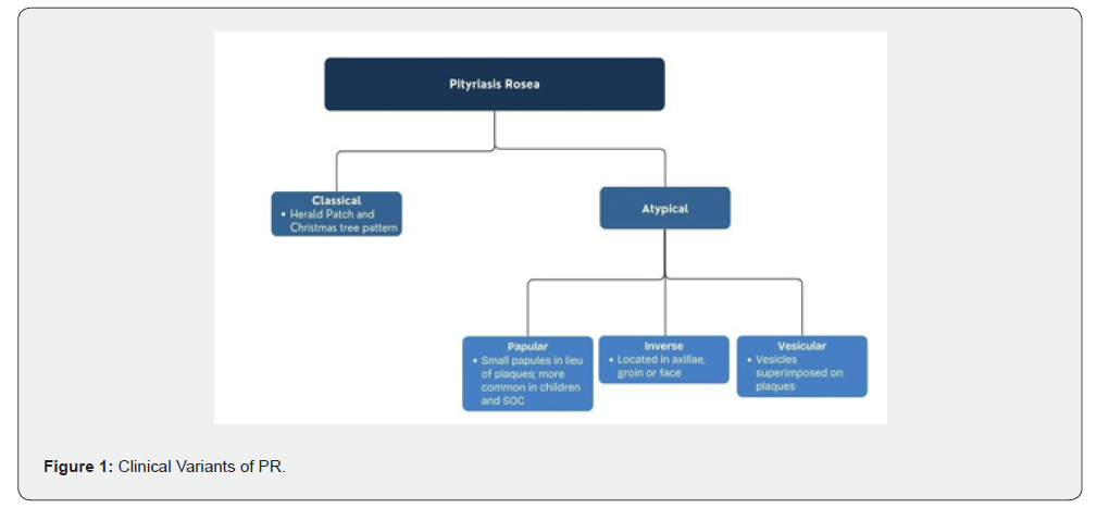

This schematic describes the numerous clinical variations of PR, highlighting the atypical presentations. Recognizing the variants is vital to avoid misdiagnosis. This high degree of clinical variability makes PR a “diagnostic chameleon” that can masquerade as many other dermatoses as possible [3,6] including tinea, syphilis, eczema, or drug eruption, increasing the risk of misdiagnosis and delayed management [7,8]. Diagnostic delays are also due to the limited representation of SOC patients in dermatologic textbooks. In a review of seven textbooks, fewer than 20% of images depicted individuals with dark skin, which can limit clinicians’ ability to use pattern recognition [9,10]. Closing these gaps has clinical and equity-related implications.
Delayed or missed recognition of PR in SOC can result in unnecessary investigations (such as biopsies and infectious serologies) and inappropriate therapies. While PR is benign, it can leave post-inflammatory hyperpigmentation (PIH) in pigmented skin, which can result in significant psychosocial distress if unanticipated and/or unmanaged. In this narrative review, we summarize the literature over the last decade on PR in SOC, with a particular focus on atypical presentations and diagnostic pitfalls, pigmentation- and culture-related factors, and lack of representation in dermatology education. We aim to provide clinicians with a more comprehensive understanding of PR in all skin types and share tips to enhance diagnostic accuracy, patient counseling, and equity in dermatologic care.
Methods
A detailed literature review was conducted to identify articles related to PR, with a focus on SOC and atypical presentations. PubMed/MEDLINE, Scopus, and Google Scholar databases were searched for English-language articles published from 2015 to 2025 using keywords: “pityriasis rosea,” “skin of color,” “dark skin,” “atypical pityriasis rosea,” and “hyperpigmentation”. Reference lists from the included articles were also hand-searched to identify additional articles, including seminal older articles used for background information. The initial search yielded approximately 210 articles across databases. After removal of duplicates and screening of titles and abstracts for relevance, approximately 80 full-text articles were assessed. Of these, approximately 55 studies were ultimately included in the final analysis. Study selection was based on relevance to clinical presentation, diagnostic features, and management of pityriasis rosea in skin of color, with prioritization of studies providing detailed clinical descriptions, dermoscopic or histologic correlation, or discussion of atypical presentations and pigmentary sequelae.
Included studies were randomized controlled trials, cohort and case-control studies, cross-sectional studies, case series, and single case reports that described clinical information regarding PR in pigmented skin. Articles were also included if they addressed epidemiology, clinical presentation, histologic or dermoscopic findings, differential diagnosis, management, and/or outcomes of PR. Studies were excluded if they lacked specific discussion of skin of color, did not provide clinically relevant or original data, were duplicate publications, or were not available in English. Since this was a narrative review, no quality assessment was done, and no formal meta-analysis was conducted. Following narrative review guidelines, systematic and broad general reviews were primarily used in the Introduction, whereas the Results and Discussion focused on original studies and case reports published between 2015 and 2025. The review was formatted to focus thematically on clinical presentation and diagnostic challenges, as well as cultural and pigmentation-specific influences and implications for dermatology education and equity.
Results
Epidemiology and Prevalence in Diverse Populations
A recent cross-sectional analysis of over 300,000 individuals from the federally funded U.S. population-representative All of Us cohort found a PR prevalence of 0.21% without any meaningful difference between the four largest racial groups.10 These results corroborate what is observed in the literature globally, with a uniform incidence in all populations [11]. Reports from Nigeria and India similarly reported rates like those in Europe and North America [12]. Seasonality has been reported across the world, but the pattern is not dissimilar among skin types [13]. Although incidence is the same across all groups, there may be differences in clinical expression. A series from India, including patients with phototypes IV-V, described herald patches in only 35% of cases (vs 50-90% in classic series) and 22.5% with atypical morphology (i.e., papular, vesicular, or inverse variants) [5]. Similarly, a Turkish series reported ~20% of cases presented with vesicular, purpuric, or urticarial variants [14]. These reports suggest that clinicians who care for patients with darker phototypes will be more likely to encounter non-classical variants, particularly papular PR, which is most prevalent in SOC [7].
Atypical Clinical Presentations in Skin of Color
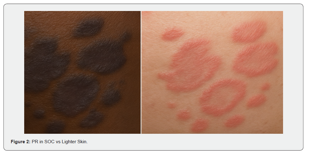
In skin of color, pityriasis rosea lesions often lack visible erythema and instead appear gray, brown, or violaceous rather than the classic salmon-pink seen in lighter phototypes, which may make recognition more challenging [3,4,15]. The initial presentation of SOC in Black patients can present as a dusky, violaceous plaque, with fine scaling, as opposed to the classic erythematous herald patch in lighter skin (Figure 2). Violaceous patches and plaques in SOC (left image), in contrast to the salmonpink erythematous patches and plaques seen in lighter skin (right image). Distribution and morphologic variants are more common in SOC as well. Papular PR is one of the variants that is much more common, with up to one-third of Black American children having the papular variant at presentation, compared to a relatively lower rate in Caucasian cohorts [16].
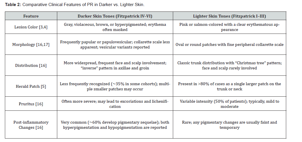
Papulovesicular or lichenoid variants have been described in Asian and African patients as well [6,17], and rare vesicular variants seem to affect children with darker skin preferentially [1]. These variants can be easily misdiagnosed if the physician is not familiar with atypical variants. Distribution is often atypical in SOC, with less prominent truncal “Christmas-tree” patterns and more frequent involvement of the face, scalp, and extremities; facial lesions occur in up to 30% of Black pediatric patients and scalp involvement in approximately 8%, both uncommon in White patients [16]. Inverse (axillae, groin, or extremities) and unilateral variants, like pityriasis rosea unilateralis, have been described [13,18,19] and rare cases of palmoplantar lesions can mimic secondary syphilis or hand-footmouth disease [20]. These differences are listed in Table 2.
Diagnostic Challenges and Misdiagnosis
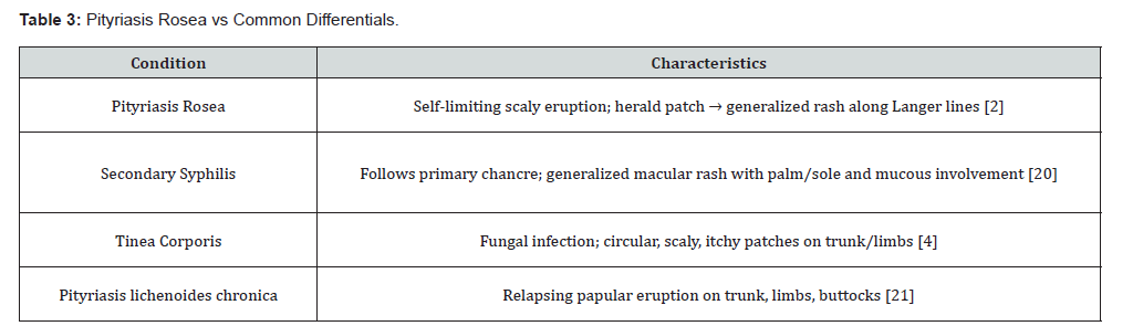
Establishing a diagnosis of PR in SOC is challenging since erythema is the main visual clue. Melanin often masks erythema, so lesions lack the typical salmon-pink color and appear dark brown, violaceous, or hyperpigmented. The herald patch is less common, and the eruption may not be recognized, especially since its appearance is easily confused with tinea corporis [4]. Dermoscopy can be helpful: the finding of collarette scaling, coppery-brown pigmentation, and peripheral vessels, in the absence of erythema, can increase diagnostic confidence [12,22-24]. Failure to diagnose PR without dermoscopy leads to frequent errors (tinea, syphilis, pityriasis lichenoides, eczema, drug eruptions), unnecessary tests, and inappropriate treatments [2,23]. These diagnostic challenges prompted a comparison with common mimickers, summarized in Table 3.
Post-Inflammatory Hyperpigmentation and Sequelae
One of the most common long-term consequences of PR in SOC is post-inflammatory hyperpigmentation. Post-inflammatory hyperpigmentation is common in SOC and may persist after lesion resolution, occasionally presenting with hypopigmented macules [26]. Almost 50% of Black children with PR develop residual hyperpigmentation, compared to only a minority of Caucasian patients, where changes are usually mild and temporary [27]. This makes surveillance for pigmentary changes critical. In addition to cosmetic sequelae, PIH can have major psychosocial effects. Discoloration may last months, causing embarrassment and lowered self-esteem [28,29]. Early treatment may help limit this: in a retrospective case series of 56 PR patients treated with valacyclovir, disease duration and severity were significantly shorter, potentially reducing cumulative inflammatory insult and pigmentary sequelae [30]. The treatment of PIH is often challenging, with agents such as topical retinoids, azelaic acid, glycolic acid peels, and careful use of hydroquinone being potential options with variable success [31].
Rigorous photoprotection, gentle skin care, and avoidance of harsh scrubbing are crucial, as sunscreen use is essential in preventing the worsening of PIH. Notably, decreased sunscreen use among SOC patients has been reported, which may contribute to the persistence of pigmentary changes and highlights the importance of patient education on photoprotection [28]. Hypopigmented sequelae, which may be more distressing to patients with darker skin types due to the contrast, are usually associated with papular or vesicular PR and may merit dermatologist referral for additional therapies and counseling in the realm of pigmentary disorders [27]. Patient counseling is also essential. Clinicians should reassure most patients that pigmentary changes will be temporary, as well as discourage unsafe remedies such as steroidor mercury-containing creams [25]. Education of the patient with visual aids can also help set realistic expectations. Early diagnosis and culturally sensitive counseling can help prevent unnecessary treatments and assist patients in avoiding further skin damage from misattributed rashes or unnecessary therapies.
Cultural and Psychosocial Factors
Beyond biological differences, the patient’s cultural belief systems can also have a major impact on how PR is understood and treated in SOC. In many cultures, rashes are thought to be caused by dietary changes, contagion, or spiritual imbalance. This belief can result in delays in seeking medical care, as well as the application of traditional remedies that may be harmful or exacerbate inflammation, leading to an increased risk of PIH [25,32]. Language can also be a barrier to care, as some languages lack specific vocabulary to describe changes in redness or pigmentation [33]. Patients may be more concerned with the presence of persistent dark marks than with the acute rash itself.
They may experience embarrassment or distress due to the impact on their social or dating life, which may be more pronounced in adolescent patients [23]. Clinicians can provide culturally sensitive care by asking about the use of traditional or home remedies, acknowledging the validity of the patient’s cultural belief system, and providing clear and comprehensive education about the condition. Clinicians should also address any concerns patients have about pigmentary changes, including reassuring them that PIH often resolves over time and may be treated with specific agents, such as azelaic acid or hydroquinone derivatives [34]. Setting expectations for pigmentary changes and recovery times can help build trust with the patient.
Underrepresentation in Education and Training
Diagnostic uncertainty of pityriasis rosea in SOC is exacerbated by the lack of representation in dermatology education. Images in dermatology texts were found to be 80-90% skin types I-IV, and some conditions, such as PR, may not be depicted at all [9]. Print and online learning modules for dermatology trainees also remain limited in representation of SOC (Fitzpatrick types IVVI). Consequently, trainees may not be adequately exposed to the breadth of clinical variants they encounter, nor become comfortable or prepared to detect PR in SOC [8,9]. Despite the availability of resources such as the Atlas of Dermatological Conditions in Populations of African Ancestry [35] and Dermatology for Skin of Color [36], broad curricular adoption has been a barrier.
PR in lighter skin is often described as salmon-colored plaques with an associated herald patch, but in darker skin, it is frequently hyperpigmented, purple, or gray, brown. It may or may not have a herald patch. In individuals with darker skin, appearance may mimic or be mistaken for tinea corporis, lichen planus, or cutaneous T-cell lymphoma, leading to diagnostic delays [6]. Dermatology trainees with appropriate exposure to skin conditions in the SOC during their training are more likely to identify them and provide expedient care. Facilities, and the dermatologists within them, are being increasingly provided with SOC image libraries and training curricula from organizations such as the Skin of Color Society and the American Academy of Dermatology. Practical tools at the bedside include: “reverse side” (comparison of affected with unaffected area bilaterally) to determine unilateral rash, and increased emphasis on palpation to reveal psoriatic plaques, lichenified eczematous lesions, and secondary or residual changes, such as hyperpigmentation, when erythema is not apparent or is subtle [7]. Efforts to formally integrate such tools in medical education and training would help mitigate diagnostic delays
Discussion
This case review emphasizes that pityriasis rosea in skin of color presents unique diagnostic challenges. While the pathogenesis of PR is consistent across skin types, its manifestations can vary by racial and ethnic backgrounds: erythema may be masked, lesions may appear gray, violaceous, or brown, and papular variants are more common [6,37,38]. These atypical patterns contribute to its characterization as a “diagnostic chameleon.” PR is frequently misdiagnosed; hyperpigmented plaques can be mistaken for tinea, and papular variants for syphilis, leading to unnecessary testing and treatments [8,38]. Enhanced training with diverse clinical images and instruction on PR beyond its “salmon-colored” description is essential. Therapeutic management should address sequelae, as more than 50% of patients with SOC experience distressing pigmentary changes, with pruritus often more pronounced [23,27].
Early counseling about the rash’s benign nature and treatments like antihistamines or topical steroids can enhance the patient experience. Education must be culturally sensitive, addressing patients’ concerns to mitigate stigma and build trust [25]. PR also illustrates broader issues in dermatology, such as delayed diagnosis in SOC due to underrepresentation in educational materials. Misdiagnosis can lead to unnecessary interventions and diminished confidence in healthcare [9,23,39]. Solutions include better representation of darker skin tones in training resources and epidemiological studies, as well as practical approaches like palpation and dermoscopy. A diagnostic algorithm has been included in Figure 3 to guide clinical testing in SOC [2,40,41].
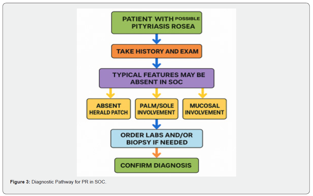
This figure outlines key diagnostic steps, emphasizing deviations in SOC (e.g., absent herald patch, palm/sole or mucosal involvement) and when to order labs or biopsy before confirmation. Future directions may include early antiviral therapy, such as high-dose acyclovir, to minimize the risk of pigmentary sequelae [2]. AI-assisted diagnostic tools could also aid in recognition for less experienced clinicians. In conclusion, PR in SOC underscores the necessity for inclusive diagnostic algorithms, consideration of atypical variants, patient counseling on pigmentary risks, and greater representation in educational materials and research to reduce misdiagnosis and advance equity in dermatologic care [6,23]. PR exemplifies how common conditions can present atypically, highlighting the importance of flexibility, cultural sensitivity, and inclusive training for accurate diagnoses and equitable care.
Conclusion
In conclusion, pityriasis rosea can present with diverse and atypical features in SOC. Lesions may be hyperpigmented or papular, involve the face, scalp, or flexures, and lead to pigmentary sequelae. Recognition requires a high index of suspicion, dermoscopy, and confirmatory testing when needed. In addition to diagnosis, clinicians must anticipate sequelae, provide culturally sensitive counseling, and discourage harmful selftreatments. Systemic gaps remain in clinical practice and research, including limited representation in educational resources and few SOC-focused studies. Addressing these challenges requires expanding training materials, diversifying research cohorts, and incorporating inclusive imagery. By recognizing PR’s unique aspects in SOC and addressing these gaps, misdiagnosis can be reduced and equity in dermatologic care advanced.
- Mashoudy KD, Kim S, Farhadi L, Scott A Elman (2025) Beyond the herald patch: exploring the complex landscape of pityriasis rosea. Am J Clin Dermatol 26(2): 237-250.
- Villalon-Gomez JM (2018) Pityriasis rosea: diagnosis and treatment. Am Fam Physician 97(1): 38-44.
- Mitchell CR, Love PB (2021) Inflammatory disorders: psoriasis, lichen planus, pityriasis rosea, and sarcoidosis. In: Li BS, Maibach HI, eds. Ethnic Skin and Hair and Other Cultural Considerations. Springer 2021: 91-101.
- Chhabra N, Prabha N, Kulkarni SS, Ganguly S (2018) Pityriasis rosea: clinical profile from central India. Indian Dermatol Online J 9(6): 414-417.
- Urbina FZ, Das A, Sudy E (2017) Clinical variants of pityriasis rosea. World J Clin Cases 5(6): 203-211.
- Ludwig R, von Stebut E (2023) Entzündliche dermatosen auf pigmentierter Haut. Dermatologie 74(2): 84-89.
- Pathak A, Perry M (2022) Dermatological conditions in skin of colour. Dermatol Ther (Heidelb) 15(8): 450-455.
- Ferdinand J (2024) Recognising clinical signs and symptoms on Black, Asian and minority ethnic (BAME) skin types. Atomic Article Preprint J.
- Adelekun A, Onyekaba G, Lipoff JB (2021) Skin color in dermatology textbooks: an updated evaluation and analysis. J Am Acad Dermatol 84(1): 194-196.
- Joshi TP, Calderara GA, Lipoff JB (2022) Prevalence of pityriasis rosea in the United States: a cross-sectional study using the All of Us database. JAAD Int 8: 45-46.
- Mahajan K, Relhan V, Kochhar Relhan A, Garg VK (2016) Pityriasis rosea: an update on etiopathogenesis and management of difficult aspects. Indian J Dermatol 61(4): 375-384.
- Nwako-Mohamadi MK, Masenga JE, Mavura D, OF Jahanpour, et al. (2019) Dermoscopic features of psoriasis, lichen planus, and pityriasis rosea in patients with skin type IV and darker attending the Regional Dermatology Training Centre in Northern Tanzania. Dermatol Pract Concept 9(1): 44-51.
- Leung AKC, Lam JM, Leong KF, Hon KL. Pityriasis rosea: an updated review. Curr Pediatr Rev. 2021;17(3):201-211.
- Kilinç F, Akbaş A, Sener S, Aktaş A (2017) Atypical pityriasis rosea: clinical evaluation of 27 patients. Cutan Ocul Toxicol 36(2): 157-162.
- Litchman G, Nair PA, Syed HA, et al. (2025) Pityriasis rosea. In: StatPearls [Internet]. Updated March 1, 2024. Treasure Island (FL): StatPearls Publishing.
- Amer A, Fischer H, Li X (2007) The natural history of pityriasis rosea in Black American children: how correct is the “classic” description? Arch Pediatr Adolesc Med 161(5): 503-506.
- Gupta N, Levitt J (2017) Unique clinical presentations of pityriasis rosea: aphthous ulcers, vesicles and inverse distribution of lesions. Dermatol Online J 23(2): 1303.
- Yadav A, Sanke S, Yadav J, Chander R (2020) Pityriasis rosea unilateralis: a rare presentation in two females. Indian Dermatol Online J 11(5): 823-825.
- Bukhari I, AlKhater SA (2015) Atypical pityriasis rosea: a case report and a review of literature. Int J Dermatol Venereol 2(2): 46-51.
- Apple A, Lin A, Kim A, Rozenberg SS, Kushner R (2020) Palmoplantar pityriasis rosea. SKIN J Cutan Med 4(6): 571-574.
- Jindal R, Chauhan P, Sethi S (2021) Dermoscopic characterization of guttate psoriasis, pityriasis rosea, and pityriasis lichenoides chronica in dark skin phototypes: an observational study. Dermatol Ther 34(1): e14631.
- Gavvala M, Gavvala M (2021) Diagnosis of pityriasis rosea using a triple light source dermoscope. J Med Allied Sci 11(1): 51-55.
- Sen S, Mridha K, Datta A (2021) Skin of color – an enigma: a systematic review. Turk J Dermatol 15(3): 45-54.
- Song J, Chandy S (2019) Atypical pitiriasis rosea: a review of the literature. Eur J Pharm Med Res 6(5): 145-149.
- Kannan RK (2021) Cultural dermatoses: a review. Dermatol Ther (Heidelb) 11: 1-7.
- Shah M, Adeel S (2024) Pityriasis rosea. InnovAiT 17(11-12): 483-487.
- Mondal A, et al. (2022) Pityriasis rosea among pediatric patients: a cross-sectional study. J Pak Assoc Dermatol 32(1): 96-102.
- Kaufman BP, Aman T, Alexis AF (2018) Postinflammatory hyperpigmentation: epidemiology, clinical presentation, pathogenesis and treatment. Am J Clin Dermatol 19(4): 489-503.
- Naik P (2022) Getting to the core of contemporary therapies for post-inflammatory hyperpigmentation. J Drugs Dermatol 21(3): 276-283.
- Ben Youssef S, Bhagavathi V, Wassef C, Miller JH (2023) What’s old is new: valacyclovir for the treatment of pityriasis rosea, a retrospective case series. JAAD Case Rep 37: 98-102.
- Mahé A (2023) Skin of color dermatology as a milestone toward diversity medicine. JEADV Clin Pract 2(2): 85-87.
- Chiang CL, Ward M, Gooderham M (2022) Dermatology: how to manage acne in skin of colour. Drugs Context 11: 1-10.
- De La Garza H, Lipoff JB, Daneshjou R (2022) Reducing language barriers in dermatology: a step toward equitable care. J Am Acad Dermatol 87(5): e189-e190.
- Wardhani PH, Rahmadewi R (2017) Treatment options for post inflammatory hyperpigmentation in color skin. Indian J Dermatol Venereol Leprol 28(3): 243-250.
- Donkor C, Aryee-Boi J, Osazuwa Itohan R, Afflu FK, Alexis AF (2021) Atlas of Dermatological Conditions in Populations of African Ancestry. Springer.
- Taylor SC, Lim HW (2025) Taylor and Kelly’s Dermatology for Skin of Color. 3rd ed. McGraw-Hill Education.
- Onalaja AA, Taylor SC (2021) Defining skin color. In: Li BS, Maibach HI, eds. Ethnic Skin and Hair and Other Cultural Considerations. Springer 2021: 10.
- Drago F, Herzum A, Varesano S, Serviddio G, Broccolo F, et al. (2025) Herald patch as the only evidence of pityriasis rosea: clinical, laboratory and pathogenetic features. Viruses 17(1): 119.
- Okeke CAV, Malik AMHU, Atwater AR, Powell DL, Wu PA (2022) American Contact Dermatitis Society position statement: dermatitis and skin of color. Dermatitis 33(1): 3-9.
- Punchihewa N, Lee S, Tan CG, Foley P (2024) Pityriasis rosea-like drug eruption secondary to deucravacitinib. JAAD Case Rep 53: 63-65.
- Shende AA, Chikhalkar SB (2024) A cross-sectional study of epidemiological and clinical aspects of pityriasis rosea along with dermoscopic analysis and histopathology correlation. Indian J Dermatol 69(2): 145-151.

























