




 JOJDC Home



Abstract
Objectives: this study was conducted on healthy skin using a fractional CO2 laser and a 532 nm laser system equipped with a fractional scanner to evaluate the synergistic interaction between the two wavelengths.
Methods: A 52-year-old male forearm was selected. Three areas were treated: CO₂ laser (Area A), 532 nm laser (Area B), and combined treatment (Area C). Healing was evaluated by LC-OCT at baseline, 24 hours, and 10 days, assessing crater morphology, inflammation, edema, and vasodilation. Edema/vasodilation were graded (0-2). Daily dermoscopy (25 days) assessed erythema (0-3) and fibrin plug expulsion, using maximum values as references for comparative analysis.
Results: The 532 nm laser (Area B) produced non-ablative sub-millimetric thermal damage without significant inflammation, with keratinocyte desquamation visible at 10 days and slow microscab expulsion; erythema, edema, and vasodilation were mild and resolved within 10 days. The CO₂ laser (Area A) generated characteristic ablation craters with coagulation zones, showing marked early edema, vasodilation, and inflammatory infiltrate, persisting at 10 days with fibrin plugs; microscab expulsion occurred around 2 weeks, with pronounced erythema resolving in 2-3 weeks. The combined CO₂ + 532 nm treatment (Area C) maintained CO₂-like ablation morphology but showed reduced early vasodilation and accelerated healing, with near-complete epidermal restoration at 10 days; microscab expulsion was faster than CO₂ alone, and although initial erythema was slightly higher, it resolved more rapidly with reduced edema and vasodilation.
Conclusion: The combination of fractional CO₂ and 532 nm lasers represents a promising approach that modulates post-treatment inflammation.
Keywords: Fractional CO2 Laser; 532 Nm Laser; Inflammation; Dermoscopic Evaluation; Coherence Tomography
Abbreviations: LC-OCT: Confocal Optical Coherence Tomography
Introduction
In CO2 laser treatments for skin rejuvenation and the reshaping of atrophic and hypertrophic scars, a crucial first step in managing healing times was the transition from total air ablation methods to the use of fractional emission techniques [1]. These fractional methods introduce areas of micro thermal damage surrounded by healthy tissue. The surrounding and uninvolved tissue serves as a reservoir of substances for faster healing. Although there is a reduced involvement of the ablated tissue, an activation of cytokine cascades has been observed, which promote in a three-dimensional way the entire dermo-epidermal compartment to obtain an adequate stimulation of fibroblastic activity [2]. A further step towards optimizing the treatments was subsequently to combine ablative light sources with non-ablative ones in the same fractional emission [3].
This synergistic laser approach has been shown to reduce the overall energy levels emitted, while enhancing the mutual effects of the two sources, thereby amplifying their efficacy [4], such as an increase in reversible or irreversible thermal heating zones and an increase in cellular turnover [5]. This strategy enables improved control of side effects and to benefit from different stimulation plans linked to the different penetration depths of different wavelengths [6]. Current evidence describes the synergistic or individual action of laser sources with fractional emission that use water as the primary chromophore to develop a selective photothermal effect. A new frontier of application, however, is the combination of an ablative source and one whose chromophore is the vascular component.
The choice of these two different contributions lies in the possibility of once again exploiting the benefits of increased cellular turnover while simultaneously reducing the diffuse vascular component of the skin, thus reducing the presence of post-treatment edema and erythema. It is well established that mitigating post-procedural vasodilation in the minutes immediately following ablative laser surgery can improve the healing process, reduce the sensation of itching and edema, and limit the risk of chronic healing [7]. For this reason, tests were conducted on healthy skin using a fractional CO2 laser and a 532 nm laser system equipped with a fractional scanner to evaluate the synergistic interaction between the two wavelengths (an ablative wavelength and a vascular targeting one).
Materials and Methods
The forearm of a 52-year-old male patient was chosen as this anatomical site allows for a more detailed temporal evaluation of the response, in fact, the forearm represents an area that heals more slowly than the facial one. Three treatment areas were identified. Area A, which received treatment with only CO2 laser (Deka Mela Srl, Calenzano, Italy) with the following parameters: 33 mJ, DOT Spot size 0.2 mm, Spacing 500 mm, 15x15 mm scan area. Area B, who received a 532 nm laser treatment only (El.En. Group, Calenzano, Italy) with the following parameters: 20 J/cm2, 20 ms, DOT Spot size 0,8 mm, Spacing 500 mm, 15x15 mm scan area. Area C, who received delivery of both laser sources with the same protocol as the single-source areas. For the evaluation of the healing, line-field confocal optical coherence tomography (LC-OCT) imaging (DeepLive™, DAMAE Medical, France) was carried out before the procedure (T0), at 24 hours (T1), and at 10 days (T10).
This imaging tool allows for the three-dimensional reconstruction of a virtual biopsy. Specifically, the morphologies of the ablation craters were evaluated in terms of both width and depth. Furthermore, hyperreflective inflammatory infiltrate, edema, and capillary vasodilation were evaluated. In subsequent follow-ups it was possible to observe the different repair processes and the desquamation of keratinocytes in the stratum corneum [5]. A three-point scale was used to analyze the level of edema and vasodilation: 0 – No edema/vasodilation, 1 – Mild edema/vasodilation, 2-Severe edema/vasodilation. The LC-OCT image showing the greatest edema/vasodilation was identified as the maximum value and assigned the maximum score (Severe). Each image was subsequently evaluated against the image with the greatest edema/vasodilation for a comparative assessment.
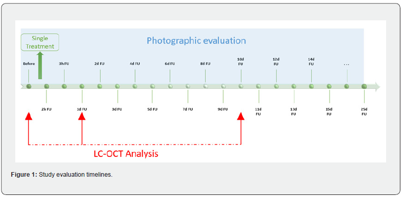
Daily thermoscopic images were also acquired on the three areas for 25 days for a quantitative assessment of the erythema intensity and for a measurement of the percentage amount of expulsion of the fibrin plugs (scabs) introduced by the CO2 laser. A four-point scale was used to analyze the level of erythema: 0-No redness, 1-Mild redness, 2-Moderate redness, 3-Severe redness. The dermoscopic image of all acquisitions showing the greatest redness was identified as the maximum value and assigned the maximum score (severe redness). Each image was then evaluated by comparing it with the one showing the greatest erythema for a comparative assessment. The evaluation timeline was indicated in Figure 1.
Results
532 nm Only = Area B
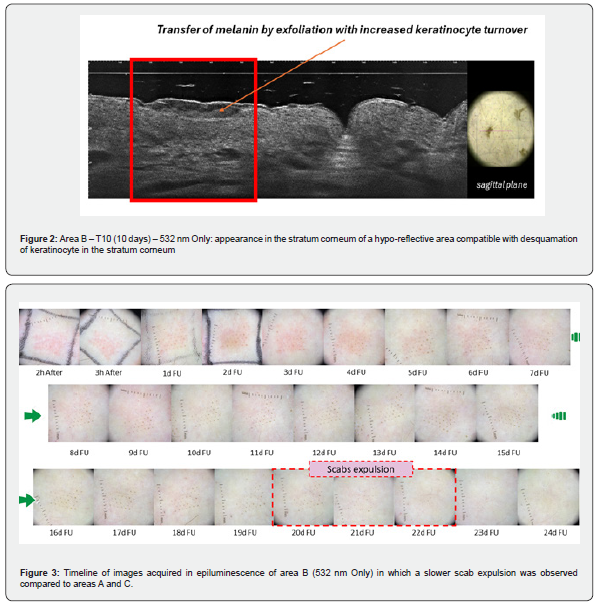
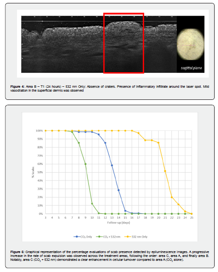
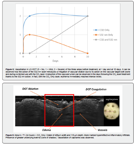
The use of the non-ablative 532 nm fractional emission source in single use (area B) determines microzones of thermal damage with sub-millimetric diameter. These can be more clearly observed by LC-OCT at 10 days in the stratum corneum, with a hypo-reflective area (Figure 2). This desquamation of keratinocyte in the stratum corneum is confirmed by epiluminescence images (Figure 3). The 532 nm wavelength does not cause an ablative action, as also confirmed by the LC-OCT in Figure 4. Furthermore, no significant inflammatory infiltrate was found around the laser spot. The epiluminescence image shows a slow expulsion of the microscabs, which occurs around the third week (Figures 3 and 5). It should be noted that the test area of the arm heals more slowly than the face. Finally, a slight appearance of erythema was observed immediately after treatment (Figures 3 and 6) confirmed by a slight presence of edema and vasodilation on the first day (Figures 7 and 8) which disappeared within 10 days of follow-up (Figure 6).

CO2 Only – Area A
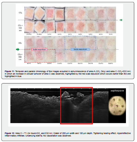
The use of the fractional emission CO2 laser source in single use (area A) determines microzones of thermal damage with a sub-millimetric diameter that present a central area where the ablation has occurred surrounded by a coagulation zone. The morphology described by LC-OCT (Figure 9) is the typical one found in histological images in H&E [8]. The image also depicted, at 1-day follow-up, significant vasodilation of the capillaries and the presence of edema. At the 10-day follow-up LC-OCT images still showed the presence of fibrin plugs, in the healing phase with the presence of inflammatory infiltrate (Figure 10) typical of a physiological remodeling of the lesion by fractional laser. The epiluminescence image showed the expulsion of microscabs, which occurs around the second week (Figures 11 and 5). It should be noted that the test area of the arm heals more slowly than the face. Area A exhibited a faster expulsion rate compared to area B when treated with the 532 nm system alone. Finally, the appearance of erythema was observed immediately after treatment (Figures 6 and 11) confirmed by a marked presence of edema and vasodilation on the first day (Figures 7 and 8) which returned to the basal condition in the second/third week of follow-up (Figure 6).
CO2 + 532 nm – Area C
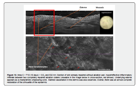
The use of the fractional CO2 laser emission combination with the 532 nm (area C) determines microzones of thermal damage with a sub-millimetric diameter characterized by a central area where the ablation occurred surrounded by a coagulation zone (Figure 12), like that observed with the CO₂ laser alone. However, at the 1-day follow-up, the image depicted the absence of significant capillary vasodilation, contrast to what was observed with CO₂ laser treatment alone. Furthermore, the beginning of a tightening healing effect, attributable to the combined use with the 532 nm, was also observed. The 10-day follow-up with LC-OCT showed that the skin was already repaired without an ablation wall. A hyperreflective inflammatory infiltrate was observed between two completely repaired ablation craters, typical of physiological lesion remodeling from fractional laser therapy. Persistent underlying edema and a marked vasodilation in the dermis were observed.
Particularly noteworthy was the complete reconstitution of the epidermal silhouette (Figure 13) in area C relative to area A. The epiluminescence image showed the expulsion of microscabs, which occurs before the beginning of the second week (Figures 5 and 11). It should be noted that the test area of the arm healed more slowly than the face. The expulsion rate of area C, which was irradiated by both sources, was faster than that of area A, which was exposed to the CO2 system alone. Finally, erythema appeared immediately after treatment (Figures 6 and 11), which was slightly more intense than with CO2 alone. However, in the following days (days 4-5), the intensity decreased more markedly compared to the use of the CO2 laser alone. This finding was confirmed by a lower presence of edema and vasodilation already evident in the first days (Figures 7 and 8).

Discussion
The LC-OCT evaluation combined with spumescence documentation allowed us to monitor over time the evolution of erythema, inflammation, dermal vasodilation, the presence of edema and the expulsion of fibrin plugs induced by the areas of controlled thermal micro-damage of the laser systems under study. The study was conducted on the forearm to have an extension of the observation times, but it can be easily recalibrated in the experiences of facial treatments which reduce the times indicated in the study by almost one and a half times. This model was created to simulate a classic skin rejuvenation treatment in which the aim is to achieve skin remodeling through renewal and stimulation of collagen fibers. The objective was to verify the possibility of containing post-treatment vasodilation, which can lead to a cascade of persistent erythema, pain, and itching, knowing that a chronicization of the wound can lead to unwanted scarring and post-inflammatory hyperpigmentation.
The use of 532 nm was therefore aimed at acting on the microcirculation to contain vasodilation and to have an alternative and complementary source that could contribute to the increase in cell turnover to guarantee safer treatment and that could reintroduce the patient to normal social activities more quickly. The reported results confirm that the combined use of the CO2 laser with the 532 nm does not introduce major critical issues in the immediate post-treatment period, while it is beneficial for containing the vasodilation found in LC-OCT which showed a reduction in erythema, therefore in inflammation, through dermoscopic monitoring in the days following the treatment. Finally, the increased healing, as indicated by delayed fibrin plug expulsion under combined treatment, may reflect a dual synergistic mechanism. The 532 nm laser certainly has a boost effect on CO2, but this paradigm can also be reversed, meaning that a CO2 laser with lower energies can facilitate the expulsion of scabs in a vascular treatment using the 532 nm laser.
Conclusion
In conclusion, the combined use of fractional CO₂ and 532 nm lasers appears to represent a promising synergistic strategy capable of modulating post-treatment inflammatory responses while preserving the efficacy of tissue remodeling. The reduction in early vasodilation and erythema, together with controlled healing dynamics, suggests a potential improvement in the safety profile and patient tolerability.
Institutional Review Board Statement
The study was conducted in accordance with the principles of Declaration of Helsinki. No activity was carried out outside the scope of the device intended purpose or that no additional invasive or burdensome procedures were carried out compared to procedure performed under the normal condition of use of the device.
Informed Consent Statement
Informed consent was obtained from all subjects involved in the study.
Conflicts of Interest
Authors Jorilda Biba, Irene Fusco and Tiziano Zingoni were employed by El.En. Group. The remaining authors declare that the research was conducted in the absence of any commercial or financial relationships that could be construed as a potential conflict of interest.
- Campolmi P, Bonan P, Cannarozzo G, Bassi A, Bruscino N, et al. (2012) Highlights of thirty-year experience of CO2 laser use at the Florence (Italy) department of dermatology. Scientific World Journal 2012: 546528.
- Prignano F, Campolmi P, Bonan P, Ricceri F, Cannarozzo G, et al. (2009) Fractional CO2 laser: a novel therapeutic device upon photobiomodulation of tissue remodeling and cytokine pathway of tissue repair. Dermatol Ther 22(Suppl 1): S8-15.
- Clementi A, Cannarozzo G, Guarino L, Zappia E, Cassalia F, et al. (2025) Sequential Fractional CO2 and 1540/1570 nm Lasers: A Narrative Review of Preclinical and Clinical Evidence. J Clin Med 14(11): 3867.
- Nisticò SP, Bennardo L, Zingoni T, Pieri L, Fusco I, et al. (2022) Synergistic Sequential Emission of Fractional 10.600 and 1540 nm Lasers for Skin Resurfacing: An Ex Vivo Histological Evaluation. Medicina (Kaunas) 58(9): 1308.
- Moro F, Fusco I, Madeddu F, Ambrosio L, Chello C, et al. (2026) Analysis with LC-OCT imaging in split face with CO2 laser and 1540 nm. A case report. Oxf Med Case Reports 2026(2): omaf296.
- Magni G, Piccolo D, Bonan P, Conforti C, Crisman G, et al. (2022) 1540-nm fractional laser treatment modulates proliferation and neocollagenesis in cultured human dermal fibroblasts. Front Med (Lausanne) 9: 1010878.
- Das A, Sarda A, De A (2026) Cooling Devices in Laser therapy. J Cutan Aesthet Surg 9(4): 215-219.
- Bonan P, Campolmi P, Cannarozzo G, Bruscino N, Bassi A, et al. (2012) Eyelid skin tightening: a novel 'Niche' for fractional CO₂ rejuvenation. J Eur Acad Dermatol Venereol 26(2): 186-193.

























