




 JOJDC Home



Abstract
Background: Polycystic Ovary Syndrome (PCOS) is the leading endocrinopathy in reproductive-age women, often presenting first with dermatological concerns. Objective: This review synthesizes the clinical implications, therapeutic strategies, and emerging biomarkers of PCOS, emphasizing the dermatologist’s role in mitigating systemic risks.
Methods: A systematic review of current literature and the 2023 International Evidence-Based Guidelines was performed. Results: Cutaneous
markers such as hirsutism, acne, and acanthosis nigricans are reliable proxies for biochemical hyperandrogenism and insulin resistance (IR).
Specifically, free testosterone (FT) and HOMA-IR serve as superior correlates of metabolic risk. Effective management requires a multidisciplinary
approach combining hormonal therapies (COCPs and spironolactone) with metabolic interventions like metformin and lifestyle modifications.
Evidence suggests that a 5–10% weight reduction significantly improves both ovulatory and dermatological outcomes. Emerging research into
the “gut-skin axis” and adipokine profiling (specifically the adiponectin-to-leptin ratio) offers a novel frontier for phenotype-specific diagnosis.
Furthermore, chronic low-grade inflammation (elevated CRP, IL-6, TNF-$\alpha$) contributes to both metabolic syndrome and impaired skin
barrier function.
Conclusion: Dermatologists are at the frontline of PCOS detection. Recognizing skin signs as indicators of systemic dysfunction-including NAFLD, type 2 diabetes, and cardiovascular disease-is essential. Transit integrate symptomatic dermatological care to integrated, multidisciplinary
models is the gold standard for reducing long-term morbidity. Future diagnosis may rely on multi-omic signatures, including microRNAs and
microbial diversity, to bridge the gap between clinical presentation and personalized therapeutic strategies. Early screening remains the most
effective tool in addressing the substantial health burden of PCOS.
Despite timely systemic management, including antivenom therapy, progressive local tissue destruction occurred. Surgical treatment consisted of staged debridement followed by definitive reconstruction using fasciotomy and skin grafting. Satisfactory functional and aesthetic outcomes were achieved. This case underscores the critical role of plastic and reconstructive surgery in the management of severe local complications of snakebite envenomation. Early multidisciplinary collaboration and appropriate surgical timing are essential to minimize morbidity and optimize outcomes.
Keywords:Polycystic Ovary Syndrome; Metabolic Syndrome; Gut-Skin Axis; Hyperandrogenism
Abbreviations:PCOS: Polycystic Ovary Syndrome; NIH: National Institutes of Health; AMH: Anti-Müllerian Hormone; IGF-1: Insulin-like Growth Factor 1; CRP: C-reactive Protein; IL-6: Interleukin-6; TNF-alpha: Tumor Necrosis Factor-alpha; DHT: Dihydrotestosterone; DHEAS: Dehydroepiandrosterone Sulfate; mFG: Modified Ferriman–Gallwey; FPHL: Female Pattern Hair Loss; AN: Acanthosis Nigricans; HOMAIR: Homeostatic Model Assessment of Insulin Resistance; HS: Hidradenitis Suppurativa; IL-17: Interleukin-17; IL-1: Interleukin-1; FT: Free Testosterone; SHBG: Sex Hormone-Binding Globulin; GnRH: Gonadotropin-Releasing Hormone; LH: Luteinizing Hormone; FSH: Follicle- Stimulating Hormone; LDL: Low-Density Lipoprotein; HDL: High-Density Lipoprotein; NAFLD: Nonalcoholic Fatty Liver Disease; BMI: Body Mass Index; OGTT: Oral Glucose Tolerance Test; HbA1c: Glycated Hemoglobin; COCPs: Combined Oral Contraceptive Pills; AUC: Area Under the Curve; miRNAs: MicroRNAs; GRADE: Grading of Recommendations, Assessment, Development, and Evaluations
Introduction
Polycystic ovary syndrome (PCOS) represents the most common endocrinopathy affecting women of reproductive age, with a global prevalence estimated at 10-13% according to the 2023 International Evidence-Based Guideline [1-2]. Recent metaanalyses have confirmed a pooled prevalence of 12.1% using Rotterdam criteria, with notable regional variation—highest in the Eastern Mediterranean (15.1%) and South-East Asian regions (14.3%) [3]. The burden of PCOS extends beyond reproductive dysfunction, encompassing metabolic, cardiovascular, and psychological sequelae that persist across the lifespan from adolescence through menopause [1-2].
Despite its high prevalence and significant health impact, women internationally continue to experience delayed diagnosis and dissatisfaction with care, highlighting persistent evidencepractice gaps in clinical management [1-2]. The diagnosis of PCOS has evolved considerably since the original 1990 National Institutes of Health criteria. Current diagnostic standards are based on the Rotterdam criteria, established in 2003 and subsequently endorsed by the 2018 and 2023 International Evidence-Based Guidelines [1-2].
These criteria require the presence of two of the following three features in adult women: (i) clinical or biochemical hyperandrogenism; (ii) ovulatory dysfunction manifesting as irregular menstrual cycles; and (iii) polycystic ovary morphology on ultrasound or, as of the 2023 update, elevated anti-Müllerian hormone levels [1-2]. Critically, other etiologies including thyroid dysfunction, hyperprolactinemia, and non-classical congenital adrenal hyperplasia must be excluded [4]. When both irregular menstrual cycles and hyperandrogenism are present, diagnosis is simplified and ultrasound or anti-Müllerian hormone testing is not required [1-2]. In adolescents, diagnostic criteria are more stringent, requiring both hyperandrogenism and ovulatory dysfunction, as ultrasound and anti-Müllerian hormone lack specificity in this age group [1-2].
Dermatological manifestations serve as critical early markers of PCOS, often representing the initial presenting features that prompt clinical evaluation. Cutaneous manifestations are usually among the first manifestations of PCOS and provide early clinical clues to underlying endocrine dysfunction [5]. An estimated 72-82% of women with PCOS present with cutaneous signs of hyperandrogenism, including hirsutism, acne vulgaris, and androgenic alopecia, while acanthosis nigricans—a marker of hyperinsulinemia—affects approximately 31-37% of patients [5- 6].
Among these dermatological findings, hirsutism and acanthosis nigricans have emerged as the most reliable cutaneous markers for distinguishing women who meet PCOS diagnostic criteria from those who do not [6]. Women meeting Rotterdam criteria demonstrate significantly higher rates of hirsutism (53.3% vs 31.2%), acne (61.2% vs 40.4%), and acanthosis nigricans (36.9% vs 20.0%) compared to those not meeting criteria [6]. Importantly, the presence of hirsutism or acanthosis nigricans correlates strongly with biochemical hyperandrogenism and metabolic abnormalities including insulin resistance, dyslipidemia, and elevated body mass index, warranting comprehensive diagnostic evaluation for metabolic comorbidities that may lead to long-term complications [6].
The characteristic distribution patterns of these cutaneous findings, particularly truncal hirsutism and axillary acanthosis nigricans, underscore the importance of comprehensive skin examination in the diagnostic workup [6]. Given that patients with PCOS are frequently first evaluated by dermatologists, recognition of these cutaneous markers represents a crucial opportunity for early diagnosis and intervention. This article provides a critical overview of the dermatological markers of PCOS under the updated Rotterdam criteria, offering clinicians a framework to optimize diagnosis and address the broader endocrine burden of the disorder.s
Pathophysiology of PCOS
The pathogenesis of PCOS is multifactorial, involving a complex interplay between intrinsic ovarian defects, systemic metabolic dysfunction, and chronic inflammation.
Hyperandrogenism and Insulin Resistance
Ovarian hyperandrogenism is a hallmark of PCOS, driven by an intrinsic functional up-regulation of theca cells. These cells demonstrate an increased expression of key steroidogenic enzymes, specifically cytochrome P450c17 and 3β-hydroxysteroid dehydrogenase, leading to excessive androgen production even in the absence of gonadotropin stimulation [7]. This is further exacerbated by systemic insulin resistance. Despite peripheral resistance to glucose uptake, the ovaries remain insulin-sensitive; hyperinsulinemia directly stimulates the P450c17 enzyme system and may act through IGF-1 receptors to augment androgen synthesis [7]. While obesity and genetic predisposition are primary drivers of this insulin resistance, hyperandrogenism itself appears to be a secondary effect rather than a primary cause [7].
Inflammation and Adipokine Dysregulation
PCOS is increasingly recognized as a state of chronic low-grade inflammation. Affected women exhibit elevated proinflammatory markers—including CRP, IL-6, and TNF-α—which correlate with increased risks of endothelial dysfunction and cardiovascular disease [8]. This inflammatory state is closely linked to adipose tissue dysfunction. Adipokines, such as leptin and adiponectin, are frequently dysregulated, with reduced adiponectin levels contributing to decreased insulin sensitivity [9]. Notably, visceral adiposity and altered adipokine profiles (e.g., decreased omentin-1) are observed even in lean patients with PCOS, providing a mechanistic explanation for the presence of insulin resistance regardless of body mass index [9].
Major Dermatological Manifestations
Cutaneous manifestations are among the most visible clinical features of PCOS, with patients frequently present with dermatologic signs such as acne, hirsutism, and androgenetic alopecia, which not only reflect underlying hormonal imbalance but may also serve as clinically relevant markers of disease severity [10].
Acne
Acne is one of the most common dermatological manifestations observed in women with polycystic ovary syndrome (PCOS), with reported prevalence ranging from approximately 40% to 70% in affected patients, particularly during adolescence and early adulthood [11]. The development of acne in this population is closely linked to androgen-mediated stimulation of sebaceous glands. Elevated levels of circulating androgens, especially testosterone and its more potent derivative dihydrotestosterone (DHT), increase sebaceous gland activity and sebum production, create a microenvironment that favors follicular obstruction, microcomedone formation, and subsequent inflammatory lesions
In addition to increased sebaceous secretion, androgen excess can alter follicular keratinization and contribute to the persistence of inflammatory acne lesions [12]. Several studies have demonstrated a relationship between acne severity and biochemical hyperandrogenism in PCOS, particularly with elevated total or free testosterone and dehydroepiandrosterone sulfate (DHEAS) levels. However, the correlation between serum androgen concentrations and acne severity is not always linear, suggesting that local androgen metabolism, receptor sensitivity in sebaceous glands, and metabolic factors such as insulin resistance may also influence clinical expression [13].
Hirsutism
Hirsutism is a common clinical manifestation of hyperandrogenism and affects approximately 10% of women worldwide, frequently representing a visible marker of endocrine dysfunction [14]. It is characterized by excessive terminal hair growth in androgen-dependent areas following a male-pattern distribution, including the upper lip, chin, chest, abdomen, back, arms, and thighs. The severity of hirsutism is typically assessed using the modified Ferriman–Gallwey (mFG) scoring system, which evaluates terminal hair growth across nine body sites.
Although diagnostic thresholds vary according to ethnic background, an mFG score greater than 4–6 is generally considered indicative of hirsutism. In clinical practice, polycystic ovary syndrome accounts for approximately 80–90% of hirsutism cases, while other etiologies include idiopathic hirsutism, nonclassical congenital adrenal hyperplasia, androgen-secreting tumors, Cushing syndrome, and other endocrine disorders [15]. From a biochemical perspective, the development of hirsutism is strongly associated with circulating androgen levels and increased peripheral androgen activity.
Local enzymatic activity within the pilosebaceous unit, including 5α-reductase and hydroxysteroid dehydrogenases, enhances the conversion of testosterone to the more potent androgen dihydrotestosterone (DHT), thereby stimulating terminal hair growth [16]. Consequently, evaluation of women presenting with hirsutism should include assessment of androgen levels and investigation of underlying endocrine causes, as the severity of hair growth may not always correlate directly with serum androgen concentrations due to variations in tissue sensitivity to androgens
Androgenetic Alopecia
Androgenetic alopecia, also referred to as female pattern hair loss (FPHL), represents another dermatological manifestation associated with hyperandrogenic conditions such as polycystic ovary syndrome. Clinically, it is characterized by progressive hair thinning that predominantly affects the central scalp and vertex while generally preserving the frontal hairline. The pattern of hair loss in women is commonly classified using the Ludwig scale, which describes the gradual reduction in hair density over the crown. The pathogenesis of androgenetic alopecia involves androgen-mediated miniaturization of hair follicles, largely driven by the local conversion of testosterone to the more potent androgen dihydrotestosterone (DHT), resulting in shortening of the anagen phase and progressive follicular regression [17].
In patients with PCOS, androgenetic alopecia may coexist with other hyperandrogenic dermatologic manifestations such as acne and hirsutism, reflecting the systemic hormonal imbalance characteristic of the syndrome. However, several studies have demonstrated that FPHL may occur even in the absence of significantly elevated circulating androgen levels, suggesting that genetic predisposition and increased sensitivity of follicular androgen receptors play an important role in its development. Consequently, the presence of androgenetic alopecia in women, particularly when accompanied by other cutaneous signs of hyperandrogenism, should prompt clinicians to evaluate potential underlying endocrine disorders including PCOS and related metabolic abnormalities [18].
Acanthosis Nigricans
Acanthosis Nigricans (AN) is a dermatosis identified by its distinctive morphological features, including dark, coarse, and thickened skin that possesses a velvet-like texture [19]. These lesions are generally symmetrical and are predominantly located in intertriginous areas such as the posterior neck, axillae, and groin. In women with PCOS, an additional frequent site is the inframammary region. AN is widely recognized as a marker of insulin resistance, particularly in endocrine and metabolic disorders [19,20].
Although the pathogenesis of AN is not fully known, it is strongly linked to chronic hyperinsulinemia. In states of insulin resistance, elevated circulating insulin levels increase insulin-like growth factor 1 (IGF-1) production in the liver, as well as stimulate receptors on keratinocytes and dermal fibroblasts. This activation promotes epidermal proliferation and dermal fibroblast growth, leading to the characteristic findings, including thickened and hyperpigmented lesions observed in AN [21].
In PCOS, insulin resistance is a central metabolic abnormality affecting approximately half to two-thirds of patients. Hyperinsulinemia not only contributes to metabolic dysregulation but also exacerbates ovarian androgen production, thereby linking dermatologic manifestations with endocrine dysfunction. AN frequently occurs in women with PCOS and may serve as an easily identifiable clinical indicator of underlying insulin resistance [22]. Studies have shown that the presence of AN in patients with PCOS correlates strongly with elevated fasting insulin levels and higher Homeostatic Model Assessment of Insulin Resistance (HOMA-IR) score [23].
The utility of AN in clinical practice lies in its role as a robust, non-invasive surrogate marker for identifying high-grade insulin resistance. While laboratory assessments such as the hyper insulinemic-euglycemic clamp remain the gold standard, their complexity and cost make them impractical. Acanthosis nigricans provides a visual diagnostic signal that can be assessed during a standard physical examination, facilitating diagnosis, especially in settings where laboratory testing may be limited [21,24].
Other Cutaneous Findings Seborrhea
Seborrhea is defined as an increase in sebum production because of increased activity of the sebaceous glands, clinically manifesting as oily, shiny skin and greasy scalp changes in the absence of primary inflammation. Sebaceous glands are highly responsive to androgens, particularly dihydrotestosterone (DHT), which stimulates sebocyte proliferation and lipid synthesis. In PCOS, hyperandrogenism represents a central pathophysiologic feature, often accompanied by insulin resistance and hyperinsulinemia, both of which further potentiate androgen production and peripheral androgen activity.
This hormonal environment promotes increased sebum secretion, making seborrhea a common cutaneous manifestation in women with PCOS. Clinical studies have demonstrated a high prevalence of seborrhea in PCOS patients, with reported rates ranging from approximately 34% to 43%, and significant associations with free testosterone, fasting glucose, and insulin levels [25,26]. Although seborrhea is a non-inflammatory condition, it can contribute to the development of seborrheic dermatitis.
Increased sebum production can alter the skin surface microenvironment, causing an increased proliferation of lipophilic yeasts, Malassezia spp. The metabolic byproducts of these organisms, in conjunction with host immune responses, contribute to epidermal disruption and inflammation characteristic of seborrheic dermatitis. In patients with PCOS, the persistent hyperandrogenic state may therefore act as an upstream driver, predisposing to seborrheic dermatitis through sustained sebaceous gland stimulation and alteration of skin surface lipids [26].
Skin Tags
Acrochordons (skin tags) are benign, pedunculated lesions typically found in intertriginous regions such as the neck, axillae, and groin. Beyond their benign appearance, they are increasingly recognized as cutaneous markers of systemic metabolic dysregulation. Their development is thought to be driven by the proliferative effects of hyperinsulinemia on keratinocytes and dermal fibroblasts, mediated in part through insulin-like growth factor-1 (IGF-1) signaling pathways [27,28].
In patients with polycystic ovary syndrome (PCOS), the presence of multiple acrochordons has been associated with higher insulin levels and adverse metabolic profiles, supporting their role as a visible indicator of underlying endocrine imbalance [28,29]. Clinically, acrochordons may therefore serve as a practical dermatologic clue to metabolic risk in PCOS, complementing other cutaneous findings without requiring additional diagnostic testing.
Hidradenitis Suppurativa
Hidradenitis suppurativa (HS) is a chronic, relapsing inflammatory disorder of the folliculopilosebaceous unit characterized by painful, deep-seated nodules, abscesses, sinus tract formation, and scarring. It predominantly affects intertriginous areas such as the axillae, inguinal region, inframammary folds, and perineum. The pathogenesis of HS is multifactorial, involving follicular occlusion, dysregulated innate and adaptive immune responses, and alterations in the cutaneous microbiome.
Pro-inflammatory cytokines, including tumor necrosis factor-α (TNF-α), interleukin-17 (IL-17), and interleukin-1 (IL-1), play a central role in disease progression [30,31]. An increasing body of evidence suggests a significant association between HS and polycystic ovary syndrome (PCOS). Both conditions share overlapping features, including hyperandrogenism, insulin resistance, and obesity, which may contribute to their coexistence.
Androgens are thought to influence follicular keratinization and sebaceous gland activity, potentially promoting follicular occlusion, a key initiating event in HS. Additionally, hyperinsulinemia may exacerbate androgen production and inflammatory signaling pathways, further linking HS to the endocrine and metabolic disturbances observed in PCOS [31,32]. Epidemiologic studies have demonstrated a higher prevalence of PCOS among patients with HS compared to the general population, supporting the concept of HS as part of a broader systemic disorder with endocrine and metabolic components [32,33].
Biochemical Correlates
PCOS is a multifaceted endocrine condition marked by hyperandrogenism, impaired ovulation, and polycystic ovarian morphology, and is closely linked to metabolic dysfunction. Its pathophysiology involves a complex interplay between insulin resistance, compensatory hyperinsulinemia, and androgen excess (elevated free testosterone), which contributes significantly to both reproductive and metabolic disturbances [34,35].
Free Testosterone
Free testosterone (FT), the biologically active fraction of circulating testosterone, is a key determinant of metabolic risk in PCOS. Elevated FT levels are strongly associated with insulin resistance, impaired glucose tolerance, and increased risk of type 2 diabetes mellitus [34-36]. Women with higher FT levels exhibit greater central adiposity, increased waist circumference, and a higher prevalence of metabolic syndrome components compared to those with normal androgen levels [34,36]. Unlike total testosterone, FT correlates more consistently with adverse metabolic outcomes due to its dependence on sex hormonebinding globulin (SHBG), which is suppressed in hyperinsulinemic states [34].
DHEAS
Dehydroepiandrosterone sulfate (DHEAS), an adrenal androgen, is elevated in a subset of women with PCOS, typically exceeding 350 to 400 μg/dL [34,37]. Elevated DHEAS reflects adrenal hyperactivity and contributes to the overall androgen burden; however, its association with metabolic abnormalities is less consistent compared to FT [34]. While some studies suggest a modest relationship between DHEAS and insulin resistance or lipid abnormalities, its clinical utility as a metabolic marker remains limited relative to ovarian androgens [34,37].
LH/FSH Ratio
Alterations in gonadotropin secretion are characteristic of PCOS, with an increased luteinizing hormone to follicle-stimulating hormone ratio (LH/FSH), often exceeding 2:1 or 3:1 [38]. This imbalance results from dysregulated hypothalamic gonadotropinreleasing hormone (GnRH) pulsatility, leading to preferential LH secretion and increased ovarian androgen production [38]. Although the LH/FSH ratio is useful in understanding the reproductive endocrinology of PCOS, it has limited predictive value for metabolic complications and is not consistently elevated in all patients [38].
Insulin and HOMA-IR
Insulin resistance is a fundamental feature of PCOS and is present in both obese and lean individuals. It is typically reflected by elevated fasting insulin levels (>15 to 20 μIU/mL) and increased homeostatic model assessment for insulin resistance (HOMA-IR) values (>2.5 to 3.0), indicating reduced insulin sensitivity [35,39]. Hyperinsulinemia enhances ovarian androgen production and suppresses SHBG synthesis, thereby increasing circulating FT levels and exacerbating hyperandrogenism [35]. This bidirectional relationship establishes a pathogenic cycle in which insulin resistance and androgen excess perpetuate each other [35,39].
Lipid Profile Abnormalities
Dyslipidemia is a common metabolic feature of PCOS and contributes to increased cardiovascular risk. The typical lipid profile includes elevated triglycerides (>150 mg/dL), increased low-density lipoprotein (LDL) cholesterol (>130 mg/dL), and reduced high-density lipoprotein (HDL) cholesterol (<50 mg/ dL) [39,40]. These abnormalities are closely linked to insulin resistance and hyperandrogenism, particularly elevated FT levels, which promote visceral fat accumulation and adverse lipid metabolism [36,39]. Consequently, women with PCOS exhibit a more atherogenic lipid profile compared to age-matched controls [40].
The metabolic and hormonal disturbances in PCOS are interconnected through a complex network of feedback mechanisms. Insulin resistance leads to hyperinsulinemia, which stimulates ovarian androgen production and reduces SHBG levels, thereby increasing FT concentrations [35]. Elevated FT, in turn, contributes to central adiposity and worsens insulin sensitivity, perpetuating a vicious cycle [34,35]. Although DHEAS and LH/FSH imbalance contribute to the endocrine phenotype, FT and insulin resistance remain the primary drivers of metabolic dysfunction in PCOS [34,35,38].
Understanding the interplay between these hormonal and metabolic factors is essential for risk stratification and management. FT and HOMA-IR are particularly valuable markers for identifying patients at increased risk of metabolic syndrome and cardiovascular disease. Early identification and targeted interventions, including lifestyle modification and insulinsensitizing therapies, may help mitigate long-term metabolic complications in women with PCOS [35,39].
Metabolic Syndrome and Dermatologic Severity
Metabolic abnormalities are prevalent among women with Polycystic Ovary Syndrome (PCOS) and contribute significantly to the severity of its clinical manifestations. A substantial proportion of affected individuals exhibit features of Metabolic Syndrome, including central obesity, dyslipidemia, impaired glucose metabolism, and increased cardiometabolic risk [41]. These disturbances are largely driven by underlying insulin resistance, which exacerbates ovarian androgen production and suppresses the hepatic synthesis of sex hormone–binding globulin (SHBG), thereby increasing the bioavailability of free androgens (Figure 1) [1,41].
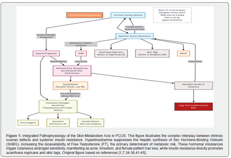
Obesity plays a central role in this metabolic dysfunction. Excess adipose tissue promotes compensatory hyperinsulinemia, further fueling the hyperandrogenic environment. This hormonal imbalance has direct pathological effects on androgen-sensitive cutaneous structures, leading to an increased prevalence and severity of acne vulgaris and hirsutism, as well as classic markers of insulin resistance such as acanthosis nigricans and skin tags [42]. Dyslipidemia in PCOS—typically characterized by elevated triglycerides and low-density lipoprotein (LDL) with reduced high-density lipoprotein (HDL) levels—is closely associated with chronic low-grade inflammation [43].
These lipid alterations may influence sebaceous gland activity and proinflammatory pathways within the dermis, potentially contributing to the persistence of inflammatory acne [43]. Furthermore, shared metabolic mechanisms, including insulin resistance and obesity, predispose women with PCOS to Nonalcoholic Fatty Liver Disease (NAFLD). This hepatic involvement mirrors systemic metabolic dysfunction and is often clinically reflected in the skin through visible indicators of severe insulin resistance [44].
Collectively, these metabolic abnormalities promote endothelial dysfunction and accelerated atherosclerosis, increasing long-term cardiovascular risk [45]. Cutaneous manifestations such as hirsutism, acne, and acanthosis nigricans serve as reliable clinical proxies for this underlying metabolic burden. Consequently, the recognition of these dermatologic markers provides a crucial opportunity for clinicians to identify high-risk patients who require early metabolic screening and targeted interventions to mitigate long-term cardiovascular complications [45].
Clinical Implications
Dermatologists occupy a critical frontline position in the early detection of PCOS, as cutaneous manifestations frequently represent the initial features prompting medical evaluation [5,46]. Given that patients often seek care for hirsutism, acne, or androgenic alopecia, these clinical markers offer a vital window for early diagnosis and systemic intervention [46]. Current international guidelines emphasize that hirsutism alone is highly predictive of biochemical hyperandrogenism and PCOS in adults [1,3]. Consequently, clinicians should perform a comprehensive physical examination using standardized tools, such as the modified Ferriman-Gallwey score (with a threshold of 4–6 depending on ethnicity), to evaluate terminal hair growth [1,47].
Effective management requires a multidisciplinary approach that addresses the syndrome’s reproductive, metabolic, and psychological sequelae [1,47]. Integrated care models should include endocrinologists, gynecologists, dermatologists, and mental health professionals to ensure a lifelong health plan focused on healthy lifestyle interventions and the prevention of excess weight gain [1,3]. Furthermore, shared decision-making is essential to address the high levels of dissatisfaction with PCOS care reported internationally [1,47]. Comprehensive metabolic screening is a cornerstone of management due to the significantly elevated risk of insulin resistance and type 2 diabetes [1,48].
Glycemic status should be assessed at diagnosis in all patients, regardless of BMI, ideally using a 75-g oral glucose tolerance test (OGTT), as fasting glucose and HbA1c demonstrate reduced accuracy in this population [1,47]. Additionally, baseline lipid profile and annual blood pressure monitoring are mandatory to mitigate cardiovascular risk [47]. Clinicians must also screen for obstructive sleep apnea, which occurs at a higher prevalence in PCOS independent of BMI [1]. Despite these clear evidencebased recommendations, metabolic screening remains largely underutilized, highlighting an urgent need for enhanced physician education and the implementation of clinical decision support tools [48-49].
Therapeutic Interactions
The management of dermatologic manifestations in PCOS requires a multifaceted approach targeting both the underlying endocrine dysfunction and the associated cutaneous symptoms.
Hormonal and Anti-androgen Therapies
Combined oral contraceptive pills (COCPs) remain a first-line pharmacological intervention for regulating menstrual cycles and mitigating hyperandrogenism. COCPs suppress ovarian androgen production and stimulate the hepatic synthesis of sex hormone–binding globulin (SHBG), which reduces circulating free testosterone and improves androgen-dependent conditions such as acne vulgaris and hirsutism [50]. In cases where COCPs alone are insufficient, anti-androgens such as spironolactone are utilized. Spironolactone functions by competitively blocking androgen receptors and inhibiting 5$\alpha$-reductase activity, significantly improving cutaneous manifestations, particularly terminal hair growth and androgenic alopecia [50].
Metabolic and Lifestyle Interventions
Insulin-sensitizing agents, primarily metformin, address the metabolic drivers of the syndrome. By improving insulin sensitivity and reducing compensatory hyperinsulinemia, metformin indirectly lowers ovarian androgen synthesis [51]. While its direct impact on skin markers is often less potent than anti-androgen therapy, it serves as a critical adjunct in patients with significant metabolic dysfunction or insulin resistance [51].
Lifestyle modification remains the cornerstone of PCOS management. Strategic weight reduction, calorie-controlled diets, and regular physical activity are essential for patients with obesity. Even a modest weight loss of 5–10% has been shown to restore ovulatory function, enhance insulin sensitivity, and ameliorate dermatologic symptoms by lowering the systemic androgen load [52].
Dermatologic-Specific Care
Direct management of cutaneous symptoms is often necessary to improve patient quality of life. This includes topical retinoids and antimicrobial agents for acne, as well as procedural interventions like laser hair removal or electrolysis for hirsutism. A comprehensive, multidisciplinary strategy—integrating endocrine, metabolic, and dermatologic expertise—is essential to effectively address the complex clinical spectrum of PCOS [50,52].
Emerging Biomarkers and Research Gaps
The search for precise diagnostic tools has shifted toward multi-omic approaches, focusing on adipokines, chronic inflammation, and the gut-skin axis as potential bridges between metabolic dysfunction and dermatological severity.
Adipokines and Inflammatory Signatures
Adipokines have emerged as intrinsic biomarkers of PCOS, independent of body mass index (BMI). Meta-analyses indicate that while adiponectin is significantly decreased, levels of chemerin, leptin, and visfatin are elevated in women with PCOS [53]. Notably, the adiponectin-to-leptin ratio has demonstrated superior discriminatory power (AUC = 0.83) in predicting the syndrome and correlates negatively with testosterone levels and insulin resistance [54]. This metabolic imbalance is compounded by chronic low-grade inflammation, characterized by a proinflammatory milieu of CRP, IL-6, and TNF-$\alpha$ [55]. These cytokines not only drive systemic insulin resistance but may also impair the skin barrier and enhance androgen bioactivity, exacerbating cutaneous symptoms [56].
The Gut-Skin Axis and Epigenetics
Emerging evidence highlights the “gut-skin axis” as a novel frontier in PCOS research. Microbial dysbiosis, specifically a reduction in alpha diversity and an increase in Bacteroides vulgatus, has been causally linked to insulin resistance and systemic inflammation [57]. This dysbiosis may influence immunemediated skin conditions, such as acne vulgaris, through the translocation of metabolites that modulate systemic inflammation [58]. Furthermore, circulating microRNAs (miRNAs) are being investigated as highly sensitive epigenetic markers; specifically, miR-29a-5p has shown a diagnostic accuracy as high as 95% in recent meta-analyses [59].
Research Gaps
Despite these advancements, significant gaps remain. Most studies on novel biomarkers lack large-scale validation across diverse ethnicities. Furthermore, while the gut microbiome’s role is compelling, standardized methodologies and high-quality longitudinal data (GRADE) are still needed to translate microbial signatures into clinical diagnostic panels or personalized therapeutic strategies [57,60].
Conclusion
Polycystic Ovary Syndrome is a multisystem disorder where the skin acts as a primary clinical mirror of internal endocrine and metabolic turmoil. This review highlights those dermatological manifestations—from classic acne and hirsutism to markers like hidradenitis suppurativa and skin tags-are intrinsically linked to a self-perpetuating cycle of hyperandrogenism and insulin resistance. The 2023 International Evidence-Based Guidelines underscore the necessity of moving beyond surfacelevel treatment toward a comprehensive diagnostic framework that includes mandatory metabolic screening for all patients. As research evolves into the gut-skin axis and epigenetic biomarkers like microRNAs, the role of the dermatologist becomes even more specialized, requiring an understanding of how systemic inflammation and microbial dysbiosis influence cutaneous health. Ultimately, the successful management of PCOS depends on early recognition and the seamless integration of dermatological, endocrine, and lifestyle interventions. By addressing the syndrome’s root metabolic drivers, clinicians can significantly improve the quality of life and long-term cardiovascular outcomes for women worldwide.
- Helena J Teede, Chau Thien Tay, Joop Laven, Anuja Dokras, Lisa J Moran, et al. (2023) Recommendations from the 2023 international evidence-based guideline for the assessment and management of polycystic ovary syndrome. Practice Guideline Fertil Steril 120(4): 767-793.
- Helena J Teede, Chau Thien Tay, Joop Laven, Anuja Dokras, Lisa J Moran, et al. (2023) Recommendations from the 2023 international evidence-based guideline for the assessment and management of polycystic ovary syndrome†. Hum Reprod 38(9): 1655-1679.
- Adriana C H Neven, Maria Forslund, Sanjeeva Ranasinha, Parneet Sethi, Raja Ram Dhungana, et al. (2026) Prevalence of polycystic ovary syndrome: a global and regional systematic review and meta-analysis. Hum Reprod Update 32(1): 14-35.
- Anju E Joham, Robert J Norman, Elisabet Stener-Victorin, Richard S Legro, Stephen Franks, et al. (2022) Polycystic ovary syndrome. Lancet Diabetes Endocrinol 10(9): 668-680.
- Stacey L Simon, Phoutdavone Phimphasone-Brady, Kathryn M McKenney, Lauren D Gulley, Andrea E Bonny, et al. (2024) Comprehensive transition of care for polycystic ovary syndrome from adolescence to adulthood. Lancet Child Adolesc Health 8(6): 443-455.
- Timothy H Schmidt, Keshav Khanijow, Marcelle I Cedars, Heather Huddleston, Lauri Pasch, et al. (2016) Cutaneous findings and systemic associations in women with polycystic ovary syndrome. JAMA Dermatol 152(4): 391-398.
- Diamanti-Kandarakis E, Nader S, Panidis D (2013) Novel insights into the pathophysiology and treatment of polycystic ovary syndrome. Endocr Connect 2(3): R12-R33.
- Ewa Rudnicka, Katarzyna Suchta, Monika Grymowicz, Anna Calik-Ksepka, Katarzyna Smolarczyk, et al. (2021) Chronic low grade inflammation in pathogenesis of PCOS. Int J Mol Sci. 22(7) :3789.
- Nagandla K, Banerjee I, Ismail NBH (2023) Adipocytokines in polycystic ovary syndrome (PCOS): a systematic review and meta-analysis. AIMS Med Sci 10(2):178-195.
- Artar G, Tas B, Turan G, Uckan HH (2022) Evaluation of androgen-dependent skin findings of polycystic ovary syndrome (PCOS). Gynecol Endocrinol 38(12): 1104-1108.
- Damoulaki E, Sioutis D, Sarli V, et al. (2025) Polycystic ovary syndrome-associated acne: the interplay of hyperandrogenism, insulin resistance, and therapeutic strategies. Cureus 17(11): e98103.
- Benyaminpour S (2024) PCOS and acne a systematic review. Int J Clin Stud Med Case Rep 37(2).
- Leanne Cussen, Tara McDonnell, Gillian Bennett, Christopher J Thompson, Mark Sherlock, et al. (2022) Approach to androgen excess in women: clinical and biochemical insights. Clin Endocrinol (Oxf) 97(2): 174-186.
- Oliveira TF, Comim FV (2024) Understanding hirsutism in PCOS. Expert Rev Endocrinol Metab 19(2): 103-110.
- Cebeci Kahraman F, Savaş Erdoğan S (2022) Grading of hirsutism: a practical approach to the modified Ferriman-Gallwey scoring system. Postepy Dermatol Alergol 39(4): 744-748.
- Mohamed Anas Patni, Rajani Dube, Subhranshu Sekhar Kar, Biji Thomas George, Manjunatha Goud Bellary Kuruba, et al. (2025) Exploring hirsutism: epidemiology, associated endocrinal abnormalities, and societal challenges in GCC—a narrative review. Int J Mol Sci 26(12): 5575.
- Jiang VS, Hawkins SD, McMichael A (2022) Female pattern hair loss and polycystic ovarian syndrome: more than just hirsutism. Curr Opin Endocrinol Diabetes Obes 29(6): 535-540.
- Owecka B, Tomaszewska A, Dobrzeniecki K, Owecki M (2024) The hormonal background of hair loss in non-scarring alopecias. Biomedicines 12(3): 513.
- Hughes EK, Brady MF, Rawla P (2023) Acanthosis nigricans. In: StatPearls.
- Popa ML, Popa AC, Tanase C, Gheorghisan-Galateanu AA (2019) Acanthosis nigricans: to be or not to be afraid. Oncol Lett 17(5): 4133-4138.
- Radu AM, Carsote M, Dumitrascu MC, Sandru F (2022) Acanthosis nigricans: pointer of endocrine entities. Diagnostics (Basel) 12(10): 2519.
- Tyebkhan G, Velaskar S, Yadav P, Phiske M, Shah D (2024) An Indian expert group review of acanthosis nigricans with recommendations for early detection and timely management. J Assoc Physicians India 72(6): 74-84.
- Márcio Augusto Pinto de Ávila, Lunna Perdigão Borges, Marina Silva Paez, Ricardo Vasconcellos Bruno, Antonio Egídio Nardi, et al. (2014) Acanthosis nigricans: metabolic interrelations inherent to the polycystic ovary syndrome. Rev Bras Ginecol Obstet 36(9): 410-415.
- Amisi CA (2022) Markers of insulin resistance in polycystic ovary syndrome women: an update. World J Diabetes 13(3): 129-142.
- Özdemir S, Görkemli H, Kiyici A, Bodur S (2010) Specific dermatologic features of the polycystic ovary syndrome and its association with biochemical markers of metabolic syndrome and hyperandrogenism. Acta Obstet Gynecol Scand 89(2): 199-204.
- Mohammad A Abusailik, Alsharif M Muhanna, Adam A Almuhisen, Abdullah M Alhasanat, Ayed M Alshamaseen, et al. (2021) Cutaneous manifestations of polycystic ovary syndrome. Dermatol Rep 13(2): 8799.
- Sari R, Akman A, Alpsoy E, Balci MK (2010) The metabolic profile in patients with skin tags. Clin Exp Med 10(3):193-197.
- Mathur SK, Bhargava P (1997) Insulin resistance and skin tags. Clinical Trial Dermatology 195(2):184.
- M Kahana, E Grossman, A Feinstein, M Ronnen, M Cohen, et al. (1987) Skin tags: a cutaneous marker for diabetes mellitus. Acta Derm Venereol 67(2): 175-177.
- Alikhan A, Lynch PJ, Eisen DB (2009) Hidradenitis suppurativa: a comprehensive review. J Am Acad Dermatol 60(4): 539-561.
- C C Zouboulis 1, N Desai, L Emtestam, R E Hunger, D Ioannides, et al. (2015) European S1 guideline for the treatment of hidradenitis suppurativa/acne inversa. J Eur Acad Dermatol Venereol 29(4): 619-644.
- Garg A, Neuren E, Cha D (2018) Evaluating patients with hidradenitis suppurativa for polycystic ovary syndrome. JAMA Dermatol 154(1): 94-97.
- Vinkel C, Thomsen SF (2018) Hidradenitis suppurativa: causes, features, and current treatments. J Clin Aesthet Dermatol 11(10): 17-23.
- Lerchbaum E, Schwetz V, Rabe T, et al. (2014) Hyperandrogenemia in polycystic ovary syndrome: exploration of the role of free testosterone and androstenedione in metabolic phenotype. PLoS One 9(10): e108263.
- Dunaif A (1997) Insulin resistance and the polycystic ovary syndrome: mechanism and implications for pathogenesis. Endocr Rev 18(6): 774-800.
- Z Dong, X Chen, L Li, J Huang, Q Yin, D Yang (2021) Free testosterone level correlated with the metabolic abnormalities dependent on central obesity in women with polycystic ovary syndrome. Exp Clin Endocrinol Diabetes 120(6): 355-360.
- Carmina E, Rosato F, Jannì A, Rizzo M, Longo RA (2006) Relative prevalence of different androgen excess disorders in 950 women referred because of clinical hyperandrogenism. J Clin Endocrinol Metab 91(1): 2-6.
- A E Taylor, B McCourt, KA Martin, EJ Anderson, JM Adams, et al. (1997) Determinants of abnormal gonadotropin secretion in clinically defined women with polycystic ovary syndrome. J Clin Endocrinol Metab 82(7): 2248-2256.
- Legro RS, Kunselman AR, Dodson WC, Dunaif A (1999) Prevalence and predictors of risk for type 2 diabetes mellitus and impaired glucose tolerance in polycystic ovary syndrome. J Clin Endocrinol Metab 84(1): 165-169.
- Wild RA, Painter PC, Coulson PB, Carruth KB, Ranney GB (1985) Lipoprotein lipid concentrations and cardiovascular risk in women with polycystic ovary syndrome. J Clin Endocrinol Metab 61(5): 946-951.
- Fu L, Xie N, Qu F, Zhou J, Wang F (2023) The association between polycystic ovary syndrome and metabolic syndrome in adolescents: a systematic review and meta-analysis. Front Endocrinol (Lausanne) 30(1): 28-40.
- Jena P, Tiwari M, Panda SR, Samantroy S, Panda J (2023) Correlation of cutaneous manifestations with body mass index (BMI) in polycystic ovary syndrome (PCOS) patients in a tertiary care centre: an observational study. Cureus 13(12):e20695.
- Tehrani FR, Behboudi-Gandevani S, Yarandi RB, Saei Ghare Naz M, Carmina E (2021) Prevalence of acne vulgaris among women with polycystic ovary syndrome: a systemic review and meta-analysis. Gynecol Endocrinol 37(5): 392-405.
- Ramiro Manzano-Nunez, Marta Santana-Dominguez, Jesus Rivera-Esteban, Clara Sabiote, Elena Sena, et al. (2023) Non-alcoholic fatty liver disease in patients with polycystic ovary syndrome: a systematic review, meta-analysis, and meta-regression. J Clin Med 12(11): 3813.
- Chau Thien Tay, Aya Mousa, Aadhya Vyas, Loyal Pattuwage, Fahimeh Ramezani Tehrani, et al. (2024) 2023 International Evidence-Based Polycystic Ovary Syndrome Guideline update: insights from a systematic review and meta-analysis on elevated clinical cardiovascular disease in polycystic ovary syndrome. J Am Heart Assoc 13(16): e033572.
- Housman E, Reynolds RV (2014) Polycystic ovary syndrome: a review for dermatologists: Part I. Diagnosis and manifestations. J Am Acad Dermatol 71(5): 847.e1-847.e10.
- Helena J Teede, Chau Thien Tay, Joop Laven, Anuja Dokras, Lisa J Moran, et al. (2023) Recommendations from the 2023 international evidence-based guideline for the assessment and management of polycystic ovary syndrome. Fertil Steril 120(4): 767-793.
- Amy S Dhesi, Katie L Murtough, Jonathan K Lim, Jay Schulkin, Peter G McGovern, et al. (2016) Metabolic screening in patients with polycystic ovary syndrome is largely underutilized among obstetrician-gynecologists. Am J Obstet Gynecol 215(5): 579.e1-579.e8.
- Leslie Cho, Melinda Davis, Islam Elgendy, Kelly Epps, Kathryn J Lindley, et al. (2020) Summary of updated recommendations for primary prevention of cardiovascular disease in women: JACC state-of-the-art review. J Am Coll Cardiol 75(20): 2602-2618.
- Shamim H, Jean M, Umair M, et al. (2024) Managing acne in individuals with polycystic ovarian syndrome. Int J Med Sci Clin Res 4(4): 22-31.
- Humaira Shamim, Marie Jean, Muaaz Umair, Pratyusha Muddaloor, Michelle Farinango, et al. (2023) Role of metformin in the management of polycystic ovarian syndrome-associated acne: a systematic review. Cureus 15(11): e48358.
- Rohit Gautam, Pratibha Maan, Anshu Jyoti, Anshu Kumar, Neena Malhotra, et al. (2023) The role of lifestyle interventions in PCOS management: a systematic review. Front Endocrinol (Lausanne) 17(2): 310.
- Kainan Lin, Xiaoting Sun, Xiao Wang, Hanchu Wang, Xia Chen (2021) Circulating adipokine levels in nonobese women with polycystic ovary syndrome and in nonobese control women: a systematic review and meta-analysis. Front Endocrinol (Lausanne) 12: 651553.
- Sameh Sarray, Samira Madan, Layal R Saleh, Naeema Mahmoud, Wassim Y Almawi (2015) Validity of adiponectin-to-leptin and adiponectin-to-resistin ratios as predictors of polycystic ovary syndrome. Fertil Steril 104(2): 460-466.
- Hongxia Deng, Yan Chen, Jilong Xing, Nannan Zhang, Liangzhi Xu (2024) Systematic low-grade chronic inflammation and intrinsic mechanisms in polycystic ovary syndrome. Front Immunol 15: 1354256.
- Singh I, Moar K, Maurya PK (2025) Diagnostic and prognostic biomarkers in polycystic ovary syndrome. Clin Chim Acta 576: 120425.
- Yue-Lian Yang, Wei-Wei Zhou, Shan Wu, Wen-Li Tang, Zong-Wei Wang, et al. (2021) Intestinal flora is a key factor in insulin resistance and contributes to the development of polycystic ovary syndrome. Endocrinology 162(10): bqab118.
- Britta De Pessemier, Lynda Grine, Melanie Debaere, Aglaya Maes, Bernhard Paetzold, et al. (2021) Gut-skin axis: current knowledge of the interrelationship between microbial dysbiosis and skin conditions. Microorganisms 9(2): 353.
- Deswal R, Dang AS (2020) Dissecting the role of micro-RNAs as a diagnostic marker for polycystic ovary syndrome: a systematic review and meta-analysis. Fertil Steril 113(5): 1037-1046.
- Navjot Kaur, Nisha Yadav, Sarika Sachan, Priya Sharma, Preeti Khetarpal (2025) Dysbiosis in PCOS: a systematic review of microbiome alterations across body sites with GRADE assessment of evidence quality. Physiol Genomics 57(12): 667-681.

























