Case Studies in Physiological Renormalization of Skin in Traumatic, Irradiation, Autoimmune, and Aging Conditions using S2RM Stem Cell Released Molecules Enhances Healing and Reduces Pain
Greg Maguire1,7*, Steven McGee1, Linda Green1, Holly Brown2, Geralyn O Brien3, Tracy Lacina4, Donna Glazer5, Debra McCarthy7 and RN Valentin Isacescu6
1NeoGenesis Inc, USA
2Looking and Feeling FAB, Inc, USA
3Integrative Cancer Review, USA
4Skin Deep Salon & Spa, USA
5Faceit London, UK
6North County Clinical Research, Oceanside, USA
7California Physiological Society, USA
Submission: October 27, 2021;Published: November 12, 2021
*Corresponding author: Greg Maguire, NeoGenesis Inc, California Physiological Society, USA
How to cite this article: Maguire G, McGee S, Green L, Brown H, O’ Brien G, et al. Case Studies in Physiological Renormalization of Skin in Traumatic, Irradiation, Autoimmune, and Aging Conditions using S2RM Stem Cell Released Molecules Enhances Healing and Reduces Pain. JOJ Dermatol & Cosmet. 2021; 4(3): 555640. DOI: 10.19080/JOJDC.2021.04.555640
Abstract
Wounds, aging, and autoimmune conditions of the skin involve a disruption of skin homeostasis, especially a disruption of proteostasis. In this study we used S2RM technology, a proprietary combination of stem cell released molecules from multiple types of skin stem cells, to renormalize homeostasis of the skin, including a renormalization of proteostasis. Dramatic reductions in scarring, pain, redness, and inflammation, more rapid and complete wound healing, and an overall enhancement of the appearance of the skin were achieved in a number of skin conditions. Prevention of radiation dermatitis was achieved by concurrent topical administration of S2RM during radiation treatment. The current study demonstrates that simple topical application of S2RM technology is a powerful means to renormalize homeostasis of the skin and remediate and prevent a number of skin indications.
Keywords:Stem Cells; Skin Trauma; Skin Disease; Proteins
Introduction
Multiple types of stem and progenitor cells are resident in the skin to maintain the skin’s physiology and structure [1,2]. Recently the importance of proteostasis in the skin [3] and the molecules, such as PDGF, that stem cells release in healing tissue has been recognized [4], particularly in the skin [5] for the development of therapeutics that mimic and facilitate protein circuits for wound healing [6]. In 1975, Rheinwald and Green at MIT showed for the first time that identified cells, what he called human keratinocytes, and what we now define as stem cells, could be passaged clonally for hundreds of generations without losing their diploidy or their ability to make tissue [7]. Later recognized was that stem cells could be passaged in the laboratory in such a manner as to collect the molecules that they release for means of therapeutic development for tissue repair [8], including the reset of the innate and adaptive immune systems from a pro-inflammatory state to an anti-inflammatory, pro-healing state [9]. In this way, the major therapeutic benefit of stem cells, acting through the many molecule types that they release [4], could be applied directly to the patient without the vagaries associated with using the stem cells themselves [8]. The conditioned medium containing growth factors, cytokines, and other molecules, secreted, not extracted, by stem cells has demonstrated efficacy in rescuing cells from a variety of stress factors [10], enhancing hair growth [11], alteration of matrix metalloproteinase (MMP) expression in dermal fibroblasts that have been exposed to ultraviolet radiation [12], controlling collagen production, modulating endothelial cell, fibroblast, and keratinocyte migration [13], and improving scarless wound healing [14,15].
These phenomena may have potential benefit in addressing aged skin conditions, cutaneous wound healing, photodamage and subsequent wrinkling and elastosis, and scarless wound healing. The choice of stem cell type is crucial for the development of a safe and efficacious secretome to be used in skin care products, such that skin derived stem cells should be used and not bone marrow stem cells [16]. Chronologically aged skin is thin, dry, and finely wrinkled, while photoaged skin typically appears leathery, lax, with coarse wrinkles, “broken”-appearing blood vessels (telangiectasia), and uneven pigmentation with brown spots (lentigines) and yellowish color due to advanced glycation end products [17]. Histologically, both aged and photoaged skin display epidermal differences compared to normal skin. Effects of natural aging and photoaging on the dermis are also profound and involve deleterious alterations to the collagenous extracellular matrix. Evidence indicates that accumulation of fragmented dermal extracellular matrix (ECM) is a key factor that mediates many of the characteristic features of aged human skin [18]. In aging skin, facial rhytides are common and are of concern to many patients seen at dermatology clinics. The cause of facial wrinkles is multifactorial involving accumulated photodamage, muscular hyperactivity, age-related volume loss, and diminution of stem cell function in aged skin. Complex II activity within mitochondria is significantly decreased with age in fibroblasts, but not in keratinocytes [19]. Complex II in mitochondria are a powerful regulator of reactive oxygen species (ROS) and have a two-fold greater activity in skin compared to liver [20]. Ultraviolet radiation causes increased oxidative stress leading to DNA damage, tissue damage, and the accumulation of reactive oxygen species, resulting in reduced procollagen synthesis in human skin fibroblasts [21]. Damage to skin connective tissues derived from fibroblasts is responsible for the characteristic aged appearance of photodamaged skin [22].
Wound repair is an elaborate and continuous process that can be thought of, in an oversimplified manner, as three overlapping phases, the inflammatory, proliferative, and remodeling phases. Age-related changes in wound repair have been described in each of these phases [23,24]. Reduced expression of the epidermal stem cell markers melanoma chondroitin sulfate proteoglycan and integrin β1 has been observed in human skin, while some stem cell numbers remained constant, suggestive of disruption of proteostasis in aged skin and not a simple diminution of skin cell numbers [25]. Thus, targeting the disruption of proteostasis with a proteostatic renormalization strategy may be warranted. Our studies here showed that the stem cell released molecules (S2RM) from multiple types of stem cells derived from human skin, used as a means to renormalize proteostasis of the skin, were well tolerated in human subjects as demonstrated by a Human Insult Repeat Patch Test (HIRPT), and proved highly efficacious in treating or preventing a number of skin conditions, including aged skin, dermatitis, including radiation dermatitis, trauma, acne, and autoimmune related issues.
Methods
Consent was obtained from our local Ethics Committee and written consent was obtained from all subjects in the study
Stem Cell released Molecules
A proprietary collection of stem cell lines derived from human skin were cultured using no penicillin/streptomycin under hypoxic conditions. When cultures reached confluence, they were passaged for a limited number of times before disuse. Total conditioned medium from the multiple cell types, containing a soluble fraction and an exosome fraction, was harvested at each passage and the passages combined into one batch for product development. The stem cell released molecules were then formulated into a product (NeoGenesis Recovery) for topical application to the affected area of skin. The formula contained 70% conditioned media along with an aqueous thickener and preservative. Parts of our stem cell technology used here are covered by US patents: 9545370; 9446075; 20140205563; 20130302273. The safety of these molecules has been previously described [26].
Case Studies
In all cases an open label application of NeoGenesis Recovery was used to treat the skin condition. Simple topical administration to the affected area without other products or procedures was utilized. All photographs were acquired in real-world conditions with no editing of the photographs. All subject conditions were evaluated by a physician and/or an esthetician, and each condition was compared within subjects using a before and after treatment paradigm. Simple “regression to the mean” (see discussion) healing was considered, and ruled out in the data presented here, based on the known sequela of the condition being evaluated and improvement thereof with Recovery application.
Results
Batch Reproducability-Total protein
The method of Bradford was used in all protein determinations [27]. The Bradford protein assay is a spectroscopic analytical procedure used to measure the concentration of protein in a solution. Samples, performed in duplicate, from a total of 15 batches of S2RM were analyzed. The variation between duplicates in all cases was less than 10%. Five samples of different S2RM batches, and 5 samples of the individual batches of SRM from the individual cell lines that make-up the S2RM, were analyzed for total protein. All samples were taken from frozen aliquots of batches previously used to make skin care products with demonstrated efficacy. All batches produced at least 500 ug/ml of total protein, with a high value in one batch of 630 ug/ml. The mean value of all batches was 554 ug/ml. The variability in all the batches was 20% or less. With the exception of one batch displaying a high protein count (630ug/ml), the rest of the batches had a variability of 12% or less.
Skin Safety Testing, Irritation
Ninety-one (91) qualified subjects, male and female, ranging in age from 18 to 66 years, were selected for this evaluation. Fifty (50) subjects completed the study. The remaining subjects discontinued their participation for various reasons, none of which were related to the application of the test material. Of the 50 subjects completing the study, all were rated at 0 during both the induction phase and the challenge phase, indicating that the S2RM induced no immediate or long term irritation, or allergic reaction.
Skin Testing Efficacy, Case Studies
We used topical application of the stem cell based S2RM technology to treat a number of skin conditions involving trauma, autoimmunity, and aging. All procedures used an open label topical application of NeoGenesis Recovery, containing 70% of the S2RM material.
Traumatic Wounds
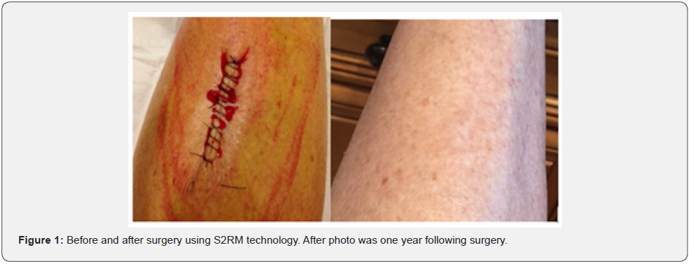
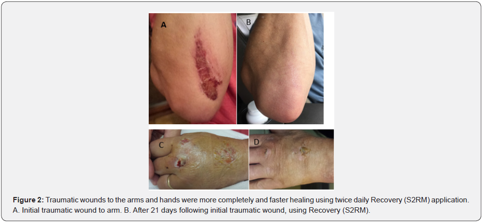
For analysis of traumatic wounds, we treated patients with scalpel surgery or wounds from traumatic accidents. In Figure 1 a patient had a tumor about the size of a ping pong ball (about 40mm in diameter) removed from her leg, leaving a large void and a very large incision. The patient was upset by the appearance of the wound, and her physician recommended using NeoGenesis’ Recovery to aid the closure and for anti-scaring benefit. One year post-surgery there was complete healing, normal remodeling of the tissue and extracellular matrix, with no scarring. In another subject (Figure 2) who had experienced traumatic wounds to his head, legs, torso, arms and hands due to a bicycle accident, the patient used Recovery for more complete and faster healing. As a competitive bicycle racer, he had been in previous like accidents, and compared his healing using Recovery to past experiences. He also suggested himself to compare the healing effects of Recovery versus no treatment by using Recovery on one hand, but not the other. Both arms and hands were lacerated and bleeding to a similar degree. Twice daily application of Recovery to the arm was compared to the left hand control. An obvious faster and more complete wound healing was observed using Recovery on the arm (Figure 2A & 2B) compared to not using Recovery on the hand (Figure 2C&2D).
Radiation Treatment and Chemical Peel
Prior to radiation treatment, a plastic surgeon removed the scar tissue from the patient’s breast that had resulted from a lumpectomy. Unfortunately, the patient simultaneously opted to include an elective procedure of a laser and chemical peel, resulting in severe blistering and burning (Figure 3). About 3 weeks later the patient began radiation treatment for cancer. Recovery was used twice daily and no peeling, blistering, or scaring was observed. Her skin darkened for about 10 days, and then dissipated. The results were especially surprising to the attending physician and nurses given the patient also suffers from Lupus, making her very sensitive to these treatments.
Post LASER Recovery
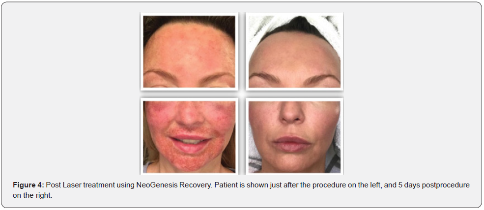
LASER treatment for chronologically aged skin and scarring is common. Fractional ablative lasers create zones of ablation at variable depths in the layers of skin with subsequent induction of collagen production, wound healing, and collagen remodeling. Recent studies suggest that the ablative zones in the skin may also be used in the immediate postoperative period to enhance delivery of drugs and other substances such as platelet-rich plasma or stem cell released molecules [28]. We therefore used the NeoGenesis stem cell released molecule-based product during the immediate postoperative period as a twice daily topically applied procedure.
The inflammation, pain, and redness were dramatically reduced in three patients using the Recovery product. Those practitioners administering the procedure also observed an increased efficacy with coadministration of the S2RM product (Figure 4).

Atopic Dermatitis (Eczema)
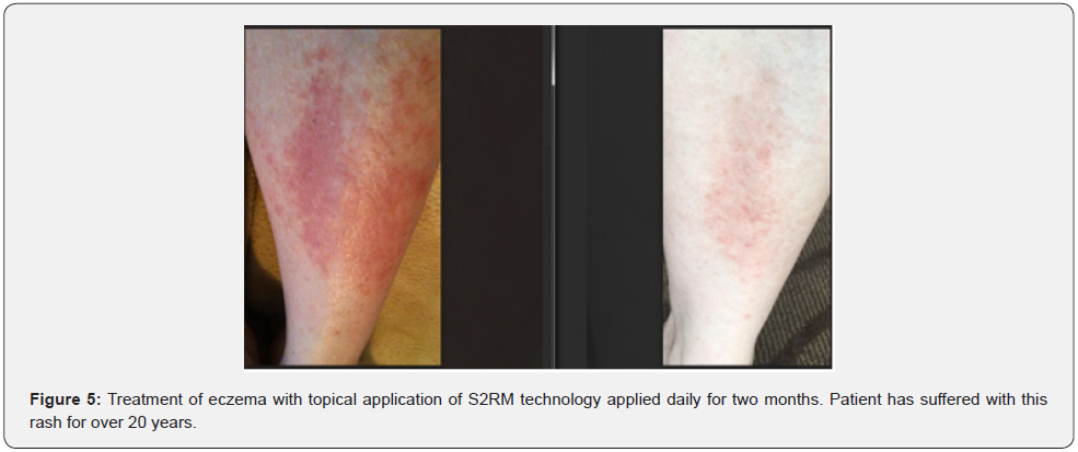
Atopic eczema, also known as atopic dermatitis and commonly referred to as eczema, is a long-term relapsing skin condition that causes both physical and psychological suffering and can have a detrimental impact on quality of life for individuals and their family. The disease is one of the 50 most burdensome globally [29]. The mainstay treatments include regular and consistent application of topical medication, predominantly emollients and steroid preparations [30]. Treatment failure is common leading to wastage of the prescriptions [31]. We presented the subject with an easy to use topically applied product applied twice daily. One patient presented in Figure 5 had suffered with the rash as shown for over twenty years. Within 30 days of the treatment with the S2RM product, the rash and pruritis had significantly reduced in severity.
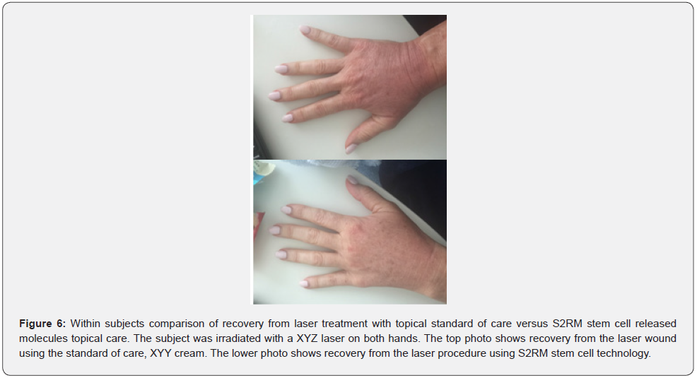
LASER treatment with postoperative Recovery versus standard of care
To test whether our S2RM-based product would provide superior postoperative care to LASER patients compared with the standard of care, we applied Recovery on one hand and the standard of care on the other hand of one patient who had both hands treated equally with LASER. The photos demonstrate a reduction in redness of the Recovery treated hand compared to the control hand treated with the standard of care. The patient also reported a significant reduction in pain using Recovery compared to the control.
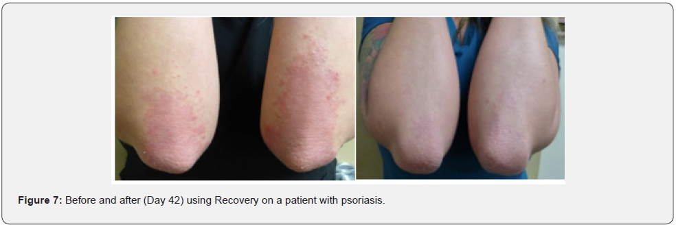
Psoriasis
Psoriasis is a chronic autoimmune condition affecting approximately 2% of the population [32]. While the cause of psoriasis is unknown, hyper-reactivity of Th1, Th17, dysregulation of Treg, and the complex relationships between immune system cells and keratinocytes and vascular endothelium play a significant role [33]. Thus in psoriasis, hyperactivity of Th1 and Th17 means that T cells are activated against viral and bacterial infection, activating macrophages, and dysregulation of Treg means that the activation of Th1 and Th17 may be misguided and uncontrolled (for a short review see Taylor [34]). Evidence also shows that plasmacytoid dendritic cells (pDC) may be central to disease development [35]. The pDCs produce about 1000 times more type I interferon (IFN-I) than any other cell type [36], and account for about 50% of all circulating IFN-I following infection [37]. Nestle et al [36] have proposed that pDCs in hereditary predisposed individuals subject to certain environmental conditions acting through the secretion of IFN-I, induce autoimmune T cells that elicit a psoriasiform phenotype. Currently there is no fully satisfactory therapy against psoriasis and patients frequently report dissatisfaction with the treatment [38]. A number of studies suggest that stem cell therapy may be useful in the treatment of psoriasis [39]. Because the efficacy of stem cell therapy is often ascribed to the molecules and exosomes released by the stem cells [4,8], we used topical S2RM technology to treat the psoriasis. Using a twice daily topical application of Recovery, the subject began to see a reduction in redness and pruritis within one week. By day 42 as shown in Figure 6, the patient had a near eradication of the psoriasis that was presenting on his arms and elbows.
Recovery From Radiation Treatment for Cancer
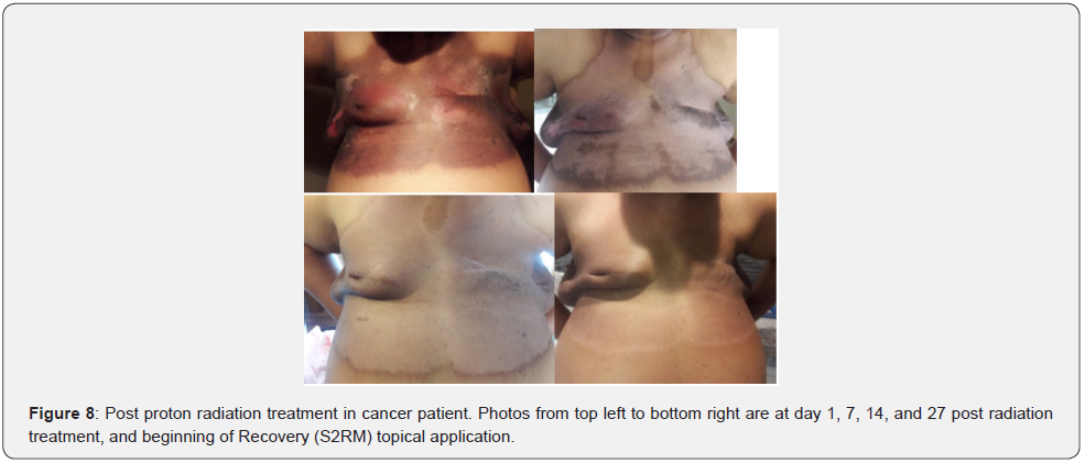
Skin color changes are key biomarker for the ill effects of irradiation on the skin during breast cancer treatment and darkening of the skin correlates in a linear manner with the maximal dosage of irradiation [40]. Irradiated skin shows a number of changes, including damage of the dermal stem cells and fibroblasts [41]. Miller et al [42] have shown the effect of 0.1% mometasone furoate (MMF) on acute skin-related toxicity in 176 patients undergoing breast or chest wall radiotherapy (Figure 7). Results showed no difference between treatment and placebo when the mean maximum grade of radiation dermatitis was used as primary endpoint, but did show a reduction in the mean grade of discomfort or burning. Because of the ability of the S2RM technology to quell inflammation and pain, and to rebuild tissue, we used a topical application of Recovery twice daily to treat radiation dermatitis. Figure 8 shows one such patient treated following the completed radiation therapy protocol. A total of 33 treatments of proton radiation were received between Aug 2017 and Sept 2017. She underwent two lumpectomies followed by a mastectomy and four rounds of chemotherapy. Within two days of topically applying Recovery, the patient began to feel a reduction in pain and sensitivity of the treated area.
Fluorouracil (5-FU) Treatment
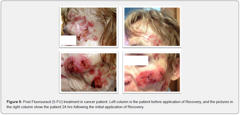
Fluorouracil is commonly given systemically for anal, breast, colorectal, oesophageal, stomach, pancreatic and skin cancers, as well as topically for some skin cancers. Frequent side effects include inflammation of the mouth, loss of appetite, low blood cell counts, hair loss, and inflammation of the skin. The patient in Figure 8 was prescribed topical fluorourcil for 30 days. Experiencing severe pruritis, pain, bleeding, and significant wounds on a large area of her face (Pictures on the left column), and having been recommended to use Vaseline by medical care team, she reached out to her esthetician for help with the itching and wounds. Recovery was applied twice daily, and as shown in the pictures on the right column, the severity of the wounds and the bleeding was greatly mitigated in 24 hours. Her pruritis and pain were self-reported to be “greatly reduced” (Figure 9).
Prevention of Radiation Dermatitis Using Recovery During Radiation Treatment
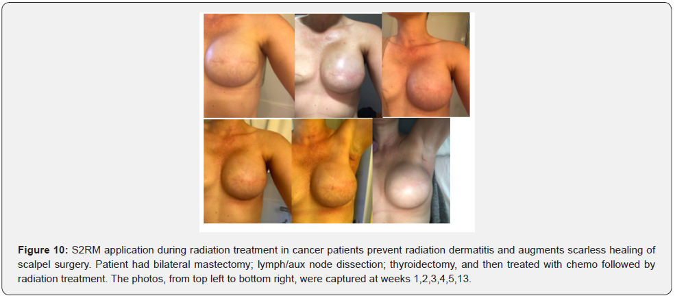
Given that post radiation treatment with Recovery was highly effective in several patients, several new patients were presented to us before their radiation treatment began in the hope that the S2RM technology would prevent, or mitigate, the induction of radiation dermatitis. Although some physicians still recommend patients not apply topical products before radiation treatment, topical application of products just before radiotherapy is safe and not contraindicated [43]. With concurrent administration of Recovery twice daily during radiation treatment the patient’s skin did not darken or become red, indicating that the Recovery was preventing radiation dermatitis. Healing of the scar from scalpel surgery of the breast was also enhanced (Figure 10).
Chemo Associated Rash
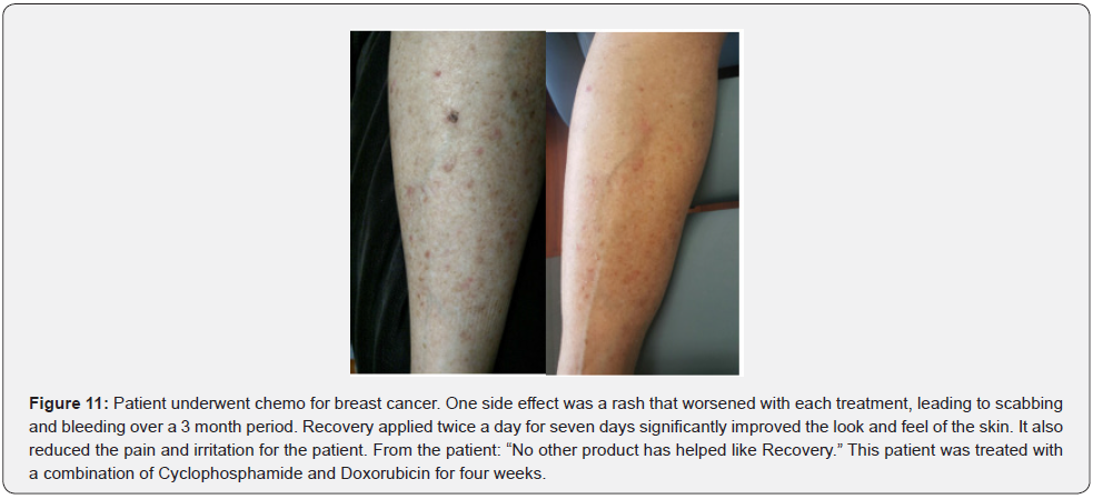
Cancer patients are often found in the hospice, and palliative care is critical to their well being. A number of drugs used in cancer treatment can cause skin problems, including rashes. One drug example is EGFR (epidermal growth factor receptor) inhibitors. These drugs are often prescribed for patients with colon, head and neck, pancreatic, non-small cell lung, and breast cancers. A number of this class of drug are available, including cetuximab (Erbitux), panitumumab (Vectibix), erlotinib (Tarceva), gefitinib (Iressa), and lapatinib (Tykerb/Tyverb). Although topical vitamin K3 was proposed to provide benefit as a prophylactic for cetuximab-induced rash in patients with metastatic colorectal cancer, a recent Phase II, placebo-controlled study of vitamin K3 cream did not support any clinical or immunohistochemical benefit [44]. Similarly, in a recent Phase II, double-blind, vehicle controlled study, vitamin K1 cream as prophylaxis for cetuximabinduced skin toxicity did not reduce the incidence of grade ≥2 skin rash [45]. One patient experiencing chemotherapy induced rash used NeoGenesis Recovery serum applied topically twice a day to the affected areas. In one week, a self reported significant improvement was achieved. The redness of the rash faded, the raised areas diminished, the blisters went away, and the pruritis completely stopped. The patient reported, “Recovery serum is a miracle product. I highly recommend it to anyone with skin issues from chemo” (Figure 11).
Old Scar Remediation
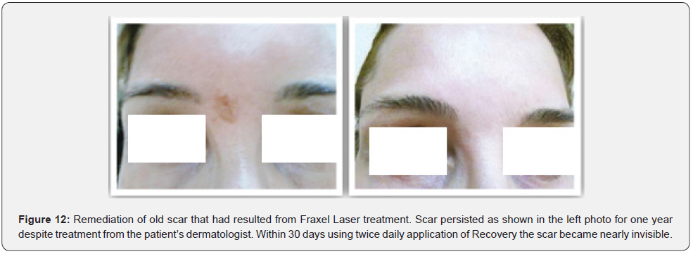
We tested Recovery in one subject who had a one year old scar that had resulted from a Fraxel Laser resurfacing procedure. The appearance of the scar had remained stable for one year. After one year, topical application of Recovery twice daily resulted in a significant visible reduction in the scar as shown in Figure 12. This was not a thick scar with a raised surface area, rather appeared as an atrophic scar with red and dark hyperpigmentation.
Chronologically aged skin
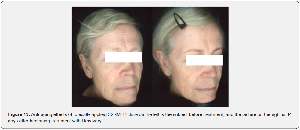
We tested the effects of Recovery on the chronological aging of the skin in twenty subjects, all between the ages of 40-75. Topical application of Recovery was done twice daily. We measured an average 67% reduction in the number of lines, with a range between 57% and 75% reduction. Measurements were made of the crow’s feet lines surrounding the eyes. Although the rater visually observed a reduction in lines in other parts of the face, no quantification was performed in those areas (Figure 13).
Acne
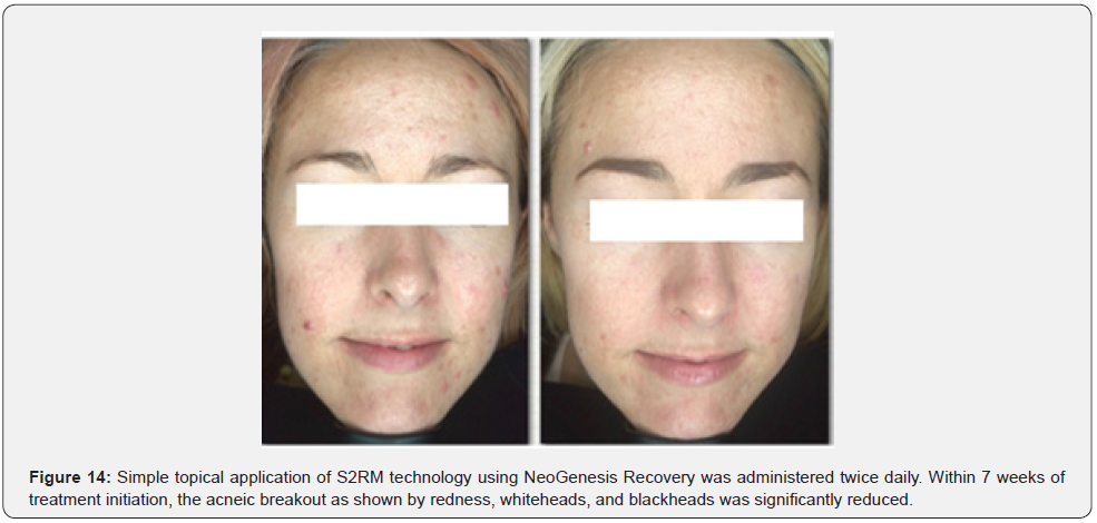
Pathogenesis of acne is characterized by androgen-stimulated overproduction of sebum, follicular hyperkeratinization, inflammatory mediators, and colonization by organisms such as Propionibacterium acnes. The etiology of acne is not well understood. While hereditary factors, including genetics may influence a patient’s risk of developing acne, the prevalence of adult acne in the US is increasing over the last few decades, where significant hereditary changes, or germline genetic variants are unlikely. Rather, environmental conditions, including diet, likely underlie the multifactorial etiology of acne [46]. In the medical setting, the standard of care for inflammatory acne is often a combination therapy including a retinoid and benzoyl peroxide. For moderate-to-severe cases, an oral antibiotic is also often prescribed, usually for no more than 4 months. Given the inflammatory and cellular perturbation in acneic skin, we used Recovery, a stem cell conditioned media product containing product topically applied to the affected area. Recovery was tested on four acne patients with results typified in Figure 14. Using a topical application of Recovery twice daily, acne breakout was shown to diminish in all 4 subjects within two months.
Discussion
Restoration of the skin’s extracellular matrix (ECM) and considering only the structural and mechanical properties of the dermal extracellular matrix, is critical to dermal maintenance and repair. In vitro studies show that fibroblasts in a mechanically stiffer environment produce more collagen and fewer MMPs than fibroblasts cultured in a more flexible collagen matrix [47-49]. Further, studies of human skin have shown that local increase in mechanical forces within the dermis can reprogram fibroblasts to up-regulate collagen production in vivo [50,51], of which the correlative effects can be observed clinically in human subjects through the addition of collagen to the dermis using dermal filler injections [52]. Although fibroblasts and mesenchymal stem cells are known to secrete procollagen and collagen [53,54], whether the effects we observed in this study were partially due to the topical application of secreted pro collagen and collagen is unknown. In favor of this suggestion, fibroblasts have been shown to secrete exosomes that contain collagen [55]. As lipophyllic structures that are known to deliver signals in the keratinocyte layer [56], exosomes are potentially an excellent vehicle to deliver transdermal signaling molecules such as collagen and pro collagen. Wound healing and maintenance of normal, unwounded skin involves multiple cell types and the specific sets of molecules that each of the multiple cell types release, including molecules from mesenchymal stem cells and fibroblasts that are resident in the skin tissue [57].
When mammalian cells are in an environment unfavorable for continued proliferation, they can exit the cell cycle in early to mid-G1 phase at the ‘restriction point’ [58] and enter a reversible, out-ofcell cycle state defined as quiescence. The increased expression of ECM proteins such as Collagen I and III in fibroblasts is partly due to changes in expression of miRNAs such as miR‐29 during their quiescent phenotype [59]. Fibroblast switch their phenotype between two distinct states, proliferating and depositing ECM. Normally the fibroblasts do not proliferate (quiescence), but during wounding, the proliferating phenotype is necessary to maintain dermal architecture, although the architecture may be abnormal (scarring). The two cellular states are balanced by a negative feedback loop between ECM deposition/remodeling and proliferation [60]. The phenotype of the fibroblasts can also be regulated by mesenchymal stem cells. Whereas the quiescent state and collagen production in fibroblasts seems to be induced by adipose-derived mesenchymal stem cells [61], leading to scarless wound healing [14], bone marrow derived mesenchymal stem cells (BMSCs) seem to induce proliferation, migration, and chemotaxis [62], and differentiation of resident stem cells to somatic cells [63] by releasing factors including Growth differentiation factor 11 (GDF11) [64]. However, adipose derived mesenchymal stem cells release molecules, packaged in exosomes [65], that have been shown to switch the ratio of collagen I to collagen III from a scarpromoting high ratio to an anti-scaring low ratio, similar to that seen in fetal wound healing [14].
Thus, for bloody, open wounds, where bone marrow stem cells are recruited to the skin, their function may be important for rapidly closing gaping wounds, inducing scars in the process, but may not be suited to finer wounds and the induction of scarless wound healing generated by endogenous mesenchymal stem cells in the skin, or derived from adipose tissue. Considering the continued use of a stem cell conditioned media based product using bone marrow stem cells (BMSCs), continued use of such a product would lead to long term over drive of the GDF11 controlled differentiation process and IGF-1[66] induced cancer-like cellular proliferation [67], and thus a deleterious effect by depletion of endogenous mesenchymal stem cells in the skin, a process called cellular exhaustion. Indeed, wound healing in skin does not increase the self-renewal capacities of progenitors as it does in the esophagus, but rather leads to a massive depletion of progenitor cells as proliferation increases [68]. This is in contradistinction to the molecules released by adipose derived stem cells that reduce the differentiation of stem cells, thus preserving the endogenous pool of stem cells and fibroblasts resident in the skin [14].
Physiological Renormalization-Why S2RM Treats Many Conditions
Physiological renormalization is a strategy recently developed for therapeutics to treat cancer. That is, renormalizing the physiology of T cells in the immune system instead of enhancement of the immune system or a direct attack on cancer cells is a new strategy in the successful development of a recently approved class of chemotherapeutics for cancer called “check point inhibitors” [69]. This is a strategy for which the 2018 Nobel Prize in Physiology or Medicine was awarded. In other words, in this new strategy, the physiology of T-cells was renormalized so that the T-cells could once again function normally to attack the cancer, doing so for many cancer types [70]. Physiological renormalization is a process that occurs naturally, for example during sleep, during which spontaneous activity renormalizes net synaptic strength and restores cellular homeostasis [71]. As a next, and broader, step in the physiological renormalization strategy, we reperfused the skin with the collective of molecules, including proteins that were normally present in the skin during a healthy state. For example, in psoriasis, more than 1200 proteins with differentially expressed peptides many with biological functions of interest in psoriasis, and many in a misfolded state were identified in a mouse model and in human psoratic skin. And, over 100 proteins were changed greater than twofold comparing psoratic to healthy skin [72]. In particular, considering the deregulation of T cells in psoriasis [33], our therapeutic contains the exosomes, and their protein cargo, that are derived from adipose mesenchymal stem cells, and have been shown to quell inflammation through regulation of T cells [73]. Thus, in this study, we present evidence that physiological renormalization of proteostasis is an excellent strategy for treating psoriasis as well as a number of other conditions. This is not unlike physiological renormalization of the immune system with “checkpoint inhibitors” to treat an array of cancer types. This explains why in both cases one therapeutic will treat a number of indications; the S2RM technology and “checkpoint inhibitors,” induce a physiological state that is renormalized such that a number of indications involving physiological perturbations can be treated.
Limitations of Our Study
All of the cases reported here used an open label application of the NeoGenesis Recovery, and only some of the cases used a placebo control or comparator product. Therefore, a direct comparison of rate and extent of the effects, such as wound closure, could not be quantified. Further, as substantiated by numerous studies, the placebo effect can be substantial, especially for indications such as pain [74]. Therefore, in our study quantification of the level of pain reduction by Recovery could not be performed. However, in our studies the patients had previously used other products, in effect serving as a placebo control by which a comparison of pain level was made. In all cases where pain was a factor, Recovery was reported by the patient to have induced substantial pain relief as compared to the “placebo product,” such as Vaseline. Regression to the mean is another statistically important way in which a therapeutic effect can be mistaken for simple self healing [75], especially for indications such as pain. In other words, if one tends to go for treatment when their condition is severe, and when their condition is at its worst or near worst, then with time, later their condition will likely improve. The remediation of the indication over time is due to simple regression to the mean, something that can be much larger than that of placebo [76]. As reviewed by the Royal Statistical Society, researchers are fooled all the time by regression to the mean, even when using blinded, randomized, placebo controlled trials [77].
In recognition of this problem, in a number of our cases, the pain was ongoing, and the statistical regression to the mean was to the state of pain. Only when Recovery was applied did the pain reduce. Upon cessation of Recovery application, the pain reappeared. Likewise, the persistence of the scar for one year and then remediation of the scar in one month with the application of Recovery, demonstrates the scar removal was not simply self healing and a regression to the mean. It is important to understand factors other than treatment that can affect outcomes, including knowing the patterns of healing for a given indication [78]. Knowing the pattern of disease will help us to determine causality [79], by answering the counter factual question: had the patient not had the S2RM administered, would she have experienced the same results? The Artus et al study showed that lower back pain diminished within many clinical trials in a given pattern, regardless of the treatment given, including placebo. In this regard, for example, radiation treatment for cancer always induces significant radiation dermatitis. In our three case studies where we administered the Recovery concurrently with the radiation treatment, we observed little or no radiation dermatitis. Thus, the Recovery treatment did not fit the pattern of the normal sequelae of skin damage and healing during and after the radiation treatment, suggesting a significant preventative effect of the Recovery, and not just a standard pattern of self healing.
In other words, answering Pearl’s counterfactual question of causality [79], “would the patient’s recovery pattern have been the same had the S2RM not been applied to the patient,” the patient would not have recovered in the observed pattern shown in this study had they not received the S2RM technology. Therefore, despite the lack of controls in most cases, the benefit of Recovery was much better than patterns of self healing, regression to the mean, or placebo effects, that a large qualitative effect of Recovery was substantiated in real world outcomes. One important method for reducing variation in a study is to construct consistent and uniform endpoint definitions [80]. In this regard, our study used easily observed, quantifiable measurements in most instances, except for measures of pain. For example, wrinkles are easily observed and objectively counted, color and darkness of skin were easily judged, and scarring could be easily judged even without rater training. Thus our data, conforming to the results of many previous studies using stem cells and their secretome, suggest that the S2RM technology, the secretome from multiple stem cell types, in a simple topical application is an effective treatment for traumatic, autoimmune, and inflammatory conditions of the skin. Our data suggest that a physiological renormalization strategy using topically applied stem cell released molecules is an important new clinical tool for a number of skin conditions.
References
- Fuchs E, Green H (1978) The expression of keratin genes in epidermis and cultured epidermal cells. Cell 15: 887-897.
- Mascré G, Dekoninck S, Drogat B, Youssef KK, Broheé S, et al. (2012) Distinct contribution of stem and progenitor cells to epidermal maintenance. Nature 489(7415): 257-262.
- Sklirou AD, Nicolas GK, Issidora P, Alexios LS, Ioannis PT (2017) 6-bromo-indirubin-3′-oxime (6BIO), a Glycogen synthase kinase-3β inhibitor, activates cytoprotective cellular modules and suppresses cellular senescence-mediated biomolecular damage in human fibroblasts. Sci Rep 7: 11713.
- Maguire G (2013) Stem cell therapy without the cells. Commun Integr Biol 6(6): e26631.
- Rivera GG (2016) PDGFA regulation of dermal adipocyte stem cells. Cell Stem Cell 4:72.
- Maguire G (2018) Protein circuits in wound healing. Science, letters, 362(6417).
- Rheinwald JG, Green H (1975) Serial cultivation of strains of human epidermal keratinocytes: the formation of keratinizing colonies from single cells. Cell 6(3): 331-343.
- Maguire G (2016) Therapeutics from Adult Stem Cells and the Hype Curve. ACS Med Chem Lett. 7(5): 441-443.
- Maguire G (2021) Stem cells part of the innate and adaptive immune systems as a therapeutic for Covid-19. Commun Integr Biol 14(1): 186-198.
- Maguire G (2019) Rescue of Degenerating Neurons and Cells by Stem Cell Released Molecules. Physiological Reports, Physiol Rep 7(9): e14072.
- Fukuoka H, Suga H (2015) Hair regeneration treatment using adipose-derived stem cell conditioned medium: follow-up with trichograms. Eplasty 15: e10.
- Son WC, Yun JW, Kim BH (2015) Adipose-derived mesenchymal stem cells reduce MMP-1 expression in UV-irradiated human dermal fibroblasts: therapeutic potential in skin wrinkling. Biosci Biotechnol Biochem 79(6): 919-925.
- Hu L, Zhao J, Liu J, Niya G, Chen L, et al. (2013) Effects of adipose stem cell-conditioned medium on the migration of vascular endothelial cells, fibroblasts and keratinocytes. Exp Ther Med 5(3): 701-706.
- Wang L, Hu L, Xin Z, Xiong Z, Zhang C, et al. (2017) Exosomes secreted by human adipose mesenchymal stem cells promote scarless cutaneous repair by regulating extracellular matrix remodeling. Scientific Reports, p. 7.
- Chicharro D, Jose MC, Rubio M, Ramon C, Belen C, et al. (2018) Combined plasma rich in growth factors and adipose-derived mesenchymal stem cells promotes the cutaneous wound healing in rabbits. BMC Vet Res 14(1): 288.
- Maguire G (2019) The Safe and Efficacious Use of Secretome From Fibroblasts and Adipose-, But Not Bone Marrow-, Derived Mesenchymal Stem Cells For Skin Therapeutics. J. Clinical and Aesthetic Dermatology 12(8): E57-E69.
- Corstjens H, Dicanio D, Muizzuddin N, Neven A, Sparacio R, et al. (2008) Glycation ssociated skin autofluorescence and skin elasticity are related to chronological age and body mass index of healthy subjects. Exp Gerontol 43(7): 663-667.
- Rittie L, Fisher GJ (2015) Natural and Sun-Induced Aging of Human Skin. Cold Spring Harb Perspect Med 5(1): a015370.
- Bowman A, Birch MA (2016) Age-Dependent Decrease of Mitochondrial Complex II Activity in Human Skin Fibroblasts. J Invest Dermatology 136(5): 912-919.
- Anderson A (2014) A role for human mitochondrial complex II in the production of reactive oxygen species in human skin. Redox Biology 2: 1016-1022.
- Quan T, He T, Kang S, Voorhees JJ, Fisher GJ (2004) Solar ultraviolet irradiation reduces collagen in photoaged human skin by blocking transforming growth factor-beta type II receptor/Smad signaling. Am J Pathol 165(3): 741-751.
- Smith J, Davidson E, Sams W, Clark R (1962) Alterations in human dermal connective tissue with age and chronic sun damage. J Invest Dermatol 39: 347-350.
- Gerstein AD, Phillips TJ, Rogers GS, Gilchrest BA (1993) Wound healing and aging. Dermatol Clin 11: 749-757.
- Gosain A, DiPietro LA (2004) Aging and wound healing. World J Surg 28(3): 321-326.
- Giangreco A et al (2008) Epidermal stem cells are retained in vivo throughout skin aging. Aging Cell 7(2): 250-259.
- Maguire G, Friedman P (202) The safety of a therapeutic product composed of a combination of stem cell released molecules from adipose mesenchymal stem cells and fibroblasts. Future Sci OA 6(7): FSO592.
- Ernst O, Zor T (2010) Linearization of the bradford protein assay. J Vis Exp (38): 1918.
- Waibel JS, Wulkan AJ, Shumaker PR (2013) Treatment of hypertrophic scars using laser and laser assisted corticosteroid delivery 45(3): 135-140.
- Weidinger S and Novak N (2016) Atopic dermatitis. The Lancet 387(10023):1109-1122.
- Santer M, Burgess H, Yardely L, Steven E, Jones SL, et al. (2012) Experiences of carers managing childhood eczema and their views on its treatment: a qualitative study. Br J Gen Pract 62(597): e261-e267.
- Smith SD, Hong E, Fearns S, Alex B, Gayle F, et al. (2010b) Corticosteroid phobia and other confounders in the treatment of childhood atopic dermatitis explored using parent focus groups. Australas J Dermatol 51(3): 168-674.
- Kurd SK, Gelfand JM (2009) The prevalence of previously diagnosed and undiagnosed psoriasis in US adults: results from NHANES 2003-2004. J Am Acad Dermatol 60(2): 218-224.
- Deng Y, Chang C, Lu Q (2016) The inflammatory response in psoriasis: A comprehensive review. Clin. Rev. Allergy Immunol 50(3): 377-389.
- Taylor AP (2017) The Ever-Expanding T-Cell World: A Primer. The Scientist.
- Wohn C, Julia LO, Haak S, Stanislav P, Cheong C, et al. (2013) Langerin(neg) conventional dendritic cells produce IL-23 to drive psoriatic plaque formation in mice. Proceedings of the National Academy of Sciences of the United States of America 110: 10723-10728.
- Nestle FO, Curdin C, Adrian TK, Homey B, Gombert M, et al. (2005) Plasmacytoid predendritic cells initiate psoriasis through interferon-alpha production. Journal of Experimental Medicine 202(1): 135-143.
- Swiecki M, Colonna M (2010) Unraveling the functions of plasmacytoid dendritic cells during viral infections, autoimmunity, and tolerance. Immunological Reviews 234(1): 142-162.
- Belinchón I, Rivera R, Blanch C, Comellas M, Lizán L (2016) Adherence, satisfaction and preferences for treatment in patients with psoriasis in the European Union: A systematic review of the literature. Patient Preference Adherence 10: 2357-2367.
- Owczarczyk SA, Magdalena K, Anna K, Placek W, Wojciech M, et al. (2017) Stem Cells as Potential Candidates for Psoriasis Cell-Replacement Therapy. Int J Mol Sci 18(10): 2182.
- Yamazaki H (2018) Comparison of radiation dermatitis between hypofractionated and conventionally fractionated postoperative radiotherapy: objective, longitudinal assessment of skin color. Sci Rep 8: 12306.
- Hur W, Yoon SK (2017) Molecular Pathogenesis of Radiation-Induced Cell Toxicity in Stem Cells. Int J Mol Sci 18(12): 2749.
- Miller RC, David J, Jeff AS, Griffin PC, et al. (2011) Mometasone furoate effect on acute skin toxicity in breast cancer patients receiving radiotherapy: a phase III double-blind, randomized trial from the north central Cancer treatment group N06C4. Int J Radiat Oncol Biol Phys 79(5): 1460-1466.
- Baumann BC, Zeng C, Bell B, Koduri S, Vachani C, et al. (2018) Assessing the Validity of Clinician Advice That Patients Avoid Use of Topical Agents Before Daily Radiotherapy Treatments. JAMA Oncology 4(12): 1742-178.
- Eriksen JG, Kaalund I, Clemmensen O, Overgaard J, Pfeiffer P (2017) Placebo-controlled Phase study II of vitamin K3 cream for the treatment of cetuximab-induced rash. Support. Care Cancer 25(7): 2179-2185.
- Hofheinz RD, Lorenzen S, Trojan J, J Ocvirk J, Schulz H, et al. (2018) EVITA-a double-blind, vehicle-controlled, randomized Phase trial II of vitamin K1 cream as prophylaxis for cetuximab-induced skin toxicity. Ann Oncol 29(4): 1010-1015.
- Mahmood, Bowe (2014) Diet and Acne Update: Carbohydrates Emerge as the Main Culprit. J Drugs Dermatol 13(4): 428-435.
- Mauch C, Hatamochi A, Scharffetter K, Krieg T (1988) Regulation of collagen synthesis in fibroblasts within a three-dimensional collagen gel. Exp Cell Res 178(2): 493-503.
- Rittié L, Berton A, Monboisse JC, Hornebeck W, Gillery P (1999) Decreased contraction of glycated collagen lattices coincides with impaired matrix metalloproteinase production. Biochem Biophys Res Commun 264(2): 488-492.
- Eckes B, Zweers MC, Zhang ZG, Hallinger R, Mauch C, et al. (2006) Mechanical tension and integrin α2β1 regulate fibroblast functions. J Invest Dermatol Symp Proc 11(1): 66-72.
- Wang F, Garza LA, Kang S, Varani J, Orringer JS, et al. (2007) In vivo stimulation of de novo collagen production caused by cross-linked hyaluronic acid dermal filler injections in photodamaged human skin. Arch Dermatol 143(2): 155-163.
- Quan T, Wang F, Shao Y, Rittié L, Xia W, et al. (2013) Enhancing structural support of the dermal microenvironment activates fibroblasts, endothelial cells, and keratinocytes in aged human skin in vivo. J Invest Dermatol 133(3): 658-667.
- Turlier V, Delalleau A, Casas C, Rouquier A, Bianchi P, et al. (2013) Association between collagen production and mechanical stretching in dermal extracellular matrix: In vivo effect of cross-linked hyaluronic acid filler. A randomised, placebo-controlled study. J Dermatol Sci 69: 187-194.
- Goldberg B, Sherr CJ (1973) Secretion and extracellular processing of procollagen by cultured human fibroblasts. Proc Natl Acad Sci 70(2): 361-365.
- Amable PR, Teixeira MV, Carias RB, Jose M, Borojevic R (2014) Protein synthesis and secretion in human mesenchymal cells derived from bone marrow, adipose tissue and Wharton's jelly. Stem Cell Res Ther 5(2): 53.
- Hong J, Erfani N, Bow JT, Kapp AE, Hong JZ, et al. (2008) Difference gel electrophoresis analysis of Ras‐transformed fibroblast cell‐derived exosomes. Electrophoresis 29(12): 2660-2671.
- Lo Cicero A, Cedric D, Floriane GM, Loew D, Dingli F, et al. (2015) Exosomes released by keratinocytes modulate melanocyte pigmentation. Nature Communications 6: 7506.
- Rodriguez ML, Marcela S, Ford D, Badiavas E (2012) Stimulation of Skin and Wound Fibroblast Migration by Mesenchymal Stem Cells Derived from Normal Donors and Chronic Wound Patients. Stem Cells Translational Medicine 1(3): 221-229.
- Pardee AB (1974) A restriction point for control of normal animal cell proliferation. Proc Natl Acad Sci U S A 71(4): 1286-1290.
- Suh EJ, Matthew YR, Miller AL, Johnson El, Talia RC, et al. (2012) A microRNA network regulates proliferative timing and extracellular matrix synthesis during cellular quiescence in fibroblasts. Genome Biol 13(12): R121.
- Rognoni E, Pisco AO, Toru H, Kalle HS, Julio M, et al. (2018) Fibroblast state switching orchestrates dermal maturation and wound healing. Molecular Systems Biology 14(8): e8174.
- Liu X, Wang S, Wu S, Hao Q, Li Y, et al. (2018) Exosomes secreted by adipose-derived mesenchymal stem cells regulate type I collagen metabolism in fibroblasts from women with stress urinary incontinence. Stem Cell Research & Therapy 9: 159.
- Smith AN, Willis E, Vincent TC, Lara AM, Isik FF, et al. (2010) Mesenchymal stem cells induce dermal fibroblast responses to injury. Exp Cell Res 316(1): 48-54.
- Maguire G, Friedman P (2015) Systems biology approach to developing S(2)RM-based "systems therapeutics" and naturally induced pluripotent stem cells. World J Stem Cells 7(4): 745-756.
- Williams G, Zentar MP, Gajendra S, Sonego M, Doherty P, et al. (2013) Transcriptional basis for the inhibition of neural stem cell proliferation and migration by the TGFβ-family member GDF11. PLoS One 8(11): e78478.
- Maguire G (2016) Exosomes: Smart Nanospheres for Drug Delivery Naturally Produced by Stem Cells. In: Fabrication and Self-Assembly of Nanobiomaterials. Elsevier, Amsterdam, Netherlands, pp. 179-209.
- Chen L, Tredget EE, Philip YG, Wu Y (2008) Paracrine Factors of Mesenchymal Stem Cells Recruit Macrophages and Endothelial Lineage Cells and Enhance Wound Healing. PLoS One 3(4): e1886.
- Renehan AG, Zwahlen M, Minder C, O Dwyer ST, Shalet SM, et al. (2004) Insulin-like growth factor (IGF)-I, IGF binding protein-3, and cancer risk: systematic review and meta-regression analysis. Lancet. 363(9418): 1346-1347.
- Aragona M, Sophie D, Rulands S, Lenglez S, Simons BD, et al. (2017) Defining stem cell dynamics and migration during wound healing in mouse skin epidermis. Nat Commun. 8: 14684.
- Sanmamed MF, Chen L (2018) A Paradigm Shift in Cancer Immunotherapy: From Enhancement to Normalization. Cell 175(2): 313-326.
- Zappasodi R, Taha M, Wolchok JD (2018) Emerging Concepts for Immune Checkpoint Blockade-Based Combination Therapies. Cancer Cell 33(4): 581-598.
- Tononi G, Cirelli C (2014) Sleep and the Price of Plasticity: From Synaptic and Cellular Homeostasis to Memory Consolidation and Integration. Neuron 81(1): 12-34.
- Lundberg KC, Fritz Y, Johnston A, Foster M, Baliwag J, et al (2015) Proteomics of Skin Proteins in Psoriasis: From Discovery and Verification in a Mouse Model to Confirmation in Humans. Molecular & Cellular Proteomics 14(1): 109-119.
- Blazquez R, Francisco, M, Rosa O, Dalemans W, Alvarez V, et al. (2014) Immunomodulatory Potential of Human Adipose Mesenchymal Stem Cells Derived Exosomes on in vitro Stimulated T Cells. Front Immunol 5: 556.
- Ellingsen DM, Wessberg J, Eikemo M, Liljencrantz J, Leknes S, et al (2013) Placebo improves pleasure and pain through opposite modulation of sensory processing. Proc Natl Acad Sci U S A. 110(44): 17993-17998.
- McDonald CJ, Mazzuca SA, McCabe GP (1983) How much of the placebo effect is really statistical regression? Statistics in Medicine 2(4): 417-427.
- O’Connell NE, Moseley GL, James HM, Wand B, Herbert RD (2015) Interpreting Effectiveness Evidence in Pain: Short Tour of Contemporary Issues. Phys Ther 95(8): 1087-1094.
- Senn S (2011) Francis Galton and regression to the mean. Focus, pp. 124-126.
- Artus M, Jordan P, Hay M, Danielle A (2010) Low back pain symptoms show a similar pattern of improvement following a wide range of primary care treatments: a systematic review of randomized clinical trials. Rheumatology (Oxford) 49(12): 2346-2356.
- Pearl J (2018) The Book of Why. The New Science of Cause and Effect. Basic Books, New York, USA.
- Evans (2011) Fundamentals of clinical trial design. J Exp Stroke Transl Med 3(1): 19-27.


