




 JOJCS Home



Abstract
Background: Maternal perception of decreased foetal movements (DFM) is an important symptom of possible foetal compromise. Assessment is challenging when cardiotocography (CTG) is performed early in gestation. True umbilical cord knots are uncommon, often undetected antenatally, and variably associated with adverse outcomes.
Case: A nulliparous woman with a body mass index of 45kg/m2 reported a new, sustained reduction in foetal movements at 29 weeks' gestation. Ultrasound showed a live foetus with growth above the 97th centile, a posterior placenta, mildly increased amniotic fluid, and normal umbilical artery Doppler indices. The CTG showed periods of reduced or absent variability, apparent decelerations, and intermittent sinusoidal-like morphology. Maternal full blood count, antibody testing, and testing for foetomaternal haemorrhage were unremarkable. Persistent DFM and a similarly abnormal CTG the following day prompted consultation with maternal-foetal medicine, antenatal corticosteroids, and in utero transfer.
At the tertiary center, ultrasound again showed limited movement but no structural explanation or Doppler evidence of foetal anaemia; middle cerebral artery peak systolic velocity was normal. Continued CTG surveillance became more concerning, prompting an emergency Caesarean birth at 29+3 weeks. A true umbilical cord knot with documented compression was found. A 1667g liveborn infant was delivered safely. There was no evidence of neonatal anaemia.
Conclusion: Reassuring ultrasound and Doppler findings do not negate the significance of persistent DFM with an unexplained abnormal CTG. Prematurity alters CTG interpretation but does not make significant abnormalities physiological. When clinical signals remain discordant, senior review, continued surveillance, and timely transfer may be more important than assigning a definitive antenatal diagnosis.
Keywords: Decreased foetal movements; Antenatal cardiotocography; Sinusoidal pattern; True umbilical cord knot; Foetal anaemia; Maternal obesity
Introduction
Maternal awareness of foetal movement is one of the oldest and most immediate forms of foetal surveillance. A sustained reduction or change from the usual pattern may precede stillbirth or be associated with foetalgrowth restriction, placental dysfunction, foetomaternal haemorrhage and other causes of foetal compromise. Contemporary guidance therefore emphasizes the woman's perception of altered movement [1,2].
No single investigation reliably excludes all important causes of DFM. Antenatal CTG assesses foetal heart-rate patterns over a limited interval, while ultrasound provides a contemporaneous assessment of foetal growth, amniotic fluid volume, movement, and, when Doppler studies are performed, foetal and placental haemodynamics. A normal ultrasound is reassuring with respect to the parameters examined but does not provide a defined period of reassurance or reliably exclude intermittent or evolving foetal compromise. Interpretation becomes more difficult at early gestations, when autonomic immaturity alters expected accelerations and variability [1,3].
This report describes persistent DFM at 29 weeks, an unusual preterm CTG, reassuring Doppler studies, and the subsequent finding of a compressed true umbilical cord knot. It illustrates the clinical challenge of responding to discordant findings when no single investigation provides a definitive diagnosis. At term, persistent uncertainty about foetal wellbeing may more readily favour delivery; at very preterm gestations, however, that decision is more difficult because the potential risks of continuing pregnancy must be balanced against the significant consequences of preterm birth.
Case Presentation
A nulliparous woman received routine antenatal care. Maternal body mass index was approximately 45kg/m2. The pregnancy had been uncomplicated until the index presentation. The placenta was posterior. A growth ultrasound at 27+2 weeks showed an estimated foetal weight of 1419g, above the 97th centile, an amniotic fluid index (AFI) of 15.6cm, and normal umbilical artery Doppler indices.
At approximately 29+1 weeks, she presented to the maternity assessment unit reporting a reduction in foetal movements over the preceding days. There had been no preceding trauma, abdominal pain, vaginal bleeding, membrane rupture, or other apparent precipitant. Maternal observations were stable, and clinical assessment did not suggest placental abruption.
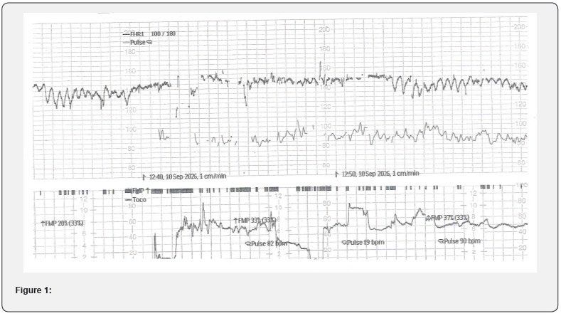
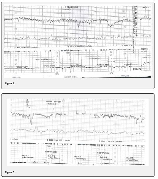
CTG monitoring was commenced (Figure 1 & 2). Obtaining a clear foetal heart-rate recording was technically challenging because of maternal habitus. Early gestation also complicated interpretation of the trace. The trace was recognized as abnormal but was not readily classified. It contained periods of reduced variability, apparent prolonged or shallow decelerations, and segments with a sinusoidal appearance, interspersed with periods of greater variability. Maternal full blood count and red-cell antibody testing were unremarkable, and testing for foetomaternal haemorrhage was negative.
Ultrasound demonstrated foetal cardiac activity and movements, with a biophysical profile of 8/8. The foetus was large for gestational age. The posterior placenta showed no sonographic evidence of abruption. AFI was approximately 25cm, above the 95th centile, and umbilical artery Doppler indices were normal. Middle cerebral artery (MCA) Doppler was not performed. Given the apparently reassuring ultrasound and uncertainty about the significance of the CTG at 29 weeks, expectant management was chosen, with instructions to return if movements remained reduced and reassessment planned for the following day.
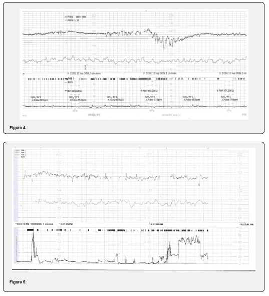
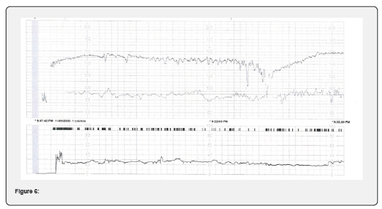
She returned the next day with ongoing DFM. Repeat CTG remained abnormal, with reduced or absent variability, recurrent shallow decelerations, and intermittent sinusoidal-like patterns (Figure 3 & 4). The clinical significance remained difficult to determine, but prematurity and maternal obesity were no longer considered sufficient explanations for the combination of persistent symptoms and repeated abnormal surveillance. Following discussion with a tertiary maternal-foetal medicine service, betamethasone was administered, and in-utero transfer was arranged. Magnesium sulfate was considered for foetal neuroprotection given the likelihood of early delivery.
At the tertiary center, CTG monitoring was continued (Figure 5 & 6). Detailed ultrasound demonstrated a cephalic foetus, posterior placenta, AFI 19.5cm, and deepest vertical pool 7.0cm. Estimated foetal weight was approximately 1700g, above the 97th centile, with abdominal circumference also above the 97th centile. Some spontaneous movement was observed, and additional movement occurred with transducer pressure, but movements remained limited and foetal breathing was not seen. Umbilical and other reported Doppler indices were normal. MCA peak systolic velocity was 1.14 multiples of the median, with no sonographic evidence of significant foetal anaemia. Continuous CTG surveillance remained abnormal. In the absence of a definitive ultrasound abnormality and given the consequences of delivery at 29+3 weeks, close observation with continuous monitoring was continued. Approximately five hours after admission, further CTG deterioration prompted an emergency lower-segment Caesarean section.
A liveborn infant weighing 1667g was delivered with good Apgar scores and required minimal resuscitation. Cord bloods were normal, and the initial hemoglobin was 162g/L (145 -240). CRP was normal and blood group was A RhD negative with no antibodies. Operative findings documented a three-vessel cord with a single, true knot associated with cord compression. The placenta weighed 348g (75-90th percentile for gestation) and appeared normal with no evidence of abruption. Histopathology confirmed a 3-vessel cord with no evidence of significant abnormality in the placenta or membranes. The infant was admitted to neonatal intensive care and progressed well with referral back to the regional hospital on Day 12.
Discussion
Foetal movement as surveillance
Formal foetal-movement counting was introduced into antenatal care in the 1970s, building on the observation that reduced activity could precede foetal death. Methods such as fixed-period counting and "count-to-ten" charts attempted to standardize subjective awareness. Current practice has moved away from a single normal number because movement patterns vary among foetuses, gestational ages, and individuals. Australian guidance now advises women to become familiar with their baby's usual pattern and to seek reassurance when movements become fewer, weaker, or otherwise different [1,4].
Movement reflects intact neuromuscular function, adequate oxygenation, and the foetal behavioural state. Transient reductions may occur during foetal sleep or after sedating medication. Important associations include foetal growth restriction and placental insufficiency, oligohydramnios, acute hypoxia, placental abruption, foetomaternal haemorrhage and foetal anaemia, infection, congenital or neuromuscular abnormality, and foetal demise. Trauma, including a motor vehicle collision, may be relevant through abruption or foetomaternal haemorrhage. A history should therefore establish the onset and degree of change and seek pain, bleeding, membrane rupture, trauma, medication exposure, and maternal disease [1,2].
DFM is clinically important, but evidence that universal counting programs prevent stillbirth is less certain. Randomized evidence has not shown a clear reduction in perinatal mortality from formal counting alone. The large AFFIRM stepped-wedge trial tested a package of movement-awareness education and standardized management. It did not significantly reduce stillbirth but increased interventions, including induction of labour and Caesarean birth. These findings do not make a report of DFM unimportant. Rather, they show that population screening and subsequent intervention can produce both benefit and harm [4,5].
PSANZ guidance, endorsed by RANZCOG, provides a defined assessment pathway starting at 28 weeks. Initial assessment generally includes confirmation of foetal viability, a focused history and examination, and CTG. Ultrasound for growth, amniotic fluid, and Doppler assessment is indicated when movements remain reduced, risk factors exist, the CTG is abnormal, or growth is clinically concerning. Tests such as maternal full blood count, blood group and antibody screen, and investigation for foetomaternal haemorrhage are not mandatory for every presentation but are appropriate when anaemia, bleeding, trauma, or an unexplained abnormal CTG is suspected, and importantly, when maternal concern persists [1].
Antenatal CTG at 29 weeks
There is no single gestational age below which CTG loses clinical value. In Australian practice, CTG is routinely incorporated into the assessment of DFM from 28 weeks, while surveillance before 28 weeks is individualized. Nevertheless, gestational age alters normal foetal heart rate behaviour. Between approximately 26 and 32 weeks, accelerations may be smaller or less frequent, cycling is less mature, and reduced variability and decelerations are more common than at term. Standard term criteria can therefore over-classify preterm tracings as abnormal [1,3].
This limitation has boundaries. Prematurity may explain absent reactivity or modestly reduced variability; it should not automatically explain persistent absent variability, recurrent significant decelerations, or convincing sinusoidal morphology. A true sinusoidal pattern is a smooth, regular oscillation of the foetal heart-rate baseline, usually 2-5 cycles per minute with an amplitude of approximately 5-15 beats per minute, absent conventional variability, and no accelerations, persisting long enough to distinguish it from transient oscillation. It is classically associated with severe foetal anaemia and is treated as a high-risk finding. Pseudo-sinusoidal or sinusoidal-like patterns are generally shorter and less regular and may be preceded or followed by normal variability or accelerations. They may occur transiently with foetal behavioral states, rhythmic foetal movements such as sucking or mouthing, and following maternal administration of some medications, particularly opioids [6,7].
Antenatal CTG has limited specificity: abnormal or non-reassuring features may occur in foetuses who are not significantly compromised, particularly at earlier gestations, when normal foetal heart rate characteristics differ from those at term. An abnormal CTG therefore does not, in isolation, establish foetal compromise but must be interpreted within the broader clinical context, including gestational age, presenting symptoms, other investigations, and changes in the trace over time. In this case, the intermittent sinusoidal-like pattern did not meet the criteria for a continuous true sinusoidal pattern. Nevertheless, its occurrence alongside persistent DFM, reduced variability, and decelerations was concerning and warranted further investigation and escalation. Ultimately, assigning a precise label to the pattern was less important than recognizing that, in the context of DFM, the tracing was repeatedly not reassuring [3,6,7].
Ultrasound and other clinical information
Normal ultrasound, biophysical profile, and umbilical artery Doppler findings reduced the likelihood of established chronic placental insufficiency but could not exclude intermittent cord obstruction or an evolving acute process. Ultrasound is a snapshot of foetal behaviour, and observing movement during scanning does not invalidate a mother's report of a sustained change from normal. Similarly, normal umbilical artery Doppler indices primarily assess placental resistance and may remain normal when compromise has a different mechanism [1,8].
The tertiary MCA peak systolic velocity of 1.14 multiples of the median argued against moderate or severe foetal anaemia at that time; a threshold above 1.5 multiples of the median is commonly used to identify foetuses at risk. The negative foetomaternal haemorrhage test and normal antibody screen provided further reassurance. MCA Doppler is a screening test rather than a direct haemoglobin measurement, but the subsequently reported normal neonatal haemoglobin provided additional evidence against significant foetal anaemia [9].
Maternal body mass index
Maternal obesity can make abdominal palpation, external foetal heart-rate recording, and ultrasound technically more difficult. Its effect on maternal perception of movement is less straightforward. Available evidence does not support the assumption that women with obesity cannot perceive meaningful changes in foetal activity. A posterior rather than an anterior placenta also made placental cushioning a less persuasive explanation in this case. BMI should therefore be treated as a factor affecting examination and test quality, not as a diagnosis that explains persistent DFM. When technical uncertainty increases while clinical concern persists, it strengthens the case for prolonged observation, alternative assessment, and senior review rather than reassurance [10-12].
True umbilical cord knot
True knots occur in approximately 1% of births and are often unsuspected before delivery. Reported associations include a long umbilical cord, polyhydramnios, multiparity, male foetus, diabetes, and increased opportunity for foetal movement. Antenatal ultrasound diagnosis is difficult, and false-positive appearances occur. Most loose knots do not cause harm, but tightening can compress the umbilical vessels and produce intermittent or sustained impairment of blood flow [13,14].
A systematic review and meta-analysis found that true knots were associated with substantially higher odds of stillbirth, although estimates were heterogeneous and the absolute risk remained low. Cohort studies also report associations with intrapartum foetal heart rate abnormalities and emergency operative birth, but not every knot is clinically significant. Consequently, finding a knot after birth does not, by itself, establish that it caused an antenatal abnormality [13,14].
In this case, the operative description of a true knot “with compression,” together with persistent DFM and an evolving abnormal CTG, makes a physiological explanation plausible. Intermittent compression could account for fluctuating CTG abnormalities despite relatively reassuring foetal growth, amniotic fluid volume, and placental Doppler assessment. Placental and cord histopathology were subsequently reported as normal, providing no alternative pathological explanation for the antenatal findings. Nevertheless, the proposed relationship between the knot and the antenatal presentation remains an inference rather than proof of causation.
Foetal anaemia and DFM
Foetal anaemia can reduce foetal activity and may be associated with reduced variability or a sinusoidal CTG pattern. Important causes include red-cell alloimmunization, foetomaternal haemorrhage occurring spontaneously or following trauma, placental abruption, vasa praevia or other foetal blood loss, parvovirus B19 infection, and inherited or iatrogenic causes. The combination of DFM and sinusoidal-like CTG changes therefore appropriately raised foetal anaemia as a differential diagnosis [7,15].
The available evidence did not support significant foetal anaemia. Maternal antibody testing and the foetomaternal haemorrhage screen were unremarkable. MCA Doppler assessment was normal, and neonatal haemoglobin after delivery was reported as normal. The CTG should therefore be described as showing intermittent sinusoidal-like segments rather than being retrospectively interpreted as evidence of anaemia. Whether intermittent umbilical cord compression contributed to this morphology remains uncertain. Overall, foetal anaemia was an appropriate differential diagnosis that was investigated but ultimately not substantiated.
Conclusion
The central difficulty in this case was discordance. Maternal perception suggested a genuine behavioural change, and the CTG was repeatedly abnormal, whereas ultrasound and Doppler studies were largely reassuring, and neither foetal anaemia nor placental insufficiency was demonstrated. No single finding mandated an inevitable response, but the persistence and combination of these findings altered the overall assessment of risk.
Decision-making was further complicated by the early gestation. The central question was whether our concern was sufficient to justify exposing this baby to the consequences of very preterm birth. Continued observation risked allowing an unrecognized process of foetal compromise to progress, while delivery at 29+3 weeks carried potentially significant consequences of its own. Reassessment, maternal-foetal medicine consultation, preparation for very preterm birth, continuous monitoring, and transfer provided a means of managing this uncertainty while allowing intervention when the CTG subsequently deteriorated.
Gestation and maternal habitus complicated CTG interpretation and signal acquisition but did not negate the observed abnormalities. A technical explanation for difficulty obtaining or interpreting a test should not, by itself, serve as a clinical explanation for an abnormal result. Likewise, the observation of foetal movement during ultrasound did not negate the preceding maternal history of a significant change in movements. The compressed true umbilical cord knot identified at delivery provided a plausible mechanism for intermittent foetal compromise, although causation cannot be established from this finding alone.
The broader lesson is that no single investigation should be considered in isolation. Persistent DFM with repeatedly abnormal CTG warrants ongoing assessment and senior escalation, even when other investigations are reassuring, particularly when the findings remain unexplained. At early gestations, this requires balancing the risks of continuing pregnancy against the potentially substantial consequences of preterm delivery. An unexplained abnormality is not made reassuring by our inability to explain it. In a high-stakes setting, persistent uncertainty itself can be a reason to seek another pair of eyes.
Learning Points
1. A maternal report of a sustained change in foetal movement remains clinically meaningful even when movements are observed during ultrasound.
2. Prematurity and maternal obesity can complicate CTG interpretation, but neither should be used to dismiss persistent reduced variability, recurrent decelerations or sinusoidal-like morphology.
3. Normal umbilical artery and MCA Doppler studies are reassuring for the conditions they assess but do not exclude intermittent umbilical cord compression.
4. At very preterm gestations, persistent concern requires balancing the risks of continuing pregnancy against the consequences of preterm delivery; continued surveillance, senior review and timely transfer can help manage this uncertainty.
References
- Perinatal Society of Australia and New Zealand (2019) Care of women with decreased fetal movements: clinical practice guideline. Brisbane: Stillbirth Centre of Research Excellence.
- Frøen JF, Tveit JVH, Saastad E, Børdahl PE, Stray-Pedersen B, et al. (2008) Management of decreased fetal movements. Semin Perinatol 32(4): 307-311.
- Hurtado-Sánchez MF, Pérez-Melero D, Pinto-Ibáñez A, González-Mesa E, Mozas-Moreno J, et al. (2021) Characteristics of heart rate tracings in preterm fetus. Medicina (Kaunas) 57(6): 528.
- Mangesi L, Hofmeyr GJ, Smith V, Smyth RMD (2015) Fetal movement counting for assessment of fetal wellbeing. Cochrane Database Syst Rev (10): CD004909.
- Norman JE, Heazell AEP, Rodriguez A, Weir CJ, Stock SJE, et al. (2018) Awareness of fetal movements and care package to reduce fetal mortality (AFFIRM): a stepped wedge, cluster-randomised trial. Lancet 392(10158): 1629-1638.
- (2025) Royal Australian and New Zealand College of Obstetricians and Gynaecologists. Intrapartum fetal surveillance clinical guideline. (5th edn). Melbourne: RANZCOG.
- Modanlou HD, Murata Y (2004) Sinusoidal heart rate pattern: reappraisal of its definition and clinical significance. J Obstet Gynaecol Res 30(3): 169-180.
- Maulik D, Mundy D, Heitmann E, Maulik D (2010) Evidence-based approach to umbilical artery Doppler fetal surveillance in high-risk pregnancies: an update. Clin Obstet Gynecol 53(4): 869-878.
- Mari G, Deter RL, Carpenter RL, Rahman F, Zimmerman R, et al. (2000) Noninvasive diagnosis by Doppler ultrasonography of fetal anemia due to maternal red-cell alloimmunization. N Engl J Med 342(1): 9-14.
- Bradford B, Cronin R, McKinlay C, Thompson J, McCowan L (2020) Maternally perceived fetal movement patterns: the influence of body mass index. Early Hum Dev 140: 104922.
- Cohen WR, Hayes-Gill B (2014) Influence of maternal body mass index on accuracy and reliability of external fetal monitoring techniques. Acta Obstet Gynecol Scand 93(6): 590-595.
- Tsai PJS, Loichinger M, Zalud I (2015) Obesity and the challenges of ultrasound fetal abnormality diagnosis. Best Pract Res Clin Obstet Gynaecol 29(3): 320-327.
- Hayes DJL, Warland J, Parast MM, Bendon RW, Hasegawa J, et al. (2020) Umbilical cord characteristics and their association with adverse pregnancy outcomes: a systematic review and meta-analysis. PLoS One 15(9): e0239630.
- Hershkovitz R, Silberstein T, Sheiner E, Shoham-Vardi I, Holcberg G, et al. (2001) Risk factors associated with true knots of the umbilical cord. Eur J Obstet Gynecol Reprod Biol 98(1): 36-39.
- Prefumo F, Fichera A, Fratelli N, Sartori E (2019) Fetal anemia: diagnosis and management. Best Pract Res Clin Obstet Gynaecol 58: 2-14.

























