




 JOJCS Home



Abstract
Evans syndrome (ES) is a rare and chronic autoimmune hematological disease described by Robert Evans in 1951 characterized by sequential or concomitant development of warm autoimmune hemolytic anemia (AIHA) and immune thrombocytopenic purpura (ITP) and less frequently autoimmune neutropenia with a positive direct Coombs test in the absence of a known aetiology. No sex predilection is known. It may occur in all ethnic groups and all ages. Evan’s syndrome is a rare diagnosis of exclusion because it is diagnosed in 5-10% of warm autoimmune hemolytic anemia cases and 2-5% of ITP cases at onset. The clinical presentation can include fatigue, pallor, jaundice and mucosal bleeding, with remissions and exacerbations during the person’s lifetime, and acute manifestations as catastrophic bleeding and massive hemolysis. The first-line treatment of Evans syndrome is intravenous corticosteroids or intravenous immunoglobulins and second-line treatment with rituximab or splenectomy for those who are refractory to steroids. Here is a case of a 25-year-old- lady who presented with bleeding from the mouth and gums, epistaxis and bluish patches over the shin and trunk along with generalized weakness and dark urine. Patient indirect bilirubin was raised, Coombs test was positive, lactate dehydrogenase and reticulocyte count were raised. The Patient showed significant recovery with initial intravenous methylprednisolone and later oral prednisolone.
Keywords: Evans syndrome; Auto immune hemolytic anemia; Immune thrombocytopenia; Direct Coombs test; Prednisolone
Introduction
Evans syndrome is a rare and chronic autoimmune disease, characterized by combination (either simultaneously or sequentially) of autoimmune hemolytic anemia (AIHA) and immune thrombocytopenic purpura (ITP) rarely immune neutropenia with positive direct Coombs test [1,2]. Evans syndrome is diagnosed when possibility of other confounding disorders is excluded. In 1951, Dr. Robert Evan discovered the spectrum-like relationship between these two combined diseases after studying twenty-four cases [3]. There is evidence to support abnormalities in both cellular and humoral immunity in Evans syndrome [4]. Both CD4 and CD8 lymphocytes were reduced; increased constitutive production of interleukin-10 and interferon-y caused activation of autoreactive, antibody-producing B cells. AIHA is the most common hemolytic autoimmune disease. Antibodies attached to red blood cells are recognized and carried by macrophages in the spleen. These cells then phagocytize part of the red blood cell membrane (RBC), which then causes a change in shape to become spherocytes. Spherocytes do not have the flexibility of normal HR, thus facilitating their destruction in the reticuloendothelial system, leading to extravascular hemolysis. ITP is a condition where the platelet count is low due to an autoimmune process of antibodies against platelet membrane glycoprotein IIB-IIIA. The presence of antibodies attached to platelets makes them susceptible to opsonization and phagocytosis by macrophages in the spleen [5]. Evans syndrome is said to be a rare disease because it is only seen in 0.8-3.7% of patients with an initial diagnosis of AIHA and ITP. There is not much literature on Evans syndrome, and most of the literature is on pediatric patients so that the characteristics and outcomes of evans syndrome patients in adults are not widely known [6]. Clinical symptoms may include weakness, pallor, jaundice and mucosal bleeding. Remissions and exacerbations can occur during the patient's life, with acute manifestations of bleeding and severe hemolysis.1 In options of treatment, corticosteroids are considered as the first line therapy for the disease along with washed RBC and platelet transfusions [7-9]. Immunosuppressive agents (e.g., Ciclosporin), danazol, splenectomy, therapeutic antibodies (e.g., Rituximab), azathioprine etc. are used as the second line therapy [10]. As the third line therapy, which is usually not needed, cyclophosphamide or alemtuzumab can be prescribed [9,11-13]. Even so, after the treatment, prognosis is not satisfactory. Relapses and remissions of both ITP (more common) and AHA is seen in most cases. Follow-up showed limited data on long term survival [10].
Case Report
A 25-year-old female presented with chief complaints of shortness of breath (SOB), generalized weakness for two weeks. SOB was more marked during exertion and relieved by taking rest. She had also complaints of yellowish discoloration of skin, sclera, passing of dark color urine, gum bleeding, epistaxis and bruising over trunk for seven days. She had past history of fever with sore throat and runny nose around three weeks back. There was no history of contact with smear positive tubercular patients. There were no joint pain, photosensitivity, oral ulcer, dysuria, chest pain, and hematuria. There was no significant recent history of travelling. On general examination, the patient was severely anemic and mildly icteric with multiple bruise mark over trunk. There was no leukonychia, koilonychia, cyanosis, or clubbing. Lymphadenopathy, thyromegaly, and bony tenderness were absent. Systemic examination failed to reveal any organomegaly.
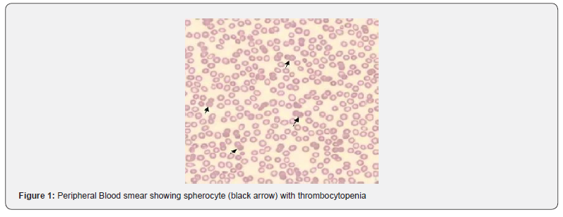
Laboratory examination results revealed haemoglobin 6.7g/dl, mean corpuscular volume (MCV) - 102f/l, mean corpuscular hemoglobin (MCH) - 28pg, mean corpuscular hemoglobin concentration (MCHC) - 33g/dl, white blood cell (WBC) - 10,400cell/cmm, and platelet - 5,000/cmm. Peripheral blood smear showed mild anisopoikilocytosis comprising normocytes and macrocytes and few spherocytes with severe thrombocytopenia (Figure 1) Reticulocyte count was raised (4.5% normal 0.5-1.5%) and lactate dehydrogenase (LDH) was elevated (496U/L- Normal up to 246U/L). Erythrocyte sedimentation rate was 108 mm at the end of first hour. Direct Coombs test was positive. Her renal function tests were normal. ANA was negative. Liver function tests showed total bilirubin of 3.7mg/dl and in indirect bilirubin of 2.9mg/dl. Her prothrombin time activated partial thromboplastin time and international normalized ratio results were normal. Ultrasonography (USG) abdomen showed no organomegaly, no ascites. 2D echocardiography (Echo) and chest X-ray showed normal study. The patient refused for bone marrow study. Anti platelet antibody test was not available in our country. Based on the Coombs positive hemolytic anemia and thrombocytopenia the patient was diagnosed with Evans syndrome.
Initially 2 units of whole blood and 2 units of platelet apharesis were transfused. Then patient was treated with methyl prednisolone 1000mg intravenously for 3 days, then oral prednisolone started from 1mg/kg/day for 3 weeks. Laboratory reports after 10 days showed hemoglobin of 9.4mg/dl, platelets of 96,000/cmm and total bilirubin of 2.1mg/dl. Her symptoms improved and she responded dramatically. Prednisolone was tapered over next 6 weeks. At the end of six weeks, she was asymptomatic with hemoglobin of 10.6mg/dl, platelets of 134,000/cmm and total bilirubin of 1.1mg/dl. She was advised to regular outpatient door follow up.
Discussion
Evans syndrome is an uncommon condition defined by the combination (either simultaneously or sequentially) of immune thrombocytopenia (ITP) and autoimmune hemolytic anemia (AIHA) with a positive direct antiglobulin test (DAT) in the absence of known underlying aetiology. Evans Syndrome was first described by Robert Evans in 1951 [3]. It is a rare diagnosis, and the exact frequency is unknown. No sex predilection is known. It may occur in all ethnic groups and all ages. There is evidence to support abnormalities in both cellular and humoral immunity in Evans syndrome. Despite the frequency of haemopoietic cell-specific autoantibodies in patients with Evans syndrome, there is very little information about the identity of target antigens.
Evans syndrome can be primary (or idiopathic) or secondary associated with autoimmune disorders like systemic lupus erythematous, Sjögren’s syndrome, anti-phospholipid syndrome and autoimmune lymphoproliferative syndrome. In a study done in 2009, Evans syndrome was found to be "primary" in 34 individuals (50%) but was linked to an underlying condition in 50% of cases, primarily systemic lupus, lymphoproliferative diseases, and common variable immunodeficiency, leukemia, lymphoma [14,15]. Even, metastatic small cell lung carcinoma was also reported in Evans syndrome [16].
Patients may present with AIHA or ITP either separately or concomitantly. Neutropenia occurs in up to 55% of patients at presentation, or pancytopenia (14%). The development of the second cytopenia may occur months to years after the first immune cytopenia and may delay diagnosis. Usual features of hemolytic anemia i.e.: pallor, lethargy, jaundice, heart failure in severe cases; and features of thrombocytopenia i.e.: petechiae, bruising, mucocutaneous bleeding may be present. The lymphadenopathy and organomegaly (hepatomegaly and/or splenomegaly) may be chronic or intermittent and in some cases may only be apparent during episodes of acute exacerbation [3,7,8,9,15]. Our patient presented with low grade intermittent fever, high color urine and generalized weakness. She had no history of joint pain, oral ulcer, photosensitivity or other systemic symptoms. She was anemic, mild icteric and had bruise on both legs. She had no bony tenderness, any lymphadenopathy or organomegaly.
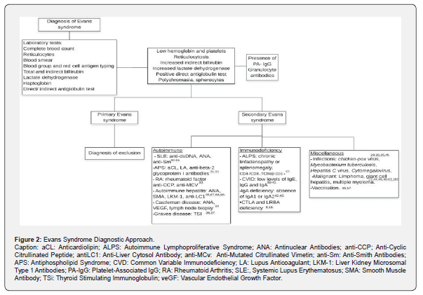
A full blood count will confirm the presence of cytopenias, and a blood film should be examined for features of AIHA (polychromasia, spherocytes) and to exclude other underlying diagnoses (malignancies, micro angiopathic hemolytic anemia, congenital hemolytic and thrombocytopenic conditions). Features of hemolysis should be sought including a raised reticulocyte count, unconjugated hyperbilirubinemia and decreased haptoglobins. The direct antiglobulin test (DAT) is almost invariably positive (although often weakly so), even in the absence of hemolytic anemia, and may be positive for IgG and/or complement (C3) [7-9,15]. The indirect antiglobulin test may also be positive in 52-83% of patients [8,17].
Assays for antiplatelet and anti-granulocyte antibodies have shown varied results [18]. In a report of 32 adult patients with AIHA, showed antiplatelet antibodies in 91% (demonstrated by thromboagglutination and indirect antiglobulin consumption tests) and leucocyte antibodies in 81% (demonstrated by a cytotoxicity test). Common variable immunodeficiency (CVID) and IgA deficiency, which have been reported to develop acquired cytopenias [12,13], and also as a baseline prior to immunomodulatory therapy.
In addition, other autoimmune conditions, particularly systemic lupus erythematosus (SLE), should be sought by measuring antinuclear antibody (ANA), double stranded DNA (dsDNA) and rheumatoid factor. The most important differential diagnosis is Autoimmune lymphoproliferative syndrome (ALPS). Therefore, measurement of peripheral blood T-cell subsets by flow cytometry is essential in all cases of Evans syndrome. The presence of double negative (CD4-/CD8-, CD3+, TCRαβ+) T cells has been found to be the most sensitive first-line screening test for ALPS (and allows differentiation from cases of Evans syndrome) [4].
In long-term follow-up most authors described more frequent episodes of ITP compared with episodes of AIHA. Causes of death were mainly related to hemorrhage or sepsis and reassuringly, given the degree of immune dysregulation seen in many patients, none of the patients described in these long-term studies (mainly of children) developed malignancy.
At best, the data suggest a role for corticosteroids +/- IVIG as first-line therapy in the acute setting, with blood product support as necessary. Second-line therapy, in the form of single agent or, for more severe cases, multi-agent, immunosuppressants will usually be required. A therapeutic trial of ciclosporin as the best second-line option for most patients, with MMF and then multi-agent therapy or rituximab for those that fail to respond. Splenectomy commonly achieves only short-term responses but may reduce the frequency of relapses and allow reduction of immunosuppressive agents. The choice of splenectomy versus rituximab may be a difficult one and will need to be made on a case-by-case basis.
Finally, high-dose therapy plus stem cell support provides hope for long-term cure in patients with a matched family donor; the success of a reduced intensity conditioning regimen is an encouraging approach for this group of patients who have usually been extensively pretreated and are often in poor clinical condition prior to referral for SCT. Evans syndrome is characterized by recurrent episodes of relapses and remission of both ITP and AIHA. In some patients it seems likely that long-term cure may only be achievable with SCT [19,20].
Conclusion
Evans syndrome is a very rare autoimmune disorder typically characterized by the simultaneous or sequential occurrence of two or more cytopenias. The two that occur most frequently are ITP purpura and autoimmune hemolytic anemia. The exact cause of this condition is unknown. The best treatment options for Evans syndrome depend on many factors, including the severity of the condition; the sign and symptoms present; and each person's response to certain therapies. It is difficult to diagnose and treat. Typically, corticosteroids and immunosuppressive medications are used to manage the syndrome. The response, nevertheless, is unpredictable and inconsistent. Evans syndrome patient’s prognosis is guarded; those who respond well to treatment get positive results. However, as with many other rare disorders, progress may depend upon the acquisition of detailed information through national/international databases and international multicenter randomized trials to accrue sufficient numbers of patients; long-term follow up is also essential for the chronic relapsing nature of this condition.
References
- Jaime-Perez JC, Aguilar-Calderon PE, Salazar-Cavazos L, Gomez-Almaguer D (2018) Evans syndrome: clinical perspectives, biological insights and treatment modalities. J Blood Med 9: 171-184.
- Audia S, Grienay N, Mounier M, Michel M, Bonnotte B (2020) Evans' Syndrome: From Diagnosis to Treatment. J Clin Med 9(12): 3851.
- Evans RS, Takahashi K, Duane RT, Payne R, Liu C (1951) Primary thrombocytopenic purpura and acquired hemolytic anemia; evidence for a common etiology. AMA Archives of Internal Medicine 87(1): 48-65.
- Teacheyet DT, Manno CS, Axsom KM, Andrews T, Choi JK, et al. (2005) Unmasking Evans syndrome: T-cell phenotype and apoptotic response reveal autoimmune lymphoproliferative syndrome (ALPS). Blood 105(6): 2443-2448.
- Yoshida H, Ishida H, Yoshihara T, Oyamada T, Kuwana M, et al. (2009) Complications of Evans’ syndrome in an infant with hereditary spherocytosis: A case report. Journal of Hematology and Oncology 2: 40.
- Dave P, Krishna K, Diwan AG (2012) Evan’s syndrome revisited. The Journal of the Association of Physicians of India 60: 60-61.
- Pui CH, Wilimas J, Wang W (1980) Evans syndrome in childhood. Journal of Pediatrics 97(5): 754-758.
- Mathew P, Chen G, Wang W (1997) Evans syndrome: results of a national survey. Journal of Pediatric Hematology/Oncology 19(5): 433-437.
- Wang WC (1988) Evans syndrome in childhood: pathophysiology, clinical course, and treatment. American Journal of Pediatric Hematology/Oncology 1988 10(4): 330-338.
- Norton A, Roberts I (2006) Management of Evans syndrome. British Journal of Haematology 132(2): 125-137.
- Oda H, Honda A, Sugita K, Nakamura A, Nakajima H (1985) High-dose intravenous intact IgG infusion in refractory autoimmune hemolytic anemia (Evans syndrome). Journal of Pediatrics 107(5): 744-746.
- Willis F, Marsh J, Bevan D, Killick S, Lucas G, et al (2001) The effect of treatment with Campath-1H in patients with autoimmune cytopenias. British Journal of Haematology 114(4): 891-898.
- Gombakis N, Trahana M, Athanassiou M, Kanakoudi-Tsakalidou F (1999) Evans syndrome: successful management with multi-agent treatment including intermediate-dose intravenous cyclophosphamide. Journal of Pediatric Hematology/Oncology 21(3): 248.
- Dave P, Krishna K, Diwan A (2012) Evan's Syndrome Revisited. Journal of the Association of Physicians of India 60(4): 60-61.
- Savasan S, Warrier I, Ravindranath Y (1997) The spectrum of Evans' syndrome. Archives of Disease in Childhood 77(3): 245-248.
- Lott A, Butler M, Leighl N, Cserti-Gazdewich CM (2015) Evan's Syndrome Associated with Pembrolizumab Therapy in Metastatic Non-Small Cell Lung Cancer. Blood 126(23): 45-43.
- Pegels JG, Helmerhorst FM, van Leeuven EF, de Plas van Dalen C, Engelfriet CP, et al. (1982) The Evans syndrome: characterization of the responsible autoantibodies. British Journal of Haematology 51(3): 445-450.
- Fagiolo E (1976) Platelet and leukocyte antibodies in autoimmune hemolytic anemia. Acta Haemtologica 56(2): 97-106.
- Hansen OP, Srrensen CH, Astrup L (1982) Evans syndrome in IgA deficiency. Episodic autoimmune haemolytic anaemia and thrombocytopenia during a 10 years observation period. Scandinavian Journal of Haematology 29(3): 265-270.
- Sneller MC, Strober W, Einstein E, Jaffe JS, Cunningham Rundles C (1993) New insights into common variable immunodeficiency. Annals of Internal Medicine 118(9): 720-730.

























