Radiological Diagnosis of Ehlers- Danlos Syndrome in an Adolescent Girl with Chronic Musculoskeletal Symptoms -- A Case Report
Saima Shan, Asim Rehman and Jovaria Ehsan*
Federal Government Polyclinic Hospital, Pakistan
Submission: February 23, 2023; Published: March 14, 2023
*Corresponding author: Jovaria Ehsan, Associate Radiologist, Federal Government Polyclinic Hospital, Islamabad, Pakistan
How to cite this article: Saima Shan, Asim Rehman and Jovaria Ehsan. Radiological Diagnosis of Ehlers- Danlos Syndrome in an Adolescent Girl with Chronic Musculoskeletal Symptoms -- A Case Report. JOJ Case Stud. 2023; 14(1): 555880. DOI: 10.19080/JOJCS.2023.14.555880.
Abstract
Ehlers-Danlos Syndrome is a very rare, genetically and clinically heterogeneous, heritable disorder characterized by abnormal collagen synthesis. There are at least thirteen subtypes with variable inheritance patterns, majority being autosomal dominant. The cardinal features include joint hypermobility, skin hyperextensibility, and soft connective tissue fragility. Radiologically, hemarthrosis, precocious osteoarthritis, arachnodactyly and kyphoscoliosis are seen involving the skeleton. We present a case of an 18-year-old girl with joint hypermobility, easy bruisability, joint pain and lax skin. She was undiagnosed until her adulthood. Radiological investigations revealed characteristic arachnodactyly, kyphoscoliosis and precocious osteoarthritis which led to her diagnosis of Ehlers Danlos syndrome.
Keywords: Ehlers- danlos syndrome; Skin laxity; Kyphoscoliosis
Introduction
Ehlers-Danlos syndrome is a hereditary disorder of connective tissue [1]. It is an umbrella term for a group of clinically and genetically heterogeneous connective tissue disorders [2]. The patient with EDS suffers from chronic pain, which may be associated with disability, poor wound healing, depression, anxiety, or physical and mental health-related quality of life impairment. The current EDS classification includes 13 subtypes [3]. EDS classical type is estimated to affect 1 per 10000 to 1 per 20000 [4]. Thirteen different types of EDS are currently recognized. These can be differentiated on a variable pattern of symptoms like skin hyperextensibility, joint hypermobility and internal organ and vessel fragility. Classical EDS involves an autosomal dominant inheritance pattern. The clinical hallmarks of EDS are tissue fragility, joint hypermobility, and skin laxity [5]. Complications of this disease include arterial and organ rupture, joint dislocation, chronic pain, fatigue and bone metabolic complication, among many others [6].
EDS is a complex illness with a multitude of symptoms. The abnormal collagen can affect virtually any body system. Patients usually present with hypermobility of joints and an increased bleeding tendency [7]. The presentation and severity of EDS range from undetectable or very mild symptoms to severe or even life threatening disease. This heterogeneity in presentation can make diagnosing EDS a clinical challenge because it is based mostly on patient’s complaints, subtle clinical findings and a family history [8].
Case Report
An 18 years old, unmarried, short statured girl came in radiology department for skeletal radiographs, referred from rheumatology department with complaints of both hand and left ankle deformity since birth,
joint pain of two weeks duration, hyperflexibilty of joints, lax skin and easy bruisabilty. She had been having these symptoms off and on for last few years.
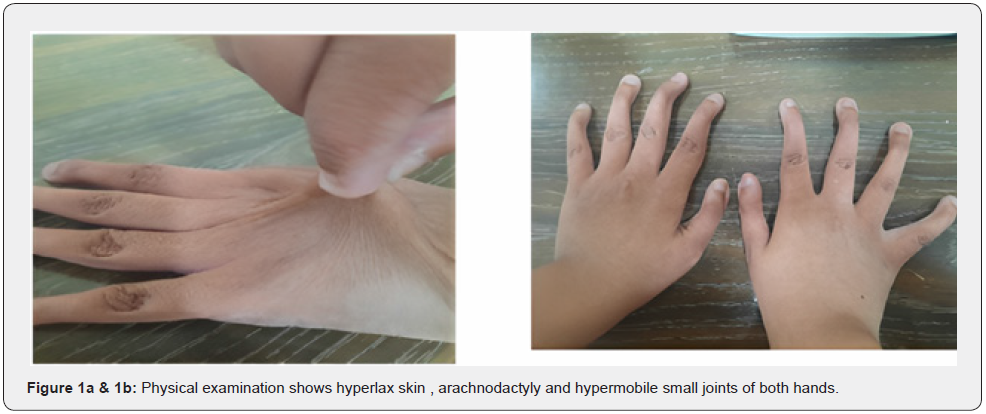
On examination, her epicanthal fold thickness was increased with attached ear lobes, bilateral genu valgus deformity and an externally rotated left ankle. She had multiple deformities of both hands and feet with hypermobility of metacarpophalangeal and interphalangeal joints. She had few bruises over her right forearm which occurred after a fall from stairs. She had lax skin and hypermobile small joints of hand and was assigned 8/9 on Beighton score (Figure 1a & 1b).

She was born from non-consanguineous parents, by spontaneous vaginal delivery and achieved developmental milestones normally. She had normal intelligence and appropriate school performance. Her
routine blood investigations were carried out which revealed that she was anemic with an unremarkable renal and hepatic function tests. Her echocardiography was normal.
She was referred to Radiology department for musculoskeletal radiographs and abdominal ultrasound. X-ray both hands revealed clino-arachnodactyly (Figure 2).

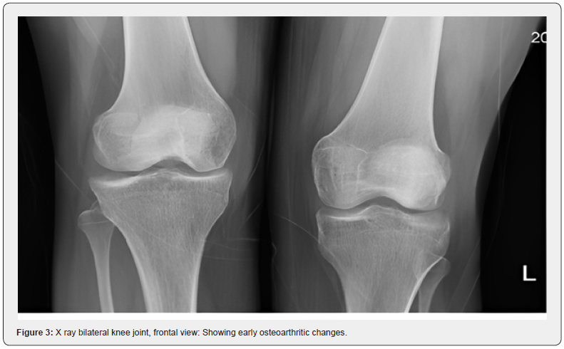
Radiographs of both knees showed sclerosis of tibial plateau and mildly reduced medial tibio-femoral joint spaces on both sides suggestive of precocious osteoarthritic changes (Figure 3).
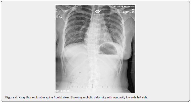
Cervical spine radiograph revealed straightening of curvature and osteophytosis along superior vertebral end plates at multiple levels. There was kyphoscoliosis deformity as seen on radiograph of thoracic and lumbosacral spine with osteophyte formation at multiple levels in thoracic and lumbar spine (Figure 4).


There was no evidence of diaphragmatic hernia or any other pathology on chest Radiograph. Skull radiographs was normal. Her ultrasound of abdomen was normal. No evidence of dissection or any other
vascular anomaly was seen on Doppler studies of both limbs and abdomen.
On the basis of the radiological features and clinical assessment, a diagnosis of Ehler Danlos Syndrome (Classic variant – cEDS) was made. This patient has had multiple hospital visits since her early age due to joint pain and easy bruisability but she had never been investigated or diagnosed earlier. Genetic testing is recommended for EDS types with a known molecular cause to confirm the diagnosis. However, she didn’t undergo any genetic testing due to unavailability in Pakistan and patient was non affording.
Discussion
EDS is a hereditary disorder of connective tissue [1]. Patients are affected with joint hyper mobility, skin hyper extensibility, and skin fragility leading to atrophic scarring and significant bruising [1,9]. EDS classical type is estimated to affect 1 per 10000 to 1 per 20000 [4]. Complications of joint hyper mobility, such as dislocations usually resolve spontaneously or are easily managed by the affected individual. Other features include hypotonia with delayed motor development, fatigue and muscle cramps [10]. The patients with classic EDS are usually diagnosed early in life. However our patient, despite having symptoms since early childhood, was always treated symptomatically and was never diagnosed.
A study conducted by Tewari S et al. [11] revealed that chronic widespread musculoskeletal pain is a cardinal symptom in EDS which can be both physically and psychologically disabling.11 Our patient too had chronic arthralgias, myalgias and easy bruisability for quite a long time resulting in frequent hospital visits.
A study by Liang M et al. [12] showed that Ehler Danlos syndrome is a disease with more than one affected person in the family [12]. Siblings and family members of our patients were normal with no similar complaint. None of the family members has ever been diagnosed with any other connective tissue disorder.
Studies have shown that facial dysmorphism commonly affecting midface /orbital areas with epicanthal folds and infraorbital creases more commonly observed in young patients [13]. Our patient had thickened epicanthal folds with attached ear lobes.
Ehler Danlos syndrome is usually diagnosed clinically and by genetic analysis. We diagnosed our patient radiographically in conjunction with her clinical findings. Clinically our patient was scored as 8/9 on Beighton score which is a standard method of assessment of the hypermobility syndromes [13].
On X-ray of both hands clino-arachnodactyly was observed. X-ray of both knees showed precocious osteoarthritic changes. Cervical spine radiograph revealed cervical spondylosis. All of the musculoskeletal
findings found in our patient were in consistence with those studied by Mazziotti G et al. [14] who described the skeletal manifestations of EDS and its complications [14].
The challenge observed in this case was diagnostic limitations due to unavailability of genetic screening in Pakistan. Genetic screening is being used as diagnostic tool in the world [3] however patient’s affordability and scarce resources were a major limiting factor. However, her radiological features and clinical manifestations were a major contribution towards her diagnosis and it will help in her further management and rehabilitation. She was referred to the spinal surgeon in the hospital to consult on her progressing kyphoscoliosis deformity.
Conclusion and Recommendation
Ehlers-Danlos syndrome (EDS) is a genetic disorder affecting collagen formation and function. It affects virtually every organ system, which can result in significant morbidity and mortality. Proper diagnosis of EDS is essential to improving the overall health and well-being of affected patients as well as mitigating these complications.
References
- Bowen JM, Sobey GJ, Burrows NP, Colombi M, Lavallee ME, et al. (2017) Ehlers–Danlos syndrome, classical type. American Journal of Medical Genetics Part C: Seminars in Medical Genetics 175(1): 27-33.
- D'hondt S, Van Damme T, Malfait F (2018) Vascular phenotypes in nonvascular subtypes of the Ehlers-Danlos syndrome: a systematic review. Genetics in Medicine 20(6): 562-573.
- Ritelli M, Colombi M (2020) Molecular genetics and pathogenesis of Ehlers–Danlos syndrome and related connective tissue disorders. Genes 11(5): 547.
- Miklovic T, Sieg VC (2021) Ehlers danlos syndrome. In: StatPearls [Internet]. StatPearls Publishing.
- Miller E, Grosel JM (2020) A review of Ehlers-Danlos syndrome. JAAPA 33(4): 23-28.
- Formenti AM, Doga M, Frara S, Ritelli M, Colombi M, et al. (2019) Skeletal fragility: an emerging complication of Ehlers–Danlos syndrome. Endocrine 63(2): 225-230.
- Van Camp N, Aerden T, Politis C (2020) Problems in the orofacial region associated with Ehlers- Danlos and Marfan syndromes: a case series. Br J Oral Maxillofac Surg 58(2): 208-213
- Zhou Z, Rewari A, Shanthanna H (2018) Management of chronic pain in Ehlers-Danlos syndrome: Two case reports and a review of literature. Medicine (Baltimore) 97(45): e13115.
- Ghali N, Sobey G, Burrows N (2019) Ehlers-Danlos syndromes. BMJ 366: l4966.
- Malfait F, Wenstrup R, De Paepe A (2018) Classic ehlers-danlos syndrome. GeneReviews® [Internet].
- Tewari S, Madabushi R, Agarwal A, Gautam SK, Khuba S (2017) Chronic pain in a patient with Ehlers–Danlos syndrome (hypermobility type): the role of myofascial trigger point injections. J Bodyw Mov Ther 21(1): 194-196.
- Liang M, Chen C, Dai Y, Chang Y, Gao Y (2022) Two closely spaced missense COL3A1 variants in cis cause vascular Ehlers-Danlos syndrome in one large Chinese family. Journal of Cellular and Molecular Medicine 26(1): 144-150.
- Malek S, Reinhold EJ, Pearce GS (2021) The Beighton Score as a measure of generalised joint hypermobility. Rheumatology International 41(10): 1707-1716.
- Mazziotti G, Dordoni C, Doga M, Galderisi F, Venturini M, et al. (2016) High prevalence of radiological vertebral fractures in adult patients with Ehlers-Danlos syndrome. Bone 84: 88-92.


