Extra Medullary Multiple Myeloma in Breast
Ravi Shankar B1*, Madhavi BV2, Pradeep Bhaskar3 and Prithviraj B4
1Consultant, Queens NRI Hospital, India
2Department of Pathology, GITAM Medical College (GIMSR), India
3Senior Resident, Queen's NRI Hospital, India
4Department of Medico, India
Submission: February 28, 2018; Published: March 12, 2018
*Corresponding author: Ravi Shankar B, Consultant, Queens NRI Hospital, India, Email: ravi_bellala@yahoo.co.in
How to cite this article: Ravi Shankar B, Madhavi BV, Pradeep Bhaskar, Prithviraj B. Extra Medullary Multiple Myeloma in Breast. JOJ Case Stud. 2018; 6(3): 555689. DOI: 10.19080/JOJCS.2018.06.555689
Introduction
Multiple myeloma is a disease of plasma cells and accounts for 1% of all 10% of haematological malignancies [1]. It is a systemic disease in the elderly. The primary site of involvement in the disease is the skeletal system which accounts for 97% of the cases. Multiple Myeloma may involve extra osseous sites. About 3% of the cases involve the soft tissues [2]. Its incidence in patients younger than 40 years is rare. Only in rare cases it is observed in the breast. First case of myeloma breast was reported in the year 1925 [3]. Since then only 20 cases of myeloma Breast had been reported till now. More than half of the cases reported had bilateral breast involvement. We are reporting one such rare case of bilateral myeloma breast with a brief review of literature.
Case Presentation
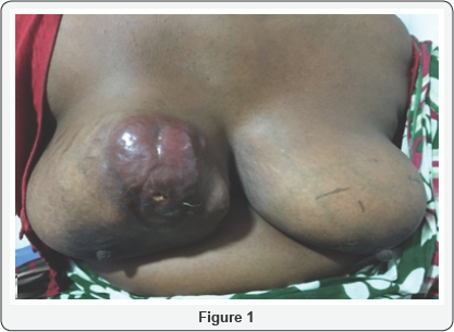
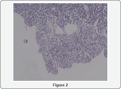
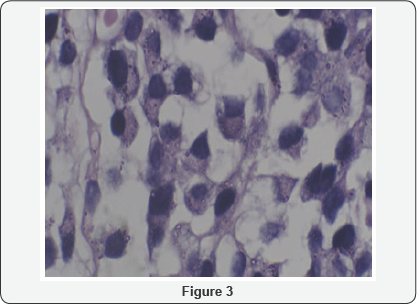
A 32yr multiparous premenopausal female presented to our OPD at Queens NRI Hospital, Visakhapatnam, with bilateral breast lumps of 4 months duration. It started as a small lump in Right breast 4 months ago which gradually progressed in size. A similar small lump showed up in the Left breast 3 months later. Both the lumps were not associated with pain. There was no associated fever, weight loss. She had no history of (H/o) trauma in the past, no H/o Tuberculosis, no significant Family History. She was a diagnosed case of Multiple Myeloma treated for 3yrs (from 2012-2015) with Thalidomide and Dexamethasone but had stopped taking medication since 2015. On examination a single 15X15cm firm lobulated mass occupying almost the entire Right breast with infiltration of the overlying skin in the upper outer quadrant was seen with normal looking Nipple Areola Complex (NAC). Left breast showed multiple discrete firm mobile lumps in the upper outer and inner quadrants with the largest measuring 6X5cm with normal NAC and overlying skin (Figure 1). Bilateral axillae and supraclavicular fossae were normal. Tru-cut biopsy from right as well as left breast lump came out as plasma cell myeloma. Microscopic pictures of the specimen showed multiple plasma cells arranged in sheets with eccentrically placed nuclei and abundant cytoplasm confirming the diagnosis of plasma cell myeloma (Figure 2 & 3).


Discussion
Multiple myeloma is a primary malignancy of bone marrow characterized by clonal proliferation of plasma cells and production of monoclonal immunoglobulins. Soft tissue involvement is often secondary to skeletal involvement. Isolated involvement of soft tissues is much rare. The differential diagnosis in this case can be a primary breast cancer as there is literature with the presentation of breast carcinoma in patients with a known history of lymphoma or multiple myeloma [4].
The clinical course of patients with breast myeloma depends on whether the lesion is solitary or part of disseminated myeloma [5]. The overall prognosis in primary breast plasmacytoma is excellent [6]. In contrast, metastatic tumours have poor prognosis. Plasmacytomas are often a late manifestation of the disease and their appearance in a known case of multiple myeloma indicates failure of apparently successful therapy [7].Patients with a low beta2-microglobulin level tend to have a better prognosis. The presence of a chromosome 13 deletion has been shown to have a significant negative impact on outcome and also an elevated lactate dehydrogenase level is associated with a very bad prognosis [8].
Conclusion
Diagnosis of breast myeloma should always be considered in a patient with multiple breast masses, especially when bilateral, more so in a known case of myeloma and also as a differential of metastases.
References
- Angtuaco EJ, Fassas AB, Walker R, Sethi R, Barlogie B (2004) Multiple myeloma: clinical review and diagnostic imaging. Radiology 231(1): 11-23.
- Innes J, Newall J (1961) Myelomatosis. Lancet 1: 239-245.
- Cutler C, Plasma C (1925) Plasma cell tumour of the breast with metastasis. Ann Surg 100: 392-395.
- Khalbuss WE, Fischer G, Ahmad M, Villas B (2006) Synchronous presentation of breast carcinoma with plasmacytoid cytomorphology and multiple myeloma. Breast J 12(2): 165-167.
- Alverez JV, Gomez MM, Prats MDG, Agorreta MA, Lopez JIL, et al. (2003) Extramedullary plasmacytoma presenting as primary mass in the breast. A case report. Acta Cytol 47(6): 1107-1110.
- Kumar PV, Owji SM, Talei AR, Malekhusseini SA (1997) Extramedullary plasmacytoma: Fine needle aspiration findings. Acta Cytol 41(2): 363368.
- Pai RR, Raghuveer CV (1996) Extramedullary plasmacytoma diagnosed by fine needle aspiration cytology. A report of four cases. Acta Cytol 40(5): 963-966.
- Fassas AB, Tricot G (2004) Chromosome 13 deletion/hypodiploidy and prognosis in multiple myeloma patients. Leuk Lymphoma 45(6): 1083-1091.


