




 JGWH Home




Abstract
Objective: To evaluate the feasibility and outcomes of laparoscopic myomectomy for uterine fibroids in a sub-Saharan African tertiary hospital, highlighting surgical results and advantages compared to the conventional open approach.
Methods: We conducted a prospective descriptive study of patients undergoing laparoscopic myomectomy at a national hospital in Dakar from March 2017 to May 2019. Patient characteristics, fibroid details, operative parameters, and postoperative outcomes were recorded. Strict selection criteria were applied (predominantly intramural or sub serosal fibroids ≤8 cm and limited number) to ensure patient safety.
Results: Nine patients (mean age 34 years) were included; all presented with symptomatic fibroids (100% with pelvic pain) and 75% had infertility. A total of 8 laparoscopic myomectomies were completed (one case required conversion to laparotomy for an 18 cm fibroid with degeneration). The mean number of fibroids removed was 2 (range 1–4) with a mean largest fibroid diameter of 7.3 cm (4–11 cm). Average operative time was 155 minutes, and a notable learning-curve improvement was observed, with the per-fibroid suturing time reduced from 57 to 28 minutes between the first and last case. Mean intraoperative blood loss was modest at approximately 150 mL, and no patient required transfusion. Uterine defects were all closed in two layers with no endometrial cavity breaches. There were no major perioperative complications; uterine preservation was achieved in all cases. Postoperative recovery was favorable, with hospital stay ranging from 2 to 3 days (mean ~3 days) and minimal postoperative pain.
Conclusion: Laparoscopic myomectomy was feasible and safe in our setting, yielding outcomes consistent with the benefits of minimally invasive surgery. In selected patients, it offers significantly reduced blood loss, shorter hospitalization, and quicker recovery compared to laparotomy, without compromising surgical efficacy or patient fertility. Adherence to strict selection criteria and advanced laparoscopic skill are essential for optimal results.
Keywords: Laparoscopic myomectomy; Uterine fibroids; Minimally invasive surgery; Laparotomy; Surgical outcomes; Case series.
Introduction
Uterine fibroids (leiomyomas) are the most common benign tumors of the uterus, affecting a large proportion of women of reproductive age. The cumulative incidence rises with age and is particularly high in women of African ancestry – up to 70% of all women develop fibroids by menopause, and an estimated 70–80% of Black women will experience fibroids in their lifetime [1]. While many fibroids remain asymptomatic, a significant subset cause menorrhagia, pelvic pain, bulk symptoms, or infertility. These symptomatic fibroids often necessitate active treatment to improve quality of life and reproductive outcomes. Myomectomy, the surgical removal of fibroids with preservation of the uterus, is the standard of care for women who desire uterine conservation. Traditionally, myomectomies were performed via laparotomy (open surgery). However, the advent of minimally invasive techniques has transformed fibroid management [2]. For intramural and sub serosal fibroids of moderate size, laparoscopic myomectomy has demonstrated clear advantages over the open approach, including smaller incisions (better cosmesis), reduced intraoperative blood loss and postoperative pain, shorter hospital stays and convalescence, and lower risk of postoperative adhesions [3]. Over the past two decades, laparoscopic myomectomy has become widely adopted and is now considered the gold standard for appropriate candidates, particularly in women who wish to preserve fertility [4]. Studies have shown that in the hands of experienced surgeons and with proper patient selection, laparoscopic myomectomy yields superior perioperative outcomes and at least equivalent long-term results compared to open myomectomy. A meta-analysis of randomized trials by Jin et al. reported that laparoscopic myomectomy is associated with significantly less hemoglobin drop, lower operative blood loss, less postoperative pain, and fewer overall complications than laparotomy, albeit with a longer operative time; importantly, rates of major complications, fibroid recurrence, and subsequent pregnancy were similar between the two approaches.
Despite its benefits, laparoscopic myomectomy can be technically demanding, with limitations depending on fibroid size, number, and location. Large or numerous fibroids increase the risk of hemorrhage and may necessitate conversion to open surgery for adequate control [5]. Published guidelines and expert opinions generally recommend laparoscopic removal for fibroids up to about 8–10 cm in diameter (and a limited number of fibroids, e.g. ≤3–4) in order to ensure safe intracorporeal suturing and minimize the risk of excessive bleeding or incomplete resection. Proper patient selection is therefore critical. Additionally, surgeons must be skilled in advanced laparoscopic suturing and tissue extraction (morcellation) techniques to successfully perform the procedure. In resource-limited settings or regions where laparoscopic surgery is newly introduced, demonstrating the feasibility and reproducibility of laparoscopic myomectomy is important. Our center, the National Hospital of Pikine in Dakar, Senegal, began performing laparoscopic myomectomy with the goal of offering patients the advantages of minimally invasive surgery. Given the high prevalence and often large size of fibroids in our population, the implementation of laparoscopic myomectomy needed to be carefully evaluated. In this study, we report our initial experience with laparoscopic myomectomy, focusing on perioperative outcomes and highlighting how this approach compares to the conventional laparotomy. We aim to show that with strict selection criteria and appropriate technique, laparoscopic myomectomy can be safely and effectively conducted even in a low-resource setting, providing tangible benefits to patients.
Methods
This study was a prospective, descriptive case series of patients undergoing laparoscopic myomectomy for symptomatic uterine fibroids at the National Hospital of Pikine in Dakar, Senegal. The study period spanned from March 2017 to May 2019. Eligible patients were those with symptomatic fibroids (causing pain, abnormal bleeding, infertility, or bulk symptoms) who met criteria for a laparoscopic approach. We primarily included women with up to three or four fibroid nodules of moderate size; generally, a largest fibroid diameter of ≤8 cm was required for laparoscopy candidacy, in line with consensus recommendations for safe laparoscopic myomectomy. Patients with submucous fibroids requiring hysteroscopic resection or those with very large or numerous fibroids (for whom open surgery was deemed more appropriate) were excluded. All patients provided informed consent after thorough counseling on the risks, benefits, and alternatives of the procedure. Preoperative evaluation included pelvic examination and ultrasound to map the number, size, and location of fibroids. In some cases of very rapid fibroid growth or diagnostic uncertainty, pelvic MRI was obtained to better characterize fibroid architecture and exclude atypical pathology (e.g. suspicion of sarcoma), as rapid growth can occasionally indicate malignancy or degeneration [6]. Hemoglobin levels were optimized preoperatively to reduce anemia. Standard premedication and prophylaxis were given, including antibiotic prophylaxis at induction and thromboembolism prophylaxis with low molecular weight heparin continued for 14 days postoperatively (given the moderate-risk profile of these gynecologic surgeries).
All surgeries were performed under general anesthesia with endotracheal intubation. Patients were placed in modified dorsal lithotomy position (legs slightly flexed and abducted ~30°) with a slight Trendelenburg tilt for pelvic access. A Foley catheter was inserted for bladder drainage. The laparoscopic technique entailed insufflation of the abdomen (creation of pneumoperitoneum) via a Veress needle at the left upper quadrant (Palmer’s point) or periumbilical area, followed by placement of a primary 10 mm umbilical trocar for the laparoscope. Two or three ancillary trocars (5–12 mm) were inserted under direct vision in the lower abdomen to facilitate instrument access. We used a standard multi-port laparoscopy (there was no single-port or robotic assistance in this series). The myomectomy procedure involved four main steps:
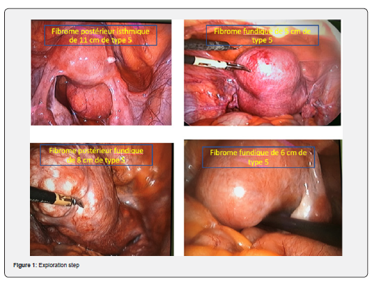
1. Exploration (figure 1), an incision was made on the uterine serosa overlying the fibroid (often using monopolar electrocautery) and extended to reach the pseudo capsule;
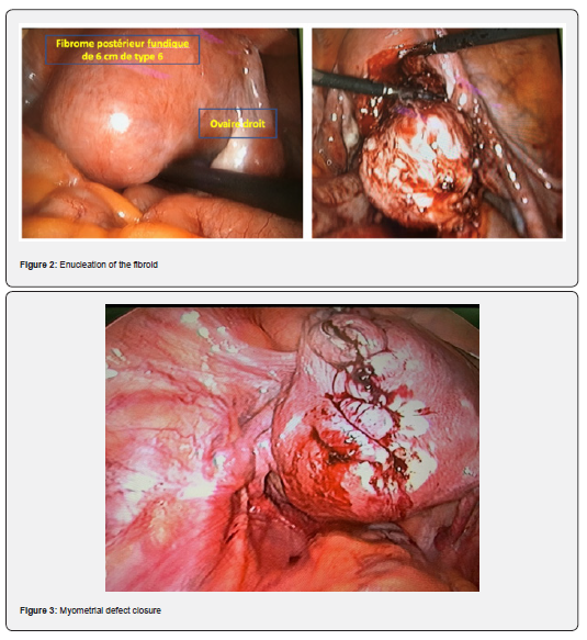
2. Enucleation – the fibroid was grasped and shelled out of the myometrium using traction and counter-traction, with judicious use of energy or hemostatic agents for bleeding control (figure 2);
3. Myometrial defect closure – the uterine incision was sutured in layers to reapproximate the myometrium and serosa. We predominantly performed two-layer closure with delayed absorbable sutures (e.g., 2-0 vicryl) to ensure a strong uterine scar, as is recommended to prevent uterine rupture in subsequent pregnancies, (figure 3). In all cases, closure was done laparoscopically using intracorporeal knot-tying techniques. No case required conversion to Mini laparotomy for suturing.
4. Specimen retrieval – the enucleated fibroid(s) were removed from the abdominal cavity. In our practice, removal was accomplished by morcellation: fibroid tissues were fragmented and extracted using an electromechanical morcellator introduced through an enlarged port site. (In one early case where morcellation was not available, a small abdominal incision was used to remove the fibroid, but this was avoided whenever possible.) Throughout surgery, measures were taken to minimize blood loss. This included use of diluted vasopressin injection into the myometrium and uterus-enclosing clamps or a temporary tourniquet in some cases, as well as the meticulous surgical technique. (No topical hemostatic agents were specifically used in this series.) If at any point bleeding was uncontrolled or visualization was lost, a low threshold for conversion to open surgery was maintained for patient safety.
Data collected for each case included patient demographics and gynecologic history (age, gravidity/parity, infertility history), fibroid characteristics (number, size, locations), operative details (duration of surgery, estimated blood loss, any need for conversion or transfusion, intraoperative complications), and postoperative recovery parameters (pain, length of hospital stay, postoperative complications). Descriptive statistics were used to summarize the findings given the small sample size. We report mean values with ranges for continuous variables. The study being primarily observational, no comparative statistical tests were applicable. However, a temporal observation of operative time was noted to assess the learning curve across cases.
Results
Patient Characteristics
Over the 26-month study period, a total of 9 women underwent an attempt at laparoscopic myomectomy that met inclusion criteria. The mean age was 34 years (range 28–40 years). Most patients were nulliparous; 62.5% had never been pregnant (nulligravidae). A history of infertility was present in 6 of the 8 women (75%) who had completed the procedure, with an average duration of infertility of about 14 months prior to surgery. All patients presented with significant pelvic pain attributed to fibroids (100% reported chronic pelvic pain as a symptom). Other symptoms included menstrual disturbances in a minority of cases, but heavy menstrual bleeding was not the leading complaint in our series. No patient had undergone prior uterine surgery.
Fibroid Characteristics
On ultrasound (and MRI when used), the women had between 1 and 4 fibroids requiring removal (mean number of fibroids per patient was 2.0). Fibroid sizes ranged from 4 cm up to 11 cm in maximum diameter. The mean diameter of the largest fibroid per patient was 7.3 cm. In five patients (62.5%), at least one fibroid was located on the posterior uterine wall (predominantly intramural posterior or subserosal posterior), which can pose additional technical difficulty due to access and visualization. The fibroids addressed were primarily intramural (within the uterine wall) or subserosal (surfacing on the uterine exterior); cases with submucous components were not included in this laparoscopic series. None of the patients had more than four fibroids targeted, per our selection criteria.
Operative Outcomes
Out of 9 intended laparoscopic myomectomy cases, 8 were successfully completed entirely by laparoscopy. One case (11%) required conversion to an open procedure (laparotomy) due to uncontrollable hemorrhage from a very large fibroid. In that particular patient, a bulky 18 cm fibroid with recent degeneration (necrobiosis) was encountered, which exceeded the usual size limit for laparoscopic removal. Despite initial attempt, bleeding was significant and the decision was made to convert to open surgery for safety. No patient in this series required an unplanned hysterectomy – even in the converted case, the myomectomy was completed via laparotomy and the uterus was preserved. Among the 8 fully laparoscopic cases, the mean total operating time (skin-to-skin) was 155 minutes, with individual surgery durations ranging from approximately 120 up to 180 minutes. The complexity of cases and the nascent experience of our team contributed to the longer operative times. However, a clear improvement was observed as the team gained experience. Notably, the time required for suturing the uterine incision (from the start of myometrial closure to completion) per fibroid significantly decreased over the series: for the first case, this step took 57 minutes for one fibroid, whereas by the 8th case, the suturing of a similar fibroid defect was completed in 28 minutes. This reflects the learning curve and increased dexterity in intracorporeal suturing techniques over time [7]. All uterine incisions were closed in two layers, and in no instance was there a detected entry into the endometrial cavity during suturing (no endometrial breaches), indicating careful technique in preserving the integrity of the uterine lining.
Intraoperative blood loss was low in most cases. The average estimated blood loss was approximately 150 mL (with observed losses ranging from about 100 mL to 300 mL). No patient in the laparoscopic group required a blood transfusion, which underscores the effective hemostasis achieved. By contrast, in open myomectomy literature, blood loss can be substantially higher and often blood transfusion rates of 20–30% are reported for multi-fibroid cases. The limited blood loss in our series can be attributed to both the minimally invasive approach and the use of vasoactive agents (e.g., vasopressin injection) and meticulous surgical technique. We did not routinely use adjunctive measures such as uterine artery tourniquets or intrauterine injection of TXA/oxytocin in this small series, but careful coagulation and timely suturing sufficed. No intraoperative complications such as visceral injuries (bowel, bladder, or ureteric injury) occurred in the laparoscopic cases. Aside from the one case of heavy bleeding prompting conversion, there were no other conversions and no surgical accidents. The postoperative course for all patients was generally smooth. Postoperative Outcomes: Patients resumed oral intake within the same day of surgery and mobilized early. Postoperative pain was minimal-to-moderate and effectively managed with usual analgesics; patients uniformly reported high satisfaction with the reduced pain and faster recovery compared to experiences of others undergoing laparotomies. The length of hospital stay after laparoscopic myomectomy ranged from 2 to 3 days. The mean hospitalization was ~2.8 days, which is significantly shorter than the typical 5–7 days observed with open abdominal myomectomy in our setting. All laparoscopic patients were discharged by the third postoperative day in good condition. In the single converted case, the hospital stay was slightly longer (4 days) due to the laparotomy. There were no postoperative fevers, infections, or significant wound issues in the series. We did not document any case of clinically significant postoperative adhesions in the short term (though formal assessment like second-look laparoscopy was not performed, the absence of readmissions for bowel obstruction or chronic pelvic pain suggests no severe adhesions). At follow-up visits (1–3 months post-surgery), all patients were doing well, and those with infertility were counseled to attempt conception after an adequate healing period (usually 3–6 months).
Discussion
This case series demonstrates that laparoscopic myomectomy is a feasible and beneficial surgical option for uterine fibroid treatment in our context. Despite a relatively small sample of eight completed cases, our findings align with the broader literature on the advantages of minimally invasive myomectomy. We achieved successful removal of fibroids up to ~10 cm laparoscopically with acceptable operative times and minimal blood loss, and without major complications. Importantly, all patients had their uteri preserved and were discharged quickly, reflecting the enhanced recovery associated with laparoscopy. One key observation from our experience is the importance of patient selection and adherence to inclusion criteria. We generally limited laparoscopic myomectomy to fibroids not exceeding 8 cm in diameter and to cases with a moderate total number of fibroids. These criteria mirror those suggested by experts and guidelines for ensuring safety in laparoscopic myomectomy. Fibroids beyond a certain size pose technical challenges: large tumors (>8–10 cm) can obscure visualization and are associated with increased bleeding risk and prolonged suturing time, which collectively raise the likelihood of conversion to open surgery. In our series, the only conversion to laparotomy occurred in a case with an 18 cm fibroid that in retrospect was outside the ideal laparoscopic range. This underscores that exceeding recommended limits can lead to intraoperative difficulties. Various authors have reported that the risk of conversion and intraoperative hemorrhage rises with increasing fibroid size and number [8]. We therefore stress that strict selection and preoperative planning (including imaging assessment of fibroid load) are crucial for success. As laparoscopic skills and technologies improve, these limits can be cautiously extended, but patient safety must remain the priority.
Our median operating time was on the higher side (over 2.5 hours), which is expected in the initial learning phase. Other studies have noted longer operative times for laparoscopic myomectomy compared to laparotomy, especially early in a surgeon’s experience. With increasing experience, operative duration tends to decrease markedly. In high-volume centers, laparoscopic myomectomy times can approach or even be shorter than open surgery for comparable fibroid sizes [9]. We documented a clear downward trend in the critical portion of suturing time over just 8 cases, reflecting a rapid learning curve. This suggests that with continued practice and possibly adoption of newer techniques (such as barbed sutures or automated suturing devices), our operative efficiency will further improve. We also anticipate that implementing adjuncts like preoperative GnRH analogues (to shrink fibroids) or uterotonics could shorten surgery by reducing fibroid vascularity, though these were not routinely used in our series. The average blood loss in our laparoscopic cases (approx. 150 mL) was notably low. Laparoscopic surgery inherently offers superior visualization and the ability to cauterize or coagulate bleeding vessels in a magnified field, which helps minimize blood loss. Additionally, pneumoperitoneum pressure can tamponade venous bleeding to some extent. Our results mirror findings from comparative studies where laparoscopic myomectomy consistently results in less intraoperative blood loss than open myomectomy.
For example, Jin et al. reported significantly smaller hemoglobin drops and transfusion requirements in the laparoscopic groups across multiple trials. The avoidance of blood transfusion in all our laparoscopic patients is a positive outcome, given that hemorrhage and transfusion are common concerns in fibroid surgery. To further control bleeding, literature supports the use of interventions such as intra-myometrial vasopressin injection, application of tourniquets to uterine arteries, use of hemostatic agents (e.g., gelatin-thrombin matrix), or administration of uterotonics like oxytocin and misoprostol [10]. In particular, a randomized trial by Agostini et al. showed that oxytocin infusion during myomectomy significantly reduced blood loss. A Cochrane review also indicates various effective measures to curb hemorrhage during myomectomy [11]. We employed vasopressin in some cases and found it helpful; future cases might benefit from combining these strategies to make the procedure even safer. Another advantage observed was the quick recovery and short hospitalization after laparoscopic myomectomy. Our patients stayed a median of only 2–3 days in the hospital. In contrast, open myomectomy typically mandates longer hospital stays (often around 5 days or more in our local practice, and at least 2–3 days even in developed settings) [12]. The reduced postoperative pain and faster return to normal activity with laparoscopy cannot be overstated – these enhance patient satisfaction and reduce healthcare costs. Even though our hospital stay could likely be shortened further (some protocols allow discharge within 24 hours for laparoscopic myomectomy [13]), the observed length in our context was still markedly better than with laparotomy. Minimal access surgery also likely decreases the risk of infection and wound complications, as evidenced by our absence of such events, since large abdominal incisions are avoided. From a fertility standpoint, preserving the uterus and minimizing trauma are critical for patients desiring future pregnancies. All our patients retained their uteri and can attempt conception.
An important concern historically with laparoscopic myomectomy has been the integrity of uterine scar and risk of uterine rupture in pregnancy. We addressed this by ensuring a proper multilayer closure of the myometrium in every case, which should provide a robust scar. The literature suggests that when myometrial defects are sutured securely, the risk of subsequent uterine rupture is very low, and pregnancy outcomes after laparoscopic myomectomy are comparable to those after open myomectomy [14]. Indeed, multiple studies have shown no significant differences in pregnancy rates or obstetric outcomes between laparoscopy and laparotomy for myomectomy. Some reports even suggest a possible trend toward higher pregnancy rates after laparoscopic myomectomy, potentially due to less adhesion formation and faster postoperative recovery enabling earlier attempts at conception. In our series, 75% of patients had infertility issues; while our follow-up is still short, we remain optimistic that the minimal invasive approach will facilitate positive fertility outcomes. We plan to track their pregnancy success in the coming years. Moreover, the fact that laparoscopy tends to cause fewer pelvic adhesions is advantageous for fertility, as adhesions involving fallopian tubes or ovaries can impair fertility. Research indicates that meticulous laparoscopic technique, use of anti-adhesion barriers, and avoidance of extensive tissue handling result in lower adhesion formation than open surgery. We did not formally evaluate adhesions, but none of our patients have shown signs of adhesion-related complications to date.
Comparing our findings to other published series, we note both similarities and context-specific differences. Our conversion rate of 11% (1 in 9 cases) is within acceptable ranges, especially considering one case fell outside ideal criteria. Many experienced centers report conversion rates below 5%, often near zero for fibroids <10 cm. With continued adherence to selection guidelines and growing surgeon experience, we anticipate our conversion rate will decrease further. Our mean operative time was higher than some reports (e.g., some authors report average 90–120 minutes for laparoscopic myomectomy of moderate fibroids), but again this should improve as we overcome the learning curve. Encouragingly, our mean blood loss (150 mL) and hospital stay (≈2–3 days) were very much in line with international data, confirming that even in a low-resource setting, the fundamental benefits of laparoscopy are realizable. Notably, we achieved these outcomes without access to the most advanced tools (such as robotics or newer energy devices) – indicating that standard laparoscopy, when carefully executed, suffices to attain good results. This study’s limitations include the small sample size and the lack of a direct comparison group (we did not formally compare to our open myomectomy outcomes, though qualitatively the differences are clear). Additionally, long-term outcomes such as fibroid recurrence and pregnancy success rates will require extended follow-up beyond the scope of this initial report. Nevertheless, the strength of our study lies in providing real-world data from a sub-Saharan African hospital, demonstrating that laparoscopic myomectomy can be introduced successfully. The novelty of our report is that it is among the first from our region to document the implementation of exclusive laparoscopic myomectomy and its outcomes. This is highly relevant given the historically limited access to minimally invasive surgery in many low-resource settings. Our findings serve as a proof-of-concept that with proper training and patient selection, the transition from open to laparoscopic fibroid surgery is achievable and greatly benefits patients.
Looking ahead, increasing the availability of laparoscopic myomectomy could significantly impact women’s health care in our country and similar settings. The reduced morbidity and faster recovery mean women can return to their daily activities and work sooner, and potential surgical complications are minimized. To further improve outcomes, we recommend ongoing training of gynecologic surgeons in advanced laparoscopy, investment in necessary equipment (e.g. energy devices, morcellators, quality imaging systems), and perhaps establishing protocols for preoperative optimization (including the use of GnRH analogs to shrink larger fibroids pre-surgery, when appropriate). We also emphasize the value of surgeon experience – as evidenced by our improving operative metrics, outcomes are directly tied to the surgical team’s proficiency. Partnerships with high-volume centers and mentorship can accelerate the learning process and help new programs avoid early complications.
Conclusion
Laparoscopic myomectomy is a safe and effective surgical approach for uterine fibroid removal that provides clear advantages over the traditional open method. In our case series from an emerging minimally invasive surgery program in Dakar, we achieved successful fibroid removals with minimal blood loss, no need for transfusions, and rapid postoperative recovery. The avoidance of large abdominal incisions led to markedly reduced pain and hospital stay, highlighting the patient-centric benefits of this approach. Uterine preservation was accomplished in all cases, aligning with the fertility aspirations of the majority of our patients. These outcomes underscore that, even in resource-limited environments, the adoption of laparoscopic myomectomy is feasible and can yield outcomes on par with international standards, given adherence to appropriate case selection and surgical expertise. The laparoscopic approach to myomectomy should be considered the treatment of choice for women with symptomatic fibroids who are suitable candidates, especially those wishing to maintain fertility. By leveraging the strengths of minimally invasive surgery, we can improve patient outcomes and satisfaction. Our experience adds to the growing evidence that laparoscopic myomectomy not only replicates the efficacy of open surgery in terms of fibroid clearance and symptom relief, but also significantly reduces surgical morbidity. As we continue to follow these patients, we will monitor for long-term benefits such as sustained symptom resolution, low fibroid recurrence, and successful pregnancies. In conclusion, laparoscopic myomectomy represents an important advancement in gynecologic surgery in sub-Saharan Africa. We advocate for its broader implementation, alongside continued research and training, to ensure more women can access this optimal standard of fibroid care.
References
- Igboeli P, Walker W, McHugh A, Sultan A, Al-Hendy A (2019) Burden of Uterine Fibroids: An African Perspective, A Call for Action and Opportunity for Intervention. Curr Opin Gynecol Obstet 2(1): 287-294.
- Jin C, Hu Y, Chen X, Zheng F, Lin F, Zhou K, et al. (2009) Laparoscopic versus open myomectomy – a meta-analysis of randomized controlled trials. Eur J Obstet Gynecol Reprod Biol 145(1): 14-21.
- Agostini A, Ronda I, Franchi F, Bretelle F, Roger V, Cravello L, et al. (2005) Oxytocin during myomectomy: a randomized study. Eur J Obstet Gynecol Reprod Biol 118(2): 235-238.
- Kongnyuy EJ, Wiysonge CS (2007) Interventions to reduce haemorrhage during myomectomy for fibroids. Cochrane Database Syst Rev (8): CD006421.
- Malartic C, Morel O, Akerman G, Tulpin L, Clément D, Barranger E (2007) Laparoscopic myomectomy in 2007: state of the art (in French). J Gynecol Obstet Biol Reprod (Paris) 36(6): 567-576.
- Dubuisson JB, Chapron C, Chavet X, Morice P, et al. (1995) Laparoscopic surgery of large uterine fibromas: operative technique and results (in French). J Gynecol Obstet Biol Reprod (Paris) 24(7): 705-710.
- Touboul C, Fernandez H, Deffarges JV, Boudot A, et al. (2001) Uterine rupture after laparoscopic myomectomy. Gynecol Obstet Fertil 29(6): 454-456.
- Makinen J, Brummer T, Jalkanen J, et al. (2017) Factors affecting the learning curve in laparoscopic myomectomy. J Minim Invasive Gynecol 24(2): 256-262.
- Holzer A, Jirecek S, Illievich UM, Huber J (2004) Tourniquet application during laparoscopic myomectomy to reduce blood loss. J Am Assoc Gynecol Laparosc 11(3): 317-320.
- Pournaras DJ, Richards T, Venkatesh V, Sharp D, et al. (2010) Conversion to laparotomy in gynecologic laparoscopic surgery: a retrospective analysis. J Minim Invasive Gynecol 17(4): 470-476.
- Ligia Balulescu, Simona Brasoveanu, Marilena Pirtea, Dorin Grigoras, Cristina Secos Flavius Olaru, et al. (2024) The Impact of Laparoscopic Myomectomy on Pregnancy Outcomes: A Systematic Review. J Pers Med 14(4): 340.
- Ibrahim S, Patel B, Karim M (2025) A Comparison of Clinical Outcomes Between Laparoscopic and Open Abdominal Myomectomy in Women with multiple symptomatic uterine fibroids: A Systematic Review and Meta-Analysis. Cureus 17(11): e97211.
- Hiroyuki Takeuchi, Ryohei Kuwatsuru (2003) The Indications, Surgical Techniques, and Limitations of Laparoscopic Myomectomy. JSLS 7(2): 89-95.
- Laparoscopy Myomectomy Recovery Time: 7 Essential Healing Facts. Robotic Gynecologic Surgery.