Giant Phyllodes Sarcoma of the Breast: A Case Report
Lamrani M*, F Belouazza F, Y Alami, F Tijami, H Hachi, Z El Hanchi and A Baydada
Gynaecological-Mammary Unit of the National Institute of Oncology, Sheikha Fatma Hospital, Rabat Morocco
Submission: November 11, 2024; Published: November 19, 2024
*Corresponding author: Meryem Lamrani, Department of Gynecology-Obstetrics and Endoscopy, University Hospital Center IBN SINA of Rabat, Mohammed V of Rabat University, Morocco, Email: meryemlamrani97@gmail.com
How to cite this article: Lamrani M, F Belouazza F, Y Alami, F Tijami, H Hachi, Z El Hanchi, A Baydada. Giant Phyllodes Sarcoma of the Breast: A Case Report. J Gynecol Women’s Health 2024: 27(1): 556205. DOI: 10.19080/JGWH.2024.27.556205
Abstract
Breast sarcomas are rare, constituting less than 1% of malignant breast tumors, with phyllodes tumors being the most common. This report details a case of a 50-year-old woman who presented with a rapidly growing right breast mass, initially diagnosed as a low-grade phyllodes tumor two years prior. Upon re-evaluation, the mass had significantly enlarged, exhibiting characteristics of a phyllodes sarcoma. Imaging studies revealed a large mass with axillary adenopathy. Surgical intervention involved a total right mastectomy, with the pathological analysis confirming phyllodes sarcoma due to its dual epithelial and mesenchymal components. These tumors often present similarly to benign conditions, complicating diagnosis, and require careful imaging and ss evaluation. Treatment typically involves surgical excision, with ongoing debates about the role of adjuvant therapies. The case underscores the challenges in diagnosing phyllodes sarcomas and the importance of considering them in the presence of rapidly enlarging breast masses.
Keywords: Phyllodes Sarcoma; Fibroepithelial; Histopathology; Lymphadenopath; Tumor proliferation
Introduction
Breast sarcomas are very rare tumors, accounting for less than 1% of all malignant breast tumors. The most frequent are phyllodes tumors, which can be benign or malignant (then called phyllodes sarcomas). [1] They form a special entity, as they also belong to the group of phyllodes tumors, which can be benign, borderline or malignant [2]. Phyllodes sarcomas present clinical and mammographic signs comparable to those of benign lesions. Prognosis varies according to tumor size and histological grade. The average age of onset is 50-55 years [3-5]. Dissemination is hematogenous and very rarely lymphatic. In this article, we focus on the case of a 50-year-old female patient referred to the gynecological breast unit of the Moulay Abdellah National Institute of Oncology, for a rapidly growing mass of the right breast, which subsequently proved to be a phyllodes sarcoma.
Case Presentation
We present the case of a 50-year-old patient, with no particular medical or surgical history, no family history of cancer, single, nulliparous and nulligravida. The history of the disease dates back to 2020, when a nodule was detected on the right breast, prompting consultation.A breast ultrasound was performed, followed by a biopsy revealing a fibro-epithelial tumour probably corresponding to a low-grade phyllodes tumour, with in-situ lobular carcinoma lesions within the epithelial component. The patient was subsequently lost to follow-up for 2 years. She was seen again in June 2022 for breast enlargement.
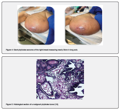
Examination of the right breast revealed a highly enlarged breast reaching to the umbilicus (30 cm long axis), completely indurated, with collateral venous circulation, ulcerated in places, and no homolateral adenopathies. The examination of the left breast was unremarkable. A thoracic CT scan revealed a mass occupying almost the entire right breast, well limited, with regular contours, extending beyond the field of exploration and measuring approximately 165 mm, with homolateral axillary adenopathies (Berg I, II and III) [6]. Presence of a few lefts, oval, infracentimetric axillary lymph nodes. Absence of metastatic lesions.


The patient was scheduled for a total right mastectomy without axillary curage.
The anatomopathological findings after surgery were as follows: the specimen weighed 6,500 g and measured 33x32x28 cm. The morphological and immunohistochemical appearance was in favour of a phyllodes sarcoma (tumour proliferation with a double contingent: an epithelial contingent and a contingent composed of mesenchymal cells of very high density), with infiltration of the surrounding skin and a non-tumourous nipple.
Discussion
Phyllodes sarcoma of the breast is rare. The clinical distinction between a phyllodes sarcoma and a fibroadenoma is difficult. In 30% of cases, the tumour is found in the upper external quadrant, varying in size from a few centimetres to several tens of centimetres in long axis. It presents as a firm, rapidly enlarging nodule, often with a doubling time of less than 3 months, polylobed, regular [7] and not attached to the skin or deep layers. Lymph node involvement is uncommon in sarcomas.
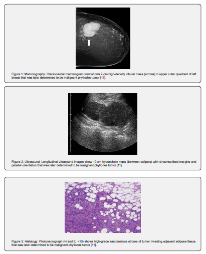
Mammography reveals significant hyperdensity compared with adjacent breast parenchyma [8], and may show a typically benign opacity without microcalcifications.
Ultrasound reveals images of low echogenicity with regular contours and no posterior shadow cone, which may be mistaken for benign tumors. These tumors are typically characterized by a large, oval mass with a long horizontal axis, parallel to the cutaneous plane. The notion of rapid growth is sometimes the only element suggestive of the diagnosis.
TAP CT should be performed for extension assessment, as these tumors have à high potential for local and/or metastatic relapse, particularly in the lung.
MRI is only performed when there is a strong suspicion of malignancy; in the case of aggressive lesions, enhancement occurs early, sometimes with a washout phenomenon. The existence of cystic remodelling is often correlated with a high degree of malignancy.
Phyllodes sarcoma is diagnosed histologically [9], by demonstration of a dual epithelial and conjunctival component on microbiopsy or macrobiopsy, or on a lumpectomy or mastectomy specimen.
Immunohistochemistry is currently an indispensable tool for confirming histological type : Ki67 and stromal P53 positivity were more often associated with high-grade GST; Ki67 expression can also help distinguish between benign and malignant phyllodes tumours in cases of difficult diagnosis.
The reference treatment is a simple mastectomy or a large lumpectomy without lymph node curage. The association of adjuvant treatment with radiotherapy or chemotherapy is still debated [10,11].
Conclusion
In conclusion, phyllodes sarcomas are malignant tumors of the breast that are difficult to diagnose. They are rare, representing less than 1% of malignant breast tumours, and pose a problem of differential diagnosis with adenofibromas, which are benign tumours. A phyllodes sarcoma should be considered in the presence of a rapidly enlarging mass of the quandrant-superolateral breast. Ultrasound, mammography and mammary MRI can help to orient the diagnosis, but only anatomopathological evidence can confirm it.
References
- Grenier J, Delbaldo C, Zelek L, Piedbois P (2010) Tumeurs phyllodes et sarcomes du sein : mise au point. Bulletin Du Cancer 97(10): 1197-
- Pollard SG, Marks PV, Temple LN, Thompson HH (1990) Breast sarcoma. A clinicopathologic review of 25 cases. Cancer 66(5):941-944.
- Zelek L, Llombart Cussac A, Terrier P, Pivot X, Guinebretiere JM, et al. (2003) Prognostic factors in primary breast sarcomas: a series of patients with long-term follow-up. J Clin Oncol 21(13): 2583-2588.
- McGowan TS, Cummings BJ, O’Sullivan B, Catton CN, Miller N, et al. (2000) An analysis of 78 breast sarcoma patients without distant metastases at presentation. Int J Radiat Oncol Biol Phys 46(2): 383-390.
- Blanchard KD, Reynolds CA, Grant CS, Donohue JH (2003) Primary non-phylloides breast sarcomas. The Am J Surg 186(4): 359-361.
- Stebbing JF, Nash AG (1995) Diagnosis and management of phyllodes tumor of the breast: experience of 33 cases at a specialist center. Ann R Coll Surg Engl 77(3): 181-184.
- Phyllodes tumors of the breast Eric Sebban (2016) Henri Hartmann Breast Institute (ISHH - Genesis N°189).
- Yilmaz E, Sal S, Lebe B (2002) Differentiation of phyllodes tumors versus fibroadenomas. Acta Radiol 43(1): 34-9.
- Taira N, Takabatake D, Aogi1 K, Shozo Ohsumi, Shigemitsu Takashima et al. (2007) Phyllodes tumor of the breast: stromal overgrowth and histological classification are useful prognosis-predictive factors for local recurrence in patients with a positive surgical margin. Jpn J Clin Oncol 37(10): 730-736.
- Parham Khosravi-Shahi (2011) Management of non-metastatic phyllodes tumors of the breast: Review of the literature. Surg Oncol 20(4): e143-e148.
- Megan Kalambo, Beatriz E Adrada, Modupe M Adeyefa, Savitri Krishnamurthy, Ken Hess, et al. (2018) Phyllodes Tumor of the Breast: Ultrasound-Pathology Correlation. AJR Am J Roentgenol 210(4): W173-W179.


