Vestibular Papillomatosis Confused to Genital Warts in A Pregnant Woman
Khadija Elboukhari*, Sara Elloudi, Ibtissam Assenhaji and Fatima Zahra Mernissi
Departement of Dermatology, University Hospital of Fez, Morocco
Submission: April 21, 2020; Published: June 09, 2020
*Corresponding author: Khadija Elboukhari, Departement of Dermatology, University Hospital of Fez, Morocco
How to cite this article: Khadija E, Asmae R, Sara E, Fatima Z M. Vestibular Papillomatosis Confused to Genital Warts in A Pregnant Woman. J Gynecol Women’s Health. 2020: 18(5): 556003. DOI: 10.19080/JGWH.2020.18.556003
Keywords: Vestibular papillomatosis; Genital warts; Dermoscopy; Pregnancy
Physiologic Papillomatosis
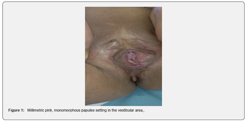
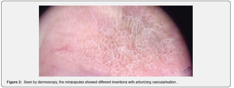
32 years old woman, pregnant in her second trimester, was followed for a bartolinitis and benefited from repetitive evacuations. This patient was referred to our department after discovering verrucous papules by her gynecologist. Considering these lesions as genital warts, the gynecologist was worried about the delivery route and referred the patient to our department for warts managements. The pregnant woman has never felt a modification in her genital organs. In dermatological examination of the vulva, we noted symmetric, filiform papules, matching the color of the adjacent mucosa, sitting between inner labia minora and vaginal orifice with distinct insertions (Figure 1). We putted our dermoscope to have better charecterisations, and we found multiple filiform projections with axial arborizing vascularisation and different bases of insertion (Figure2). This presentation is compatible with Vestibular papillomatosis. The patient and her gynecologist were reassured of the absence of genital warts and an explication of this entity was conducted.


Described for the first time by Altemeyer et al. [1], vestibular papillomatosis consists of a benign and rare anatomical variant of the vulva [2]. This condition has various appointments such as: pseudocondyloma, micropapillomatosis, hirsutoid papilloma of the vulva and squamous vulvar papillomatosis. The clinical presentation of vestibular papillomatosis is composed of several symmetrical and multiple minipapilles of 1 to 2mm in pink color (similar to the surrounding mucosa). They are symmetrical and have a soft touch and are generally limited to the area between the labia minora and the vestibular opening, inspection with acetic acid shows no bleaching, and the human papillomavirus is not implicated in this entity [3]. The dermoscope provides us with great help in supporting the diagnosis. In fact, it shows filiform papillae with separate bases [4], with irregular axial arborescent vessels [4,5]. These clinical and dermoscopic signs made the difference with genital condyloma. Vestibular papillomatosis is a normal anatomical condition of the vulva which can be considered as a female equivalent of the translucent papules of the penis. Correct diagnosis of this entity avoids unnecessary stress and laboratory tests [5].
Author’s Contributions
All the authors contributed to the conduct of this work. All authors also state that they have read and approved the final version of the manuscript
Acknowledgement
We are indebted to the patient for giving us the consent for the publication.
References
- Altmeyer P, Chilf GN, Holzmann H (1981) Pseudokondylome der Vulva. Geburtshilfe Frauenheilkd 41(11): 783-786.
- Verma S, Wollina U (2010) Vulvar vestibular papillomatosis. Indian J Dermatol Venereol Leprol 76(3): 270-272.
- Savini C, De Magnis A (2008) Multiple papillae on labia minora. CMAJ 179(8): 799-780.
- Kim SH, Seo SH, Ko HC, Kwon KS, Kim MB, et al. (2009) The use of dermatoscopy to differentiate vestibular papillae, a normal variant of the female external genitalia, from condyloma acuminata. J Am Acad Dermatol 60(2): 353-355.
- Chan CC, Chiu HC (2008) Vestibular Papillomatosis. New England Journal of Medicine 358(14): 1495.


