Bladder Stone : A Rare Cause of Dystocia
Dieng C, Biaye B*, Diouf AA, Niass A, Diallo M, Ndour MD, Mbodj A, Lo A, Diouf A and Moreau JC
Department of Gynecologist, Aristide Le Dantec Hospital, Senegal
Submission: October 09, 2019;Published: October 18, 2019
*Corresponding author: Biaye B, Department of Gynecologist, Aristide Le Dantec Hospital, Senegal
Dieng C, Biaye B, Diouf AA, Niass A, Diallo M, Ndour MD, Mbodj A, Lo A, Diouf A, Moreau JC. Bladder Stone : A Rare Cause of Dystocia. 2019: 17(1): 555952. DOI: 10.19080/JGWH.2019.17.555952
Summary
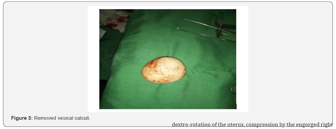
Mechanical dystocia by bladder calculus is a rare complication. We report the case of a lack of commitment by a bulky bladder calculation. The diagnosis was made in peresarean section by the palpation of a firm and mobile intra-vesical mass evoking a calculation. The cystostomy revealed an ovoid, pearly white calculus of 7cm long axis 6.5cm, 8cm in circumference, weighing 200g. The postoperative course was simple.
Keywords: Bladder calculus; Mechanical dystocia; Cystostomy
Introduction
The urinary lithiasis obeys in the pregnant woman a physiopathology different from that of the non-pregnant woman: anatomical modifications of the urinary tree, biological blood and urine (increase of lithogenic but also lithoprotective factors) leading to a new equilibrium, different from that existing outside pregnancy [1]. Mechanical dystocia by bladder calculus is a rare complication. Very few cases have been described in the literature. Diagnosis and early management of bladder stones in pregnant women avoids the complications that are the most formidable in terms of mechanical dystocia and vesico-vaginal fistula [1,2]. We report the case of a defect of engament by bladder calculation whose diagnosis was posed intraoperatively.
Observation
This is a second gesture second parish of 18 years old with a 14-month-old living child born vaginally with no personal or family history of lithiasis. Pregnancy is poorly followed with only two prenatal examination without ultrasound, moreover episodes of urinary disorders such as dysuria and pollakiuria have been noted. The patient was evacuated in a context of lack of commitment with complete dilatation, an apex presentation in occipito-iliac left anterior fixed with a hump. We also note the perception of a hard mass, smooth and regular, immobile, about 6cm, forming part of the pubic symphysis which perfectly matches the posterior face preventing the placement of the bladder catheter.
The Caesarean section was able to extract a new born female weighing 3300g with an APGAR score of 8/10. The perception of a hard bladder had motivated an intraoperative cystotomy high lighting an ovoid, pearly white 7cm of major axis on 6.5cm, 8cm in circumference, weighing 200g (Figure 1-3). The bladder was repaired in two planes with placement of a bladder catheter for 10 days. The postoperative course was simple


Discussion
The incidence of urinary calculi in developed countries has declined considerably since the 19th century due to improved nutrition and infection control. The incidence of urolithiasis during pregnancy ranges from 1/200 to 1/2500. Indeed, bladder stones account for 5% of urinary stones [1].
During pregnancy, there are physiological factors that may facilitate the formation of urinary stones. The dilation of the urinary tract, often more frequent on the right than on the left, is observed physiologically in the first trimester of pregnancy. This dilatation, found in 90% of pregnant women, appears in the first trimester (sixth to tenth week), will promote urinary stasis and aggregation of urinary crystals. She will still be present six to ten weeks after delivery. The dilation of the urinary tract results from the combined action of progesterone during early pregnancy and especially from ureteral compression by the pregnant uterus, especially during the last trimester [1,3]. This physiological dilation is peculiar since it stops at the promontory, which makes it possible to differentiate it from an expansion linked to an obstacle of another origin, such as a calculation. The right ureter will be more often concerned than the left due to dextro-rotation of the uterus, compression by the engorged right genital vein and protection provided to the left ureter by the mesosigmoid [4].
Physiological changes are observed during gestation, likely to promote the formation of stones in the urinary tract. These include increased plasma renal flux, glomerular filtration rate (30-50%), clearance of creatine, uric acid, urea, and filtration of sodium and calcium [1]. These factors add up to hypercalciuria present in pregnancy by decreasing parathyroid hormone production and elevating 1,25-dihydrocholecalciferol formed in the placenta resulting in increased gastrointestinal absorption of calcium and bone calcium mobilization. Indeed, the pregnant woman undergoes an important bone remodeling which favors this hypercalciuria [4]. All this maternal calcium mobilization is of course intended for the skeleton of the fetus.
At the same time, modifications will interest the inhibitors of the lithiasic formation. Thus, there will be an increase in urinary excretion of citrate, magnesium or glycoproteins [2,4]. In total, the simultaneous action of these antagonistic factors in the formation of urinary stones leads to a zero balance since the incidence of calculi in the pregnant woman is not higher than in the rest of the population. Clinically, bladder calculus may appear asymptomatic. However, these calculi can be manifested by macroor microscopic hematuria, hypogastric pain, urinary dysuria and repeated urinary tract infection [1]. In our patient we noted that repetitive urinary disorders.
Ultrasound and cystoscopy are the best diagnostic methods for bladder stones in pregnant women because of their non-radiating effects [1,5]. In case of diagnostic uncertainty after ultrasound, nuclear magnetic resonance imaging (MRI) is a choice remedy because of the absence of irradiation or contrast medium and its safe nature not exerting effects. teratogenic. A sensitivity of 100% has been shown in some studies [6]. In our case, the patient did not even receive obstetric ultrasound.
Frequent complications of bladder calculus during pregnancy include infection, abortion, premature labor, and urinary fistula [1]. Very rare cases of uterine rupture have also been reported [2]. The diagnosis of dystocia by bladder stones is usually easy when the calculation is large and palpable by vaginal examination, as in our case [3]. The differential diagnosis of pelvic bone tumors, vaginal neoplasms or bladder and fibroids should be kept in mind. In certain doubtful situations, ultrasound and radiography of the urinary tract may be useful, but they are always limited by the interposition of the fetal presentation.
The management of bladder computation during pregnancy should take into account the mother and the fetus knowing that 70 to 80% of patients can expel spontaneously their calculation [1,6]. Conservative treatments will therefore be considered as a priority and the therapeutic limitations imposed by pregnancy on analgesics or antibiotics should be well understood [7,8]. In the pregnant woman, the hot bath would be effective for a temporary relief of the pains.
If the calculus is not bulky and asymptomatic during pregnancy, monitoring may be recommended. Otherwise a cystolithotomy is recommended [1]. In patients presenting with mechanical dysfunction by bladder calculus, a caesarean section is recommended with a cystotomy at the same time [4]. This therapeutic option was made in our patient with setting up a urinary catheter for 10 days. The postoperative course was simple.
Conclusion
Bladder stones are a rare cause of obstructed labor. The diagnosis is usually based on the patient’s history, clinical examination and routine prenatal ultrasound. The delivery route can be planned according to the size of the calculation. Complications can be avoided by rapid diagnosis and appropriate treatment. When caesarean section is indicated, intraoperative cystotomy with removal of the computation is mandatory, even though it seems to increase the risk of urinary fistula.
References
- Saussine C, Lechevallier E, Traxer O (2008) Lithiase et grossesse. Prog Urol 18(12): 1000-1004.
- Abubakar BM, Abdulkadir A, Atterwahmie AA, Panti AA, Maina AI, et al. (2019) Prolonged Obstructed Labor Is an Uncommon Presentation of a Giant Bladder Calculus: A Case Report and Literature Review. Open Journal of Urology 9(4): 77-83.
- Sonak M, Ramteke S, Davile M (2017) Bladder Stone: A Rare Cause of Obstructed Labour. International Surgery Journal 4(7): 2352-2354.
- Escobar-del Barco L, Rodriguez-Colorado S, Duen˜as-Garcia OF, Avilez-Cevasco JC (2008) Giant intravesical calculus during pregnancy. Int Urogynecol J Pelvic Floor Dysfunct 19(10): 1449-1451.
- Benkaddour AY, Aboulfalah A, Abbassi H (2006) Bladder stone: uncommon cause of mechanical dystocia. Arch Gynecol Obstet 274(5): 323-324.
- Dave A, Mathur P, Mathuriya G (2012) Vesical Calculus: An Uncommon Cause of Obstructed Labor. J Obstet Gynaecol India 62(6): 687-688.
- Seth S, Malik S, Salhan S (2002) Vesical Calculus Causing Dystocia. Eur J Obstet Gynecol Reprod Biol 101(2): 199-200.
- Penning SR, Cohen B, Tewari D, Curran M, Weber P, et al. (1997) Pregnancy complicated by vesical calculus and vesicocutaneous fistula. Am J Obstet Gynecol 176(3): 728-729.


