Unexplained Unilateral Absence of Fallopian Tube and Ovary with Trotted Dermoid Cyst in the Remaining Ovary
Sali Z Talab and Haitham A Badr*
Security Forces Hospital, KSA
Submission: February 22, 2018 ; Published: March 07, 2018
*Corresponding author: Haitham A Badr, Security Forces Hospital, KSA, Email: sali.zt@hotmail.com
How to cite this article: Sali Z T, Haitham A B. Unexplained Unilateral Absence of Fallopian Tube and Ovary with Trotted Dermoid Cyst in the Remaining Ovary. J Gynecol Women�s Health 2018; 8(5): 555746. DOI:10.19080/JGWH.2018.08.555746
Abstract
Unilateral absence of a part or all of a fallopian tube with or without adjacent ovarian agenesis is considered a very rare clinical situation.Its etiology remains unclear. In this report, a 14 years old single female presented to Security Forces hospital (SFH) Emergency Room (ER) with acute pain in the right lower abdominal quadrant associated with nausea. Abdominal examination revealed moderate tenderness in the right iliac fossa. Investigations done were complete blood count (CBC), pelvic ultrasound (US), and Computerized tomography (CT) abdomen and pelvis. Both ovaries were not seen in pelvic ultrasound (US), however, right adnexal mass was seen with cystic structure which could be dermoid cyst.CT abdomen and pelvis showed well defined mixed lesion occupying mainly the midline and to the right paramedian aspect of the pelvis. She underwent lapatomy, which revealed adnexial torsion involving the right ovary with the adjacent tube and agenesis of the left ovary and tube. The cyst was opened and the most gangrenous area with the dermoid contents evacuated. Histopathology results revealed that the right ovary showed hemorrhagic dermoid cyst, consistent with torsion. As she was single, neither hysterosalpingogram nor hysteroscopy was performed for assessment of uterine cavity.
Keywords: Ovarian agenesis; Dermoid cyst; Torsion; laparotomy
Abbreviations: SFH: Security Forces Hospital; ER: Emergency Room; CBC: Complete Blood Count; CT: Computerized Tomography
Introduction
Unilateral absence of a part or all of a fallopian tube with or without adjacent ovarian agenesis is considered a very rare clinical situation. Its exact incidence is unknown through literature review. However, it has been reported that it encountered in a rate of one in 11, 240 females [1].
The etiology of fallopian tube and ovary agenesis remains unclear. However, two etiologic causes are possible. Asymptomatic segmental torsion of the uterine tube and/or ovarian pedicle may occur for uncertain reasons during adulthood, in childhood, or even during the fetal stages. Consequently, torsion may give rise to necrosis and autoamputation [2-4]. Alternatively, the absence of these organs may be congenital, associated with developmental alterations of the mesonephric and paramesonephric ducts [5,6].
The majority of patients is asymptomatic and can be diagnosed incidentally following a laparoscopy or laparotomy for various gynecological or obstetric complications [7].
In the present study, the literature was reviewed in order to identify possible causes of these anomalies.
Case Report
A 14 years old single female presented to Security Forces hospital (SFH) Emergency Room (ER) with acute pain in the right lower abdominal quadrant for three days which stared suddenly and not relived by analgesia. The pain was associated with nausea. The patient had no vomiting, change in the bowel habits or signs or symptoms of urinary tract infection (UTI).
She had no known medical history and no history of abdomino-pelvic surgery. Her menarche had occurred at the age of thirteen, with regular menstrual periods without dysmenorrhea.
On Examination, vitally; temperature was 38.2 °C, pulse was 101/minutes, blood pressure was 118/64 mmHg and O2 Saturation was 99%.
Abdominal examination revealed moderate tenderness in the right iliac fossa, no rebound tenderness or masses.
Investigations done were complete blood count (CBC), pelvic ultrasound (US), and Computerized tomography (CT) abdomen and pelvis. CBC showed white blood cells of 22.91 (10X9/L), heamoglobin of 132(g/l), Neutrophils of 70.5% and C-reactive protein of 102.19 (mg/l).
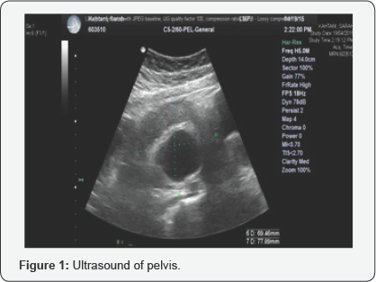
Pelvic US showed that the uterus was anteverted normal in size measures 62.56*22.85*31.92mm. Midline endometrial thickness was 5.49mm. Both ovaries were not seen. There was no free fluid within the pouch of douglas (P.O.D). Right adnexal mass was seen measured 70.42*70.48*78.64 mm, with cystic structure seen within it measures of 39.81*47.51mm, which could be dermoid cyst (Figure 1).
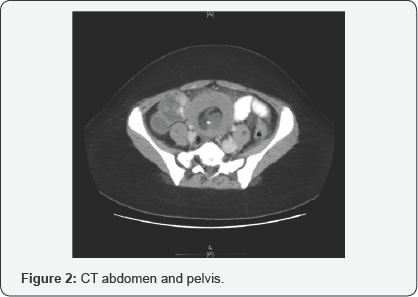
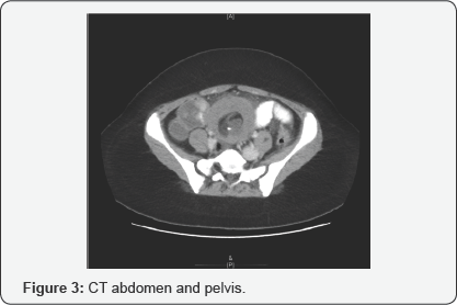
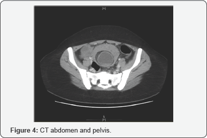
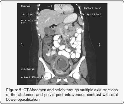
CT abdomen and pelvis through multiple axial sections of the abdomen and pelvis post intravenous contrast with oral bowel opacification showed well defined mixed lesion occupying mainly the midline and to the right paramedian aspect of the pelvis posterocranial location in relation to fundus of the uterus. The lesion showed areas of cystic changes, fat and calcification with perifocal soft tissue density. The wall dimension measures 83.8x87x99.5mm. It was associated with mild tilted uterine shadow towards the right side, associated with moderate free pelvic fluid. Picture likely represent right ovarian dermoid cyst with possible complication and quiry torsion (Figure 2-4). Apparent distended ascending colon and cecum with some contrast Gastrografin reaches the lumen was noted. Tiny mesenteric lymph nodes were noted. No significant free ascites in the upper abdomen was seen. The liver, spleen, gall bladder, pancreas, adrenal glands and both kidneys were unremarkable. No gross para-aortic lymphadenopathy seen (Figure 5).
She underwent lapatomy, which revealed adnexial torsion involving the right ovary with the adjacent tube and agenesis of the left ovary and tube. The right twisted ovary looked black gangrenous. The involved gangrenous material was the right ovary with a cyst measuring 8cm in size. The right tube was going downward was also involving the right infindbuloplevic ligaments but not the left. The left ovary and tube were not identified. The uterus was normal in shape and size. The left and right round ligaments seen. The left gonadal vessels also not identified.
The finding were discussed with 3 gynecologist consultants intraoprativly and due to the findings and history, the decision was made to conserve ovarian tissue as much as possible due to the hormonal needs as she is only 14 years old with single ovary that is torsed and twisted. The decision was to evacuate the content of the ovarian cyst as the color was improving after spending more than 20 minutes following untwisting the right ovary. The cyst was opened and the most gangrenous area with the dermoid contents evacuated. All the gangrenous dead tissue removed with conservation of the right ovary as much as possible.
Histopathology results revealed that the right ovary showed hemorrhagic dermoid cyst, consistent with torsion. Right fallopian tube showed hemorrhagic fallopian tube tissue, consistent with torsion with no evidence of malignancy.
Post-operatively, Follicular stimulating hormone level (FSH) was 68, 7(IU/L), Leutinizing hormone level (LH) was 39,18(IU/L) and Oestradil level was 94.74(PMOL/L). No periods for 6 months. Investigations after 6 months showed FSH of 5.3 (IU/L), LH of 4.61 (IU/L) and Oestradil of 307.3(PMOL/L) with regular cycles.
Then she again underwent pelvic ultrasound as normal volume and follicle count were observed for the right ovary.
As she was single, neither hysterosalpingogram nor hysteroscopy was performed for assessment of uterine cavity.
Discussion
Although it has been reported that congenital agenesis of fallopian tubes and ovaries is a very care clinical presentation, the rate may be higher than reported as it is not easy to estimate the actual number of cases since the condition is mostly asymptomatic and goes unreported. All cases reported in the literature were diagnosed incidentally following a laparoscopy or laparotomy for various obstetric or gynecological complications as in the present case. In last ten years, several cases have been reported in the literature [3,6-12]. This could be attributed to the wide utilization of laparoscopy for diagnostic purposes; however, these case are still considered very rare.
The patient in the present case had single ovary that is torsed and twisted. Therefore, the decision was to conserve ovarian tissue as much as possible due to the hormonal needs. The dangerousness of this case relied on the potential risk of subfertility. This is especially true if the right tube and or ovary were to be affected by either salphingitis or ectopic pregnancy, necessitating its removal [13].
Torsion of the ovarian pedicle or mesosalpinx can lead to avascular necrosis, separation of those tissues, and resorption.13Such torsion is most likely to present with acute abdominal pain, nausea, and vomiting if it occurs in childhood or adulthood. Jamieson & Soboleski [14] and Goktolga et al. [15] reported adnexal torsion in prepuberty. In accordance with that, the present case presented at ER with acute pain in the right lower quadrant and not relived by analgesia. The pain was associated with nausea.
The mechanism of adnexal torsion is unknown exactly. However, these torsions can occur repetitively, because of the anatomic and physiologic anomalies, which suggests that the torsions concern both adnexa. Varras et al. [16] reported that the frequency of recurrent attacks of pain interspersed with asymptomatic intervals ranged from 10% to 50% of cases.
The-torsion hypothesis that could explain the unilateral absence of fallopian tube and ovary could be supported by what has been documented by Dueck et who found separated structures in the abdominal cavity at surgery that were later histologically confirmed to be ovarian and tubal tissue.
Several authors have hypothesized that unilateral adnexal absence does not diminish female fertility, particularly when the condition is not accompanied by a uterine malformation.10 On the other hand; Uckuyu et al. [6] concluded that unilateral adenexal agenesis is a possible factor for infertility. Gursoy et al. [9] reported a patient with this condition who had four normal pregnancies that resulted in normal vaginal deliveries.
In the present case report, the uterus was considered normal in shape and structure during diagnostic US and CT. The literature was reviewed and a number of similar cases without uterine anomalies were identified [6-12,17].
Histopathological findings of the present case revealed that the right ovary showed hemorrhagic dermoid cyst, consistent with torsion. It has been documented that unilateral absence of the adnexa may reduce the probability of becoming pregnant, especially if the etiology is likely due to an ovarian cyst and torsion [18] as in our case.
Surgical detorsion applied for the present case often allows for the preservation of the affected ovary and keeping the hormonal needs.
In conclusion, unilateral ovarian and fallopian tube agenesis is a rare condition. Its true etiology remain unclear, although torsion or congenital defects may be the most likely explanations. In addition, Surgical detorsion of ovarian cyst is essential to maintain hormonal needs and the chance of pregnancy among patients.
References
- Sivanesaratnam V (1986) Unexplained unilateral absence of ovary and fallopian tube. Eur J Obstet Gynecol Reprod Med 22: 103-105.
- Eustace DL (1992) Congenital absence of fallopian tube and ovary. Eur J Obstet Gynecol Reprod Biol 46(2-3): 157-159.
- Vaiarelli A, Luk J, Patrizio P (2012) Ectopic pregnancy after IVF in a patient with unilateral agenesis of the fallopian tube and ovary and with endometriosis: search of the literature for these associations. J Assist Reprod Genet 29(9): 901-904.
- Mylonas I, Hansch S, Markmann S, Bolz M, Friese K (2003) Unilateral ovarian agenesis: report of three cases and review of literature. Arch Gynecol Obstet 268(1): 57-60.
- Sirisena LA (1978) Unexplained absence of an ovary and uterine tube. Postgrad Med J 54(632): 423-424.
- Uckuyu A, Ozcimen EE, Sevinc Ciftci FC (2009) Unilateral congenital ovarian and partial tubal absence: Report of four cases with review of the literature. Fertil Steril 91(3): 936.e5-8.
- Chen B, Yang C, Sahebally Z, Jin H (2014) Unilateral ovarian and fallopian tube agenesis in an infertile patient with a normal uterus. Experimental and Therapeutic Medicine 8(3): 831-835.
- Rapisarda G, Pappalardo EM, Arancio A, La Greca M (2009) Unilateral ovarian and fallopian tube agenesis. Arch Gynecol Obstet 280(5): 849850.
- Gursoy AY, Akdemir N, Hamurcu U, Gozukucuk M (2013) Incidental diagnosis of unilateral renal and adnexal agenesis in a 46-year-old multiparous woman. Am J Case Rep 14: 238-240.
- Muppala H, Sengupta S, Martin JE (2008) Unilateral absence of tube and ovary with renal agenesis and associated pyloric stenosis: communication. Eur J Obstet Gynecol Reprod Biol 137(1): 123.
- Elkington N, Rahman R (2008) Unexplained Unilateral Absence Of Fallopian Tube And Ovary. The Internet Journal of Gynecology and Obstetrics 11(2): 1-3.
- Barsky M, Beaulieu AM, Sites CK (2015) Congenital Ovarian-Fallopian Tube Agenesis Predisposes to Premature Surgical Menopause: A Report of Two Cases. J Androl Gynaecol 3(1): 3.
- Eustace DL (1992) Congenital absence of fallopian tube and ovary. Eur J Obstet gynecol Reprod Biol 46(2-3): 157-159.
- Jamieson MA, Soboleski D (2000) Isolated tubal torsion at menarche-a case report. J Pediat Adoles Gynecol 13(2): 93-94.
- Goktolga U, Cethan T, Ozturk H, Gungor S, Zeybek N,et al. Isolated torsion of fallopian tube in a premenarcheal 12-year-old girl. J Obstet Gynecol Res 33(2): 215-217.
- Varras M, Akrivis C, Demou A, Antoniou N (2005) Asynchronous bilateral adnexal torsion in a 13-year-old adolescent: our experience of a rare case with review of the literature. J Adolesc Health 37(3): 244247.
- Dueck A, Poenaru D, Jamieson MA, Kamal IK (2001) Unilateral ovarian agenesis and fallopian tube maldescent. Pediatr Surg Int 17(2-3): 228229.
- Barsky M, Beaulieu AM, Sites CK (2015) Congenital Ovarian-Fallopian Tube Agenesis Predisposes to Premature Surgical Menopause: A Report of Two Cases. J Androl Gynaecol 3(1): 3.


