Cervical Pregnancy: Presentation of Cases and Therapeutic Update
Illia R1*, Lobenstein G2, Mayer H3, De Anchorena M3, Urangalmaz M2, Manrique G2, Fiameni F1 and Guallan F4
1Department of Gynecology and Obstetrics, Hospital Alemán, Buenos Aires, Buenos Aires University, Argentina
2Department of Obstetrics, Buenos Aires University, Hospital Alemán, Argentina
3Department of Obstetrics, Medical staff Obstetrics service, Argentina
4Department of Gynecology, Medical staff Gynecology service, Argentina
5Chief of Residents Obstetrics & Gynecology, Argentina
Submission: July 23, 2017; Published: July 31, 2017
*Corresponding author: Illia R, Department of Obstetrics, Buenos Aires University, Hospital Alemán, Argentina, Email: rhillia@gmail.com
How to cite this article: Illia R, Lobenstein G, Mayer H, De Anchorena M, UrangaImaz M, Manrique G, Fiameni F, Guallan F. Cervical Pregnancy: Presentation of Cases and Therapeutic Update. J Gynecol Women's Health 2017; 6(3): 555687. DOI: 10.19080/JGWH.2017.06.555687.
Abstract
Among severe obstetric hemorrhages, cervical pregnancy is very infrequent entity of unknown etiology, but it is associated with significant maternal mortality. Now days the implementation of an early trans vaginal ultra-sound scan enables to try to non surgical approaches. Four cases of cervical pregnancy, which ones have been treated in our Department with different therapeutic regimens, are presented in this report. A review of the world-wide casuistic is also carried out, finding support to the present trend toward non surgical treatments.
Keywords: Ectopic pregnancy; Cervical pregnancy; Cervical bleeding; Maternal mortality
Introduction
Obstetric hemorrhage is a major cause of morbidity and maternal death [1]. Among the major obstetric hemorrhages, cervical pregnancy (CP) is a rare entity, but associated with considerable maternal morbidity and mortality if an early diagnosis and proper treatment is not done. The incidence of the CP is variable according to the authors, probably due to diagnostic variations. Giambanco et al. [2] refer the incidence ranges from 1/1000 to 1/95,000 pregnancies. Acosta et al. [3], conducted a review and reported an incidence of a CP every 2,400 deliveries, this represents less than 1% of all ectopic pregnancies. Ushakov et al. [4] quoted an incidence of a CP every 8628 births. The etiology is unknown, but there is evidence of an association with instrumental cervical maneuvers. It should also be present a likely relationship with chromosomal embryonic abnormalities and the late acquired capacity of the embryo to implantation [4]. The cervical implantation could be developed in three different ways: the gestational sac growth towards the external cervical OS, the gestational sac could theoretically reach the uterine cavity with normal development of pregnancy even with placental implantation on the inner cervical orifice, and finally the gestational sac develops fully in the cervical channel leading towards an obstetrical catastrophe [2]. The most common query is the presence of vaginal bleeding associated with menstrual delay, with or without hormonal pregnant status confirmation. The early implementation of the trans vaginal ultrasound, allows starting conservative strategies and avoiding adverse results [3]. The CP, is defined as the implantation of the blastocyst into the cervical channel [5] and the sonographic image of the gestational sac located in the cervix is required to confirm the diagnosis [4]. Around 60% of the CP contain a live fetus [4] and the majority of the patients have low rate of parity, so that the current trend is to implement conservative treatments that preserve fertility [4]. Until not long ago, the CP was treated by a total hysterectomy due to the severity of the bleeding. But the development of the trans vaginal ultrasound, has allowed an early diagnosis and chemotherapy with methotrexate, opening up the possibility of new therapeutic options [6].
Case 1
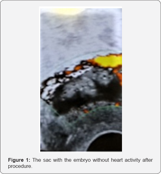
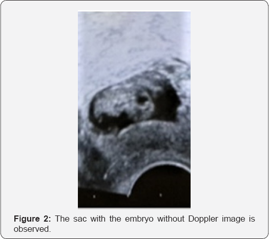
A 29-year-old patient, first pregnancy, admitted by bleeding in the first trimester with a menstrual delay of 5 weeks and 5 days. Her family doctor had indicated rest and micronized progesterone. At the vaginal examination with speculum we observed a macroscopically healthy cervix, external cervical OS closed. At the vaginal examination, these data were confirmed. The patient was obese (weight >100kg), hypothyroid medicated with T4, smoker of a daily package and with antecedent of sterility of 6 years of evolution. A trans abdominal ultrasound, which informed 85x45x46mm uterine size and endometrium of 12mm thick, adnexa without peculiarities and we received a positive beta subunit of2990mUl/ml. These data indicated a trans vaginal ultrasound scan, showing a gestational sac located in the cervical thickness with an embryo measuring 3mm, no heartbeat observable. We decide to start treatment with systemic methotrexate at a dose of 50mg/m2, which in this case reached a dose of 100mg. Four days after the administration of methotrexate, the value of beta subunit was 13.000mUl/ml. After consultation with Oncology Service, it was established therapeutic regimen of serial methotrexate, alternated with leucovorin. It was given a new dose of 100mg of methotrexate (MTX) and the next day was administered 10mg of intramuscular leucovorin. The third day was again administered MTX and the fourth day, receiving leucovorin rescue, subunit beta value was 8.600mUI/ml. On the fifth day of treatment, the value of beta subunit was 5,500, with slight increase in hepatic transaminases. After the series of MTX and leucovorin, the value of beta subunit was 4.300mUl/ml, the patient was asymptomatic and because she lived close to the Hospital, we decided to send her home in a schedule of home care. Ambulatory monitoring showed values of gonadotrophins in descent. After 15 days of discharge, a trans vaginal ultrasound informed an image in cervix of 5mm in diameter. The beta subunit continues decreasing until their final negativization60 days after discharge with trans vaginal ultrasound report of absence of cervical image (Figures 1& 2).


Case 2
A 40 years old, G4 P3, consulting to the ED of the Gynaecology at the Hospital, by menstrual backwardness and loss of scarce blood by genitals without any abdominal pain. In the vaginal exam with speculum a swollen and little open cervix was observed with clots and blood in the external cervical OS. At bimanual examination, anterior flexed uterus, increased size, irregular and firm in the background is found, while the cervix was found very increased in size and softened, with the external cervical OS permeable to the finger. Trans abdominal and transvaginal ultrasound were performed: uterus of increased size because a fundal fibroid. At cervical level, is displayed a 5mm embryo into gestational sac with positive cardiac activity. Endometrial cavity with liquid collection and thickened endometrium. There was neither adnexal abnormalities nor in Douglas cavity. Beta HCG 300.000mUl/ml. hematocrit 36%, TA 120/70mmHg, maternal heart frequency 72'. Three hours after ultrasound study, the patient started with major bleeding from genitals, with signs of de compensation, BP 70/40mmHg, heart frequency 120', pale and sweaty facies. It was decided to carry out exploration under anesthesia and there was performed an endocervical manual scraping and endometrial with Sims curette into the uterine cavity. After the procedure, the hematocrit was 17.9%, so we decided to administrate two units of whole blood, with post transfusion hematocrit of 26%. The patient continued stable, being discharged 48 hours after procedure with a HCG beta level of 33.000mUl/ml. The beta HCG, was normalized by 58 postoperative days.
Case 3
Patient of 30-years-old consulting after a metrorrhagia compatible with menstruation, but with the particularity of being more intense than usually. The gynecological exam found a globulus uterus, of increased size and normal adnexa. The next day, the beta HCG dosage was 100.000mUI/ml. The ultrasound study reports 99x47x67mm uterus and a gestational sac of 40x22x27mm that the sonographer defines as low implanted, displaying in its upper part, a 34x20x38mm probably blood collection. Not was observed embryo within the gestational sac. At 24 hours, beta HCG value was 149. 000mUI/ml. With the presumptive diagnosis of cervical pregnancy, it was decided to start treatment with MTX initial dose of 50mg administered intramuscularly. Five days after the administration of MTX, beta HCG value was 120. 000mUI/ml, with ultrasound report of a sac at isthmic level of 45x38x18mm with hematoma over the sac of 54x29x37mm. The patient was asymptomatic. Five days later, beta HCG value was 16.000mUI/ml and the ultrasound report was an isthmiccervical sac of 54x28x50mm, persisting the hypoechogenic image of 37x21x20mm. Seven days later, beta HCG level was 2,300 and the sac measures 48x30x46mm with hypoechogenic image persistence. A week later, beta HCG value was 740mUI/ml and continued falling until be negative four months after giving positive for the first time. The patient presented scant bleeding, the uterus persists increased in size with a special softening in the isthmic area. According to the ultrasound report of persistence of a sac of approximately 40x40mm, it was decided to perform an evacuator uterine curettage under ultrasound control. After the procedure, the patient presented profuse bleeding, with general status compromised. As an attempt before to perform a more radical treatment, it was placed an intra cervical Foley device and the balloon inflated, observing the cessation of bleeding. Also was decided the transfusion of two units of red blood cells. The balloon was gradually deflating until 48 hours of settled, finally it was with drew and there was no reiteration of the bleeding. The beta HCG remained negative and the patient asymptomatic. Pathology of the material sent report was ovular involutional tissue.
Case 4
Nuliparous patient of 26 year old. She presented to the ED with menstrual backwardness of 6.3 weeks and little vaginal bleeding. To the vaginal check it was proven metrorrhagia. The value of initial beta subunit was 15.300U/ml. The ultrasound reveals the presence of a gestational sac of 50x50mm within the uterine cervical canal with vital embryo inside. The patient said she refuse any invasive therapeutic option, so, despite not being indicated by the size of the gestational sac, the beta-level and size of the embryo, we began treatment with methotrexate (MTX) intramuscular 50mg/day. At 72 hours of the first dose of MTX, neither sacnor embryo conditions were modified, nor substantially modified the value of the beta, so it was indicated a second dose of MTX. At 72 hours the patient was again assessed (a week from the beginning of treatment) without modification, so we explained her clearly that we had to perform an invasive treatment and the least invasive treatment we could offer her was the de vitalization of the embryo with a needle trough cervix and administration into the sac of potassium chloride under neurol epto an algesia. Finally the patient accepts the proposal and the procedure were carried out with success, noting the devitalization of the embryo during the entry of potassium within the gestational sac. Another additional dose of MTX was administered and thereafter, both the size of the gestational sac and beta values began to diminish gradually until the betanegativization and the vanishing of the ultrasound image. This process took approximately 3 weeks, after which the patient was discharged without any sequel post treatment.
Discussion
The CP has traditionally been treated by total hysterectomy, but recent reports about the use of methotrexate and other methodologies, have raised the possibility of a less aggressive and effective treatment [7]. Treatment with MTX, may be systemic, local or combined. The combined, can be based on local and systemic MTX administration or systemic administration of MTX associated with other local alternatives such as the injection of potassium chloride in the amniotic sac or selective embolization.
One of the problems in the evaluation of treatment results, is that the low incidence of CP, generates the publication of isolated cases, as a consequence, to analyze the efficacy of treatments, we only have revisions with a few cases. Eggebo and Elve [8] report of therapeutic success with systemic administration of MTX, but is about a single case. Jurkovic et al. [9], reported two therapeutic successes with systemic administration of MTX. In his review, a total of 83 cases of CP were found. Of these cases, 40 were treated primarily with conservative surgery, 40 with medical treatment with MTX or potassium chloride and the remaining three were treated with other chemotherapeutic agents. The possibility of obtaining healing was similar in both groups (OR 1.1 CI 0.43.2). However, patients initially treated with surgery, had one increased risk of hemorrhage and requirement of hysterectomy (OR 8 CI 2.4-26.5) that those treated medically. Conclude the authors that the initial treatment should be medical, reserving surgical treatments for chemotherapy treatment failures.
Other authors share this therapeutic modality [6-11]. However, there are publications which communicate the failure of treatment with MTX. Pretzsch et al. [12] referred a case in which first they administered MTX into the gestational sac. As HCG values remained high, they decided to give systemic MTX. The HCG values decreased, but in spite of this, five weeks later, the patient presented a hemorrhage that forced them to perform a hysterectomy. A variant of treatment with MTX, is their injection in the gestational sac. Nomiyama and cols [13] reported total regression of cervical gestational sac 44 days after admission and after three MTX injections into the gestational sac. Ibghi et al. [14] reported a case solved with a single injection of 50mg of MTX in the gestational sac. Acosta (3), proposes that the therapeutic option of choice is the combined treatment with MTX. Kaminopetros et al. [15] reported two cases treated with MTX local and systemic. One eliminated the sac spontaneously and the other required a hysterectomy. Hung et al. [16] reported 11 cases treated with systemic administration of MTX with rescue of leucovorin or injection of MTX into the sac. The sac disappear satisfactorily in all cases and none required a hysterectomy They conclude that if a cervical pregnancy is diagnosed early, treatment with MTX is effective and definitive. Qasim et al. [17], also report good results associated with combination therapy of MTX, but clarify that early diagnosis may be important for therapeutic success. Hajenius and cols [18] stated that the curve of beta HCG is not helpful in the detection of problems associated with therapeutic failures of MTX. By not allow the curve of beta HCG detection of the right lower of gonadotrophins values, additional doses and not single dose of MTX should be considered in the conservative therapeutic modality of the EC. Hung et al. [19] reviewed 28 publications of 48 cases of CP.
An CP with a beta HCG value >10.000UI (OR 10.82 IC 2.5945.14), gestational age equal to or greater than 9 weeks (OR 6.44 Cl 1.46-28.52), positive cardiac activity (OR 14.29 IC 2.95-76.92) and an embryo length larger than 10mm (OR 13.33 Cl 1.46120.48) were considered to be associated with a high incidence of failure after treatment with MTX. A technique that allows the simultaneous feticide increased the effect of the treatment with MTX if embryonic heart activity was positive (OR 0.13 Cl 0.02-0.68). ln addition, the administration of high doses of MTX, was not more effective than conventional single-dose. So, it is impressed that the published failures, seem to be most associated with a poor choice of the case for conservative treatment than failures of the treatment itself. Kung et al. [20] conducted a retrospective study to examine the effectiveness of treatment with MTX and the existence of differences between patients with positive and negative embryo vitality. 62 cases were analyzed. Among the 35 cases with live embryo, 63% of patients received systemic injection of MTX or a combination of local and systemic MTX or MTX systemic associated with chloride of potassium within the gestational sac and 37% received local injection of MTX. Among the 23 cases of CP with the absence of heartbeat, 96% of patients received systemic injection of MTX. The need to implement a simultaneous surgical treatment (for example scraping, cervical tamponade or vascular embolization) was higher in cases with positive cardiac activity (43%) than in the cases without it (13%-P=0.021). Other authors have reported the administration of systemic MTX associated with the injection of potassium chloride in the gestational sac with good results [21,22] and even reported the treatment only with chloride of potassium into the sac in a case of heterotopic pregnancy [23].
Kung et al. [24] reviewed 22 patients that received treatment with MTX and analyzed the further evolution of these patients. Of the 22 cases, 18(78%) were successfully treated with MTX and 4 (22%) do not. Of 13 patients who manifested their desire to conceive again, 9 did so successfully without impacting their health or pregnancy. They conclude that in general, treatment with MTX alone or in combination with other adjuvants methods such as uterine curettage, cervical tamponade or intra cervical injection of potassium chloride, seems to be a convenient and safe treatment method for the majority of the CP less than 12 weeks, and has not been shown to generate harmful effects on subsequent reproductive capacity.
Recently, selective embolization has become a valid therapeutic alternative for the prevention and treatment of bleeding disorders [1]. Selective embolization can be used as an alternative to the failure of treatment with MTX [25,26], or as a treatment of first choice [27]. In the latter case, it was as part of treatment to preserve a simultaneous intrauterine pregnancy (Heterotopic pregnancy). lncluding, it seems that treatment with selective embolization, allows conservative treatment in CP at more advanced gestational ages than the conservative treatment with MTX.
Eblen and cols [28] report treatment using embolization of a CP of 11 weeks of gestational age, criterion not associated with a good prognosis if treatment with MTX. Creinin and Feldstein [29] reported the treatment which included scraping cervical after selective embolization of the arteries hypogastric and uterine. Cosin et al. [30] reported a case treated with MTX. After four days, the patient presented profuse bleeding controlled with arterial embolization, which avoided the need for a surgical treatment. Arterial embolization has been reported after actinomycin D systemic administration [31]. Su et al. [32] communicated a case treated only with selective embolization at 10 weeks of gestational age. Other options of conservative treatment of the CP have been reported. Spitzer et al. [33] reported three cases of CP treated by cervical evacuator curettage followed by the local administration of prostaglandins to prevent bleeding. Okeahialam et al. [34] reported a case in which the bleeding was monitored by evacuation by suction and cervical tamponade balloon. Ash and Farrell [35] made the treatment of a CP by operative hysteroscopy, with the advantage of direct visualization of the procedure. Kligman et al. [36] carried out the evacuation by curettage placing cervical suture in hour 3 and 9, with good results. Takashima et al. [37] treated a CP with two series of etoposide, local injection of potassium chloride, MTX in trophoblastic tissue and curettage of cervical tissue. For the follow-up, they used magnetic resonance imaging with gadolinium, which allowed them to control the level of blood supply to the trophoblastic zone.
Final Comment
Adequate and early diagnosis by trans-vaginal ultrasound is of vital importance for estimating the therapeutic possibilities and the opportunity to implement a conservative treatment [38]. The administration of systemic MTX and/or by instillation into the gestational sac, seem to be the best therapeutic option (21-39-40-41), being in this case good prognostic factors, the concentration of beta HCG less than 10000mUl/ml, lower than 9 weeks gestational age, absence of cardiac activity and embryo length less than 10mm [19]. The main problem of the conservative treatment is bleeding after the evacuation of the sac [4]. The early diagnosis allows consider treatment with MTX or implement selective embolization preventive or therapeutic [29]. The majority of Obstetricians will never be in front of a cervical pregnancy; those who had the need to treat one, will want to never see another again [2].
References
- Hänsch, Chitkara U, McAlpine J, El-Sayed Y, Dake MD, et al. (1999) Pelvic arterial embolization for control of obstetric hemorrage: five-year experience. Am j ObstetGynecol 180: 1454-1460.
- Giambanco L Chianchiano, Palmeri N, Catalano G, et al. (1998) Cervical pregnancy: an obstetric emergency. A clinical case Minerva Ginecol 50(7-8): 321-324.
- Acosta D (1997) Cervical pregnancy - a forgotten entity in family practice. J Am Board Fam Pract 10(4): 290-295.
- Ushakov F, Elchalal U, Axeman P, Schenker JG (1997) Cervical pregnancy: past and future. Obstet Gynecol Surv 52(1): 45-59.
- Pastorelli G, Steiner R, Haller U (1997) Cervical pregnancy. To ginecologic-obstetric emergency situation. Gynakol Geburtshilfliche Rundsch 37(4): 209-215.
- Dotters D, Katz V, Kuller J (1995) Successful treatment of a cervical pregnancy with a single low dose methotrexate regimen. EUR J ObstetGynecol Reprod Biol 60(2): 187-189.
- Marston, Dotters D, Katz V (1996) Methotrexate and angiographic embolization for conservative treatment of cervical pregnancy. South Med J 89(2): 246-248.
- Eggebo T, Elve K (1998) Cervical pregnancy treated with methotrexate. Tidsskr Nor Laegeforen 118(7): 1053.
- Jurkovic D, Hacket E, Campbell S (1996) Diagnosis and treatment of early cervical pregnancy: a review and a report of two cases treated conservatively. Ultrasound Obstet Gynecol 8(6): 373-380.
- Spaczynski M, Slomko Z, Nowak-Markwitz E (1997) Cervical ectopic pregnancy-conservative treatment with methotrexate. Ginekol Pol 68: 160-164.
- Sieck U, Hollanders J, Jaroundi K (1997) Cervical pregnancy following ultrasound-guided embryo transfer. Methotrexate treatment in spite of high beta HCG levels. Hum Reprod 12(5): 1114.
- Pretzsch G, Einenkel J, Baier D, Horn LC, Alexander H (1997) Cervical pregnancy: a case report and review of the literature. Zentralbl Gynakol 119(1): 25-34.
- Nomiyama M, Arima K, Iwasaka T, Hisashi M, Akira k, et al. (1997) Conservative treatment using a methotrexate-lipiodol emulsion containing non-ionic contrast medium for a cervical ectopic pregnancy. Hum Reprod 12(12): 2826-2829.
- Ibghi W Simon E, Castillon (1997) Medical treatment for cervical pregnancy with methotrexate on-site exclusively. J Gynecol Obstet Biol Reprod 26: 525-528.
- Kaminopetros P, Watson A, Martinez D (1996) Combined systemic and intra-amniotic treatment of cervical pregnancy by methotrexate. a report of two cases. EUR J Obstet Gynecol Reprod Biol 68: 231-234.
- Hung T, Jeng C, Yang Y (1996) Treatment of cervical pregnancy with methotrexate. Int J Gynaecol Obstet 53: 243-247.
- Qasim S, M Bohrer, Kemmann E (1996) Recurrent cervical pregnancy after assisted reproduction by intra fallopian transfer. Obstet Gynecol 87(5 Pt 2): 831-832.
- Hajenius P, Roos D, Ankum W (1998) Are serum human chorionic gonadotropin clearance curves of use in monitoring methotrexate treatment in cervical pregnancy? FertilSteril 70(2): 362-365.
- Hung T, Shau W, Hsieh T (1998) Prognostic factors for an unsatisfactory primary methotrexate treatment of cervical pregnancy: a quantitative review. Hum Reprod 13(9): 2636-2642.
- Kung F, Chang S (1999) Efficacy of methotrexate treatment in viable and nonviable cervical pregnancies. Am j Obstet Gynecol 181(6): 14381444.
- Yoshida S, Furuhashi M, Itakura A, Furuhashi Y, Suganuma N (1997) Conservative handling of the uterus in a 10-week cervical pregnancy case. Nagoya J Med Sci 60(3-4): 139-143.
- Kung F, Chang J, Hsu T, Changchien CC, Soong YK (1995) Successful management of a 10-week cervical pregnancy with combination of methotrexate and potassium chloride feticide. Acta Obstet Gynecol Scand 74(7): 580-582.
- Monteagudo A, Tarricone NJ, Timor-Tritsch IE, Lerner JP (1996) Successful transvaginal ultrasound-guided puncture and injection of a cervical pregnancy in a patient with simultaneous intrauterine pregnancy and a history of a previous cervical pregnancy. Ultrasound ObstetGynecol 8(6): 381-386.
- Kung FT, Chang SY, Tsai YC, Hwang FR, Hsu TY, et al. (1997) Subsequent reproduction and obstetric outcome after methotrexate treatment of cervical pregnancy: a review of original literature and international collaborative follow-up. Hum Reprod 12(3): 591-595.
- Yitzhak M, Orvieto R, Nitke S, Neuman-Levin M, Ben-Rafael Z, et al. (1999) Cervical pregnancy-a conservative stepwise approach. Human Reprod 14(3): 847-849.
- Nappi C, D'Elia A, Di Carlo C, Giordano E, De Placido G, et al. (1999) Conservative treatment by angiographic uterine artery embolization of a 12 week cervical ectopic pregnancy. Hum Reprod 14(4): 1118-1121.
- Honey L, A Leader, Claman P (1999) Uterine artery embolization-a successful treatment to control bleeding cervical pregnancy with a simultaneous intrauterine gestation. Hum Reprod 14(2): 553-555.
- Eblen AC, Pridham DD, Tatum CM (1999) Conservative management of an 11-week cervical pregnancy. A case report J Reprod Med 44(1): 61-64.
- Creinin MD, Feldstein VA (1995) Conservative management options for cervical pregnancy; case reports and literature review. Int J Fertil Menopausal Stud 40(4): 175-186.
- Cosin JA, Bean M, Grow D, Wiczyk H (1997) The use of methotrexate and arterial embolization to avoid surgery in a case of cervical pregnancy. Fertil Steril 67(6): 1169-1171.
- Yasuebi N, Ishikawa K, Ishizuka T (1996) Successful management of advanced cervical pregnancy by intra-arterial infusion of actinomycin D and transcatheter arterial embolization. Nihon Sanka Fujinka Gakkai Zasshi 48(10): 890-892.
- Su YN, Shih JC, Chiu WH, Lee CN, Cheng WF, et al. (1999) Cervical pregnancy: assessment with three-dimensional power Doppler imaging and successful management with selective uterine artery embolization. Ultrasound Obstet Gynecol 14(4): 284-287.
- Spitzer D, Steiner H, Graf A, Zajc M, Staudach A (1997) Conservative treatment of cervical pregnancy by curettage and local prostaglandin injection. Hum Reprod 12(4): 860-866.
- Okeahialam M, Tuffnell D, O'Donovan P, Sapherson DA (1998) Cervical pregnancy managed by suction evacuation and balloon tamponade. EUR J Obstet Gynecol Reprod Biol 79(1): 89-90.
- Ash S, Farrell SA (1996) Hysteroscopic resection of cervical ectopic pregnancy. Fertilsteril 66(5): 842-844.
- Kligman I, Adachi TJ, Katz E, McClamrock HD, Jockle GA, et al. (1995) Conserving fetility with early management of cervical pregnancy. A case report. J Reprod Med 40(10): 743-746.
- Takashima M, Yamasaki M, Fujita I, Ohashi M, Matsuo H, et al. (1995) Enhanced magnetic resonance imaging in monitoring of conservative treatment of cervical pregnancy. J Obstet Gynaecol 21(6): 545-50.
- Guerrier Wartanian R, Boblet VC et al. (1995) Cervical pregnancy. Contribution of ultra sonography to diagnosis and therapeutic management. Rev Fr Obstet Gynecol 90(7-9): 355-359.
- Uludag SZ, Kutuk MS, Aygen EM, Sahin, (2017) Conservative management of cervical ectopic pregnancy: Single-center experience. J ObstetGynaecol Res Jun 6 doi: 10.1111/jog.13362.
- Di Spiezio Sardo A, Vieira MDC, Lagana AS, Chiofalo B, Vitale SG, et al. (2017) Combined Systemic and Hysteroscopic Intra-Amniotic Injection of Methotrexate Associated with Hysteroscopic Resection for Cervical Pregnancy: A cutting-edge Approach for an Uncommon Condition. Eurasian J Med 49(1): 66-68.
- Yamaguchi M, Honda R, Erdenebaatar C, Monsur M, Honda T, et al. (2016) The treatment of cervical pregnancy with ultrasound-guided local injection methotrexate. Ultrasound Obstet Gynecol.


