Abstract
Accurate sex estimation is a cornerstone of forensic and bioarchaeological investigations, yet significant challenges remain when applying adult-based morphological criteria to subadult skeletal remains. Drawing on a comprehensive range of studies that evaluate both metric and non-metric traits-including assessments of the pelvis, cranial features, and long bones-this review assesses the effectiveness of these methods in sex determination. Research consistently demonstrates that while adult skeletal elements exhibit pronounced sexual dimorphism and yield high accuracy, the immature skeleton displays considerable overlap in morphological traits. Reported accuracy rates for subadult sex estimation frequently fall between 68% and 72%, underscoring the inherent limitations of relying solely on morphology for these age groups. A case report of skeletal remains recovered approximately one month postmortem from an individual estimated to be 9–12 years old further illustrates these challenges, as standard morphological assessments failed to provide a conclusive determination of sex. The review concludes that, compared to adults, the morphological criteria currently employed do not satisfactorily support reliable sex estimation in subadults, emphasizing the need for continued methodological refinement tailored specifically to immature skeletal populations.
Keywords: Forensic Anthropology; Subadult Sex Estimation; Morphological Analysis; Skeletal Remains; Metric and Non-Metric Methods; Developmental Variability; Forensic Limitations
Introduction
Accurate sex estimation is one of the most critical aspects in forensic anthropology and bioarchaeology, forming the foundation for constructing a biological profile from skeletal remains [1]. In forensic investigations, determining the sex of remains is essential for narrowing down the list of potential identifications and for understanding population demographics. In adult populations, morphological criteria-derived primarily from the pelvis, skull, and long bones-have been refined over decades and yield high accuracy rates due to the pronounced sexual dimorphism that develops during puberty [1-3]. Numerous studies have demonstrated that adult skeletal elements can be reliably classified with accuracies exceeding 90% using both metric and non-metric methods [1,2]. However, when these adult-based criteria are applied to subadult skeletal remains, the reliability of sex estimation decreases markedly, posing significant challenges for forensic and bioarchaeological applications [4,5]. Subadult skeletal remains present a unique set of challenges because the features necessary for accurate sex determination are still in development. Unlike adults, whose bones reflect decades of hormonal influences and biomechanical adaptations, the skeletons of children and adolescents are in a dynamic state of growth and remodeling [4]. The secondary sexual characteristics that are crucial for sex determination in adults, such as the widened pelvis or the robust cranial features, are either absent or only subtly expressed in subadults. This developmental immaturity results in a considerable overlap in the size and shape of key skeletal traits between males and females, leading to reduced classification accuracy [4,6].
Research indicates that while the morphological traits used for adult sex estimation yield clear differentiation, their application in subadult remains often results in classification accuracies in the range of 68% to 72% [4-6]. Such findings underscore the inherent limitations of extrapolating adult-based methodologies to an immature skeletal framework. The overlap in trait expression in subadults is primarily due to the fact that growth spurts, epiphyseal fusion, and other developmental processes occur at varying times in different individuals [4]. Factors such as genetics, nutrition, and overall health play a significant role in determining the pace and pattern of skeletal development [4,8]. For example, Blake et al.’s comprehensive morphometric analysis of the subadult pelvis revealed that although some pelvic traits demonstrate statistically significant differences between male and female individuals, the ranges of these measurements often overlap extensively [1]. This overlap reduces the forensic utility of pelvic parameters when applied to younger individuals. Similarly, studies examining cranial metrics-such as orbital shape, mandibular robusticity, and overall cranial proportions—have found that these features, while reliable in adults, provide ambiguous results in subadults due to their ongoing developmental changes [4,6,9]. In controlled settings, long bone diaphyseal dimensions have been used to achieve classification accuracies as high as 70% to 93% [6]; however, in forensic casework, the variability in growth rates and the quality of preservation often result in lower, less reliable accuracies. The challenges are further compounded by the significant variability in skeletal maturation among subadults. The timing of growth spurts and the gradual emergence of sexually dimorphic features differ widely among individuals, influenced by genetic, environmental, and nutritional factors [4,8]. For instance, the onset of puberty and the subsequent morphological changes in the pelvis and skull may occur earlier in some children than in others, creating a continuum of development that complicates the establishment of definitive thresholds for sex determination. As a result, thresholds and indices derived from adult populations cannot be directly transposed onto subadult samples without considerable error [4]. This variability often leads to a situation where even sophisticated statistical models yield only modest improvements in classification accuracy [6].
Another important factor is the influence of populationspecific characteristics on skeletal morphology. Morphological traits are not expressed uniformly across different populations due to variations in genetic background, environmental conditions, and cultural practices [5,10]. Methods developed and validated on one population may not directly translate to another, particularly in subadults where developmental differences are even more pronounced. For example, a study on subadult skeletal remains from one geographic region might show different patterns of growth and dimorphism compared to those from another region, leading to potential misclassifications if population-specific standards are not applied [5,10]. This variability necessitates a cautious, context-specific approach to applying adult-based criteria to subadult remains. In addition, authoritative guidelines emphasize the limitations of morphological methods when applied to subadults. The Scientific Working Group for Forensic Anthropology (SWGANTH) has issued guidelines that caution against the uncritical use of adult-based morphological criteria for subadult remains and advocate for the development of age-specific protocols [7]. These guidelines serve as an important reminder that the dynamic nature of the immature skeleton requires a tailored approach to sex estimation. They also highlight the need for integrating additional lines of evidence-such as molecular or chemical analyses-to improve the reliability of forensic determinations in subadult cases. Given these complexities, this review critically examines the fallacies associated with using adult-based morphological criteria for subadult sex estimation. By synthesizing findings from a wide range of studies-including detailed analyses of pelvic morphometrics, cranial measurements, and long bone dimensions-and integrating insights from a recent case report of subadult remains, this review aims to provide a comprehensive overview of the limitations inherent in current methodologies. The objective is to elucidate why techniques that are effective for adult remains do not seamlessly translate to subadult individuals and to underscore the need for continued methodological refinement tailored specifically to immature skeletal populations [7-9]. In summary, while adult skeletal markers yield high accuracy in sex estimation due to welldeveloped sexual dimorphism [1-3], their application to subadult remains is fraught with challenges. The dynamic processes of skeletal growth, overlapping morphological traits, and populationspecific variations collectively contribute to a significant reduction in classification accuracy [4-6]. These limitations highlight a critical gap in current forensic practice, emphasizing the urgent need for age-specific protocols and integrated approaches that can better accommodate the complexities of subadult skeletal development [7,10]. Addressing these challenges will not only enhance the accuracy of forensic sex estimation in subadults but also improve the overall reliability of biological profiles constructed from skeletal remains.
Literature Review
The determination of sex from skeletal remains has long been a cornerstone of forensic anthropology and bioarchaeology, providing essential information for establishing biological profiles and aiding in personal identification. In adult populations, wellestablished morphological markers-primarily derived from the pelvis, skull, and long bones-yield high classification accuracies due to the clear sexual dimorphism that develops after puberty [1,4,5]. However, extending these methods to subadult remains has proven challenging because the immature skeleton lacks fully developed secondary sexual characteristics. This literature review examines the various approaches to sex estimation in both adult and subadult populations, the inherent limitations of applying adult-based criteria to children, and the need for agespecific methodologies.
Adult Sex estimation methods
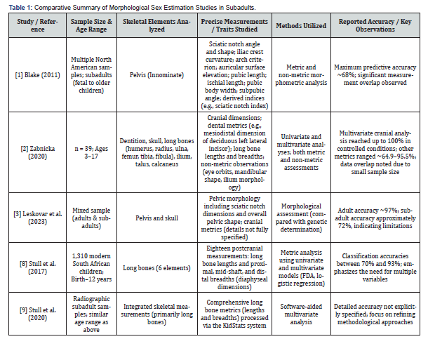
Adult skeletal analysis benefits from pronounced sexual dimorphism that arises during and after puberty. Blake et al. [1] presented a comprehensive morphometric analysis of the pelvis, demonstrating that parameters such as the sciatic notch angle, subpubic angle, and auricular surface morphology can yield classification accuracies exceeding 90%. Similarly, studies focusing on cranial structures [4,5] have shown that features like orbital shape, mandibular robusticity, and cranial proportions serve as reliable indicators in adults. Frost et al. [2] and Knickmeyer et al. [3] further emphasize that the cumulative effects of hormonal influences and biomechanical loading result in significant morphological differences between adult males and females. These robust markers, however, are the product of years of growth and remodeling, making them highly effective only when the skeletal elements have reached full maturity (Table 1).

Challenges in Subadult Sex estimation
In contrast, the subadult skeleton is characterized by ongoing growth and remodeling, which results in a continuum of morphological expression rather than distinct sex-specific traits. Zabnicka et al. [4] noted that the absence or subtle expression of secondary sexual characteristics in subadults leads to overlapping measurement ranges between males and females. Studies by Leskovar et al. [5] have reported that, when adult-based criteria are applied to immature remains, classification accuracy often drops to between 68% and 72%. These figures are indicative of the inherent limitations of using adult-derived standards for children, whose skeletal features are still in transition. The dynamic nature of skeletal development is further complicated by variability in the timing of growth spurts and epiphyseal fusion. As Malina et al. [11] discuss, factors such as genetics, nutrition, and overall health significantly influence the rate and pattern of growth. Consequently, thresholds and indices established for adult populations are often inappropriate for subadults, resulting in ambiguous and sometimes conflicting outcomes [6,12]. For example, while Blake et al.’s analysis [1] provided reliable pelvic measurements in adults, his findings also underscored the extensive overlap observed in younger individuals-a problem echoed by subsequent studies [12,13].
Pelvic Morphometrics in Subadults
The pelvis is widely regarded as the most sexually dimorphic region in the adult skeleton, yet its application in subadults is problematic. Blake et al. [1] demonstrated that while certain pelvic dimensions such as the sciatic notch, iliac crest curvature, and subpubic angle show statistically significant differences between sexes, these differences are less pronounced in children due to rapid growth and remodeling. Wilson et al. [8] reviewed the use of pelvic markers in subadults and concluded that while adult pelvic morphology may yield accuracies above 90%, the same parameters in subadults result in lower, less reliable outcomes. Furthermore, Vlak et al. [14] emphasized that local and population-specific variations in pelvic morphology add another layer of complexity, thereby necessitating cautious interpretation when applying adult-based metrics to immature remains.
?Cranial Metrics and Long Bone Dimensions
Cranial measurements have long been used in adult sex estimation; however, their reliability decreases in subadults. Gonzalez et al. [15] utilized discriminant function analysis to assess juvenile cranial morphology but found that the subtle differences between male and female subadult crania were often masked by developmental variability. Molleson et al. [9] also reported that sexually dimorphic features of the juvenile skull, although present, are not sufficiently pronounced to allow for definitive sex estimation. In parallel, long bone dimensionsparticularly diaphyseal measurements-have been explored for their potential to differentiate sex in subadult populations. Stull et al. [6] reported that multivariate models based on long bone lengths and breadths could achieve classification accuracies between 70% and 93% under controlled conditions. However, in practical forensic settings, these models are often hampered by individual variability in growth rates and by postmortem changes that affect measurement accuracy [16].
Developmental variability and environmental influences
A significant challenge in subadult sex estimation is the inherent variability in skeletal maturation. As highlighted by Zabnicka et al. [4] and further supported by Byers et al. [17], subadult skeletal development is influenced by a myriad of factors, including genetic predispositions and environmental conditions. Malina et al. [11] provide extensive evidence that nutritional status and physical activity significantly impact the timing of growth spurts and epiphyseal fusion. Such variability means that a given morphological feature may appear at different stages of development in different individuals, complicating the establishment of universal standards for sex determination. Frost et al.’s work [2] reinforces this notion by showing that the dynamic process of bone remodeling in children creates a continuum rather than discrete, sex-specific characteristics.
Population-specific variations
Another layer of complexity arises from population-specific differences in skeletal morphology. Leskovar et al. [5] have demonstrated that methods developed on one population may not directly apply to another, as genetic diversity and local environmental factors can significantly alter the expression of sexually dimorphic traits. Wilson et al. [8] and Vlak et al. [14] further emphasize that differences in pelvic morphology across populations necessitate the use of context-specific criteria. Without accounting for these variations, applying a one-size-fits-all approach to subadult sex estimation can lead to misclassification and reduced accuracy.
Contributions from juvenile osteology manuals and research
Juvenile osteology has been advanced through comprehensive manuals and field guides that provide detailed methodologies for studying subadult remains. The Juvenile Osteology Lab and Field Manual by Schaefer et al. [18] offers practical insights into the measurement and analysis of immature skeletal features, while Cunningham et al.’s work [10] on Developmental Juvenile Osteology provides an in-depth overview of the patterns of skeletal growth and development. Additionally, Molleson et al. [9] contribute valuable data on the sexually dimorphic features of the juvenile skull, which, although not as pronounced as in adults, offer important clues that can be used in conjunction with other metrics. Loth et al. [19] further elucidates the early development of mandibular dimorphism, emphasizing that even in early childhood, some differences exist, though they are not yet robust enough for forensic classification.
Methodological Limitations and Guidelines
The forensic community has increasingly recognized the limitations inherent in applying adult-based morphological criteria to subadult remains. The guidelines issued by the Scientific Working Group for Forensic Anthropology (SWGANTH) [7] explicitly caution practitioners against the uncritical use of adult standards in subadult cases. These guidelines call for the development of age-specific protocols that take into account the continuous and variable nature of skeletal growth in children. Byers et al. [17] echoes this sentiment, emphasizing the need for refined measurement protocols and integrated approaches that combine morphological data with other lines of evidence.
Synthesis and Future Directions
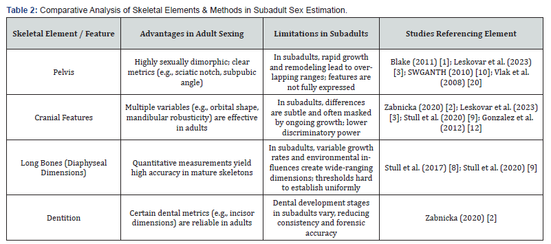
The literature reviewed here clearly indicates that while adult skeletal markers provide high accuracy in sex estimation, their direct application to subadult remains is fraught with challenges. The overlapping morphological traits observed in subadults-stemming from developmental variability, environmental influences, and population-specific differencesresult in significantly lower classification accuracies [4-6]. Moreover, the methodological limitations highlighted by studies on pelvic, cranial, and long bone metrics underscore the need for a multifaceted approach to subadult sex estimation. Future research should focus on developing normative, age-specific data for various skeletal elements [4,11,17]. Refining multivariate models using advanced computational techniques, as suggested by Stull et al. [6,16], may also improve classification accuracy. Furthermore, population-specific studies are necessary to tailor forensic criteria to the diverse genetic and environmental contexts encountered in forensic casework [5,8,14]. Finally, integrating morphological methods with complementary approaches-such as biochemical, molecular, or contextual analyses-will likely provide a more robust framework for sex estimation in subadults. The collective body of literature reveals a clear trend: the morphological markers that are highly effective in adult sex estimation do not translate seamlessly to subadult remains. The continuous and individualized nature of skeletal development, coupled with the influence of population-specific factors, creates a scenario where the overlapping ranges of morphological traits significantly impair the discriminatory power of traditional methods. The case report of skeletal remains from an individual aged between 9 and 12 years-where standard morphological assessments failed to yield a definitive sex determination-exemplifies this broader challenge. In essence, while the adult skeleton provides a reliable canvas upon which sexual dimorphism is vividly painted, the subadult skeleton remains a work in progress. The incomplete development of secondary sexual characteristics, the variability in growth trajectories, and the modulating effects of environmental and genetic factors all conspire to obscure the clear markers of sex that are evident in mature individuals. This critical review, by weaving together findings from pelvic morphometrics, cranial and long bone analyses, and case-based observations, underscores the fallacies in applying adult-based morphological criteria to subadult remains (Table 2).

Case Report
Case Background
As per the police records, the apparent cause of death was not determinable. The body was reported and subsequently referred from a nearby civil hospital to our mortuary due to its advanced state of decomposition. The hospital record indicated that the remains were highly skeletonized with an advanced stage of putrefaction, necessitating prompt forensic examination.
External Examination and Recovery Details
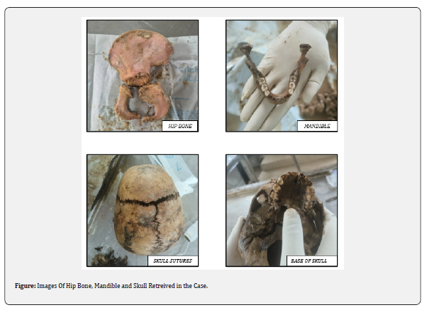
On arrival at the mortuary, the body was received as a skeletonized specimen with disorganized, mummified soft tissue remnants, all wrapped in a black plastic sheet. The clothing observed on the body included a dark-colored sleeveless vest and an elastic waist dark-colored lower garment. Additionally, a bundle of clothes and a bed sheet were found loosely draped around the remains. All garments were noticeably smudged with decomposed, greasy tissue, and the scene emitted a strong, rancid odor characteristic of putrefaction and adipocere formation. Notably, the body was in an advanced state of decomposition with significant adipocere formation. The skin was entirely absent from most areas, particularly over the face and neck, exposing the underlying bony structures. Remnants of soft tissue were scant, noted only as small adherent patches on the bilateral zygomatic areas and along the skull. Scalp hair was largely detached, with only a few tufts remaining on the skull.
Internal and Skeletal Examination:
A meticulous skeletal examination revealed the following
findings:
Skull and Cranial Structures:
• The skull was found disarticulated from its attachments
and lying loosely.
• Skull sutures were notably separated, yet there were no
signs of hemorrhage along the sutural margins.
• The scalp was completely absent, with only sporadic
tufts of hair adhering to the skull.
• Both eyeballs were missing; remnants of soft tissue
were observed adhering to the inner orbital walls, and the eye
sockets lacked normal anatomical features such as the cornea or
conjunctiva.
Facial and Dental Findings:
• The soft tissues of the face were severely decomposed.
• In the upper jaw, both sides displayed two molars and
one premolar in erupted positions; however, both canines and all
incisors were absent, with empty sockets evident.
• A similar dental pattern was observed in the lower
jaw, with the absence of canines and incisors, while molars and
premolars remained in place.
Neck and Thorax:
• The soft tissues of the neck were completely missing
beyond the bottom of the neck, exposing the bony framework of
the cervical spine.
• Structures such as the esophagus, larynx, trachea, and
thyroid gland were absent. The hyoid bone, thyroid cartilage, and
cricoid cartilage were also missing.
• The chest wall, including the sternum and ribs, was
found disarticulated. Ribs were detached both from the sternum
and vertebral column, and the pleural cavities were empty.
Upper and Lower Limbs:
• Both clavicles and scapulae were disarticulated.
• The hands and feet were not intact: the fingers were
missing, with only remnants of carpals and metacarpals present,
accompanied by a similar condition in the feet where toes were
absent and only tarsals and metatarsals were found loosely
arranged near the distal ends of the tibia and fibula.
• All joints across the body, including the pelvic joints and
the attachments of the vertebrae, were disarticulated, with many
bones found separately.
Pelvic and Lower Limb Findings:
• The pelvic bones, including both hip bones, were
disarticulated.
• The iliac crest remained unfused, and the anterior
inferior iliac spine was not apparent.
• Measurements taken from the pelvic region included a
sciatic notch width of 2.8 cm, a sciatic notch depth of 2.1 cm, and a
derived sciatic notch index of 133.
• The length of the pubis and ischium was recorded at 4
cm each, resulting in an ischiopubic index of 100.
• The total length of the femur was 26 cm, and using
established forensic formulas, the estimated stature of the
individual was approximately 122 cm.
Vertebral Column and Additional Findings:
• The cervical and thoracic vertebrae were disarticulated
and exhibited remnants of adherent soft tissue.
• The body of the sternum was unfused, and the ends of
several long bones remained unfused, corroborating the subadult
status of the remains.
• No signs of traumatic injury or perimortem fractures
were evident on any of the skeletal elements.
Forensic Analysis and Opinion:
The overall findings indicate that the remains belong to a
subadult, estimated to be between 9 and 12 years of age, based
on the state of skeletal development (unfused epiphyses in the
long bones, unfused iliac crest, and dental eruption patterns). The
advanced state of decomposition-with complete skeletonization,
adipocere formation, and significant soft tissue loss-complicates
the determination of certain vital forensic details. Notably,
the absence of soft tissue in critical regions and the extensive
disarticulation precluded any determination of the cause of death
from morphological evidence alone.The dental findings, with the
eruption of molars and premolars but the absence of canines
and incisors, further support the subadult status, although
they do not contribute directly to sex determination. The pelvic
measurements, such as the sciatic notch index and the lengths of
the pubis and ischium, were noted; however, due to the overlapping
characteristics in subadult skeletal morphology, these parameters
were inconclusive in establishing sex definitively. Consequently, the
forensic opinion states that while the body is clearly that of a child
between 9 and 12 years of age, the sex could not be determined
through morphological analysis alone. In light of these findings,
additional laboratory analyses have been recommended. Viscera
samples have been sent for chemical analysis to further explore
any potential toxicological or pathological factors that might
contribute to a determination of cause of death. Additionally, the
femur bone has been earmarked for DNA analysis to ascertain the
sex of the individual, as morphological criteria in this age group
have proven insufficient.

Discussion
The present study, alongside the reviewed literature and case report, highlights the considerable challenges associated with applying adult-based morphological criteria to subadult skeletal remains. In adults, the development of distinct sexual dimorphism facilitates highly accurate sex estimation, yet subadult remains present a host of complications that limit the reliability of traditional methods. This discussion examines the major themes arising from the literature, explores the factors contributing to ambiguity in subadult sex estimation, and considers the implications for forensic practice and future research.
Comparative Morphology: Adults Versus Subadults
Adult skeletal markers are well established as reliable indicators of sex due to the pronounced sexual dimorphism that develops during puberty. For example, Blake et al.’s morphometric analysis demonstrated that adult pelvic measurements, such as the sciatic notch angle and subpubic angle, yield classification accuracies exceeding 90% [1]. Similarly, cranial metrics-such as orbital shape and mandibular robusticity—further enhance the precision of sex estimation in mature skeletons [2,3]. However, these markers are the cumulative result of decades of hormonal influences and biomechanical adaptations, which are not fully expressed in subadult remains. In children and adolescents, the skeletal framework is still undergoing rapid change, and many of the adult characteristics have yet to appear in a distinct form [4]. As a result, the measurement ranges of key features in subadults overlap considerably between sexes, leading to lower classification accuracies that typically range between 68% and 72% [4-6]. This fundamental disparity underscores the need for approaches that specifically account for the dynamic nature of the immature skeleton.
Developmental Variability and Its Impact
One of the most significant challenges in subadult sex estimation is the inherent variability in skeletal development. Unlike the adult skeleton, subadult bones are in constant flux. Epiphyseal fusion, bone remodeling, and the gradual emergence of secondary sexual characteristics occur at different rates among individuals [4,11]. Factors such as genetic predispositions, nutritional status, and environmental influences can significantly alter growth trajectories [11]. Frost et al. [2] noted that the process of bone remodeling in children is continuous, which creates a morphological continuum rather than the discrete traits observed in adults. This continuum means that any threshold or index developed for adults might not be directly applicable to children, as the same measurement may represent different developmental stages across individuals. For instance, while Blake et al. [1] found that certain pelvic dimensions differed statistically between male and female adults, these differences are not as pronounced in subadults due to the ongoing process of growth and maturation. This variability not only complicates the direct application of adultderived thresholds but also results in significant inter-individual variability that statistical models struggle to accommodate [4,6]. The wide range of skeletal expression in subadults consequently leads to ambiguous outcomes in sex classification, even when multivariate analyses are employed [6].
Challenges in Morphological Measurements
The accuracy of morphological measurements in subadult remains is further compromised by the condition of the remains themselves. Advanced decomposition, as observed in the case report, often leads to skeletal disarticulation, loss of soft tissues, and postmortem alterations such as adipocere formation [17]. These postmortem changes can distort key landmarks and hinder the recovery of accurate measurements. For example, parameters such as the sciatic notch index or the lengths of the pubis and ischium may be affected by decomposition, leading to potential measurement errors that further reduce classification accuracy [6,8]. Moreover, while controlled studies have shown promising results-such as multivariate models achieving up to 93% accuracy under ideal conditions [6]-the variability inherent in forensic casework means that these accuracies are rarely achieved in practice [16]. Cranial metrics, although effective in adults, also present challenges when applied to subadults. Discriminate function analyses of juvenile cranial features have produced mixed results, with some studies reporting high accuracies under laboratory conditions that do not translate well in forensic scenarios [9,15]. The subtle differences in cranial morphology are often masked by the ongoing growth of the skull, leading to overlapping measurements that further reduce the reliability of sex estimation.
Population-Specific Variations
Another significant complication in subadult sex estimation arises from population-specific differences in skeletal morphology. Morphological traits are not uniformly expressed across all populations; genetic diversity, environmental factors, and cultural practices influence the development of sexually dimorphic features [5,7]. Leskovar et al. [5] illustrated that methods developed on one population may not directly apply to another, with local variations in pelvic morphology particularly notable. Wilson et al. [8] and Vlak et al. [14] further emphasize that subtle regional differences can significantly affect the accuracy of sex estimation methods if population-specific standards are not considered. This variability underscores the necessity of a context-specific approach in forensic anthropology; wherein morphological criteria are adjusted to account for the genetic and environmental background of the individuals being examined.
Implications for Forensic Practice
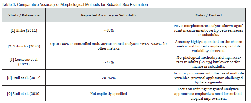
The limitations of current morphological methods for subadult sex estimation have profound implications for forensic practice. In many cases, especially those involving heavily decomposed remains, relying solely on adult-based morphological criteria can lead to indeterminate or erroneous sex determinations [12,20]. The inability to confidently assign sex to subadult remains complicates the overall process of identification and may delay investigations. As a result, forensic practitioners are increasingly recognizing the need for a multifaceted approach that integrates morphological analysis with additional lines of evidence, such as molecular or chemical methods [7,19]. Such an integrative approach can help mitigate the uncertainties inherent in subadult sex estimation and improve the overall reliability of biological profiles constructed from skeletal remains. Moreover, the challenges associated with subadult sex estimation highlight the importance of developing age-specific protocols that are tailored to the unique characteristics of the immature skeleton. Authoritative guidelines, such as those from the Scientific Working Group for Forensic Anthropology, emphasize the need for such protocols and caution against the uncritical application of adult-based methods [7]. In practice, this means that forensic laboratories should adopt standardized, age-appropriate measurement techniques and consider the incorporation of advanced statistical models that account for developmental variability [6,17]. By refining these methods and integrating complementary evidence, forensic practitioners can enhance the accuracy of sex estimation in subadult cases (Table 3).

Recommendations for Future Research
Given the challenges discussed, several avenues for future research emerge. First, there is a need to develop normative, age-specific data for various skeletal elements across subadult populations [4,11,17]. Establishing detailed growth charts and reference ranges would allow for more precise thresholds tailored to different developmental stages. Second, refining multivariate statistical models using advanced computational techniques, such as machine learning algorithms, could help improve the predictive accuracy of subadult sex estimation [6,16]. Such models must be validated across diverse populations to ensure their broad applicability.
Third, population-specific studies are critical to tailoring forensic criteria to account for regional differences in skeletal morphology [5,8,14]. Research focusing on the skeletal characteristics of specific demographic groups would help in creating localized standards, thereby reducing the likelihood of misclassification. Fourth, adopting a longitudinal approach that tracks skeletal development from childhood through adolescence could provide valuable insights into the progression of sexual dimorphism over time. Combining morphological studies with biochemical and molecular analyses would offer a more comprehensive understanding of skeletal maturation and its forensic implications [9,10]. Finally, standardizing measurement protocols across laboratories is essential to minimize interobserver variability and improve the consistency of subadult sex estimation methods [7].
Limitations
Despite the advances in our understanding of subadult sex estimation, several limitations remain. Many of the studies reviewed involve relatively small sample sizes or are based on specific populations, which may limit the generalizability of their findings [4,5]. Additionally, the quality of skeletal remains in forensic contexts is often compromised by decomposition and postmortem alterations, which can introduce measurement errors that are not fully accounted for in controlled studies [6,17]. These factors underscore the persistent uncertainty associated with morphological methods in subadult cases.
Conclusion
In conclusion, while adult skeletal markers provide high accuracy in sex estimation due to well-developed sexual dimorphism their direct application to subadult remains is problematic. The dynamic processes of growth and remodelling, combined with overlapping measurement ranges and populationspecific variations, result in a significant reduction in classification accuracy, often yielding rates of only 68% to 72%. The reviewed literature and case report collectively highlight the critical need for age-specific forensic protocols and integrated approaches that incorporate multiple lines of evidence to enhance the reliability of subadult sex estimation. Addressing these challenges is imperative for advancing forensic anthropology and ensuring that biological profiles based on skeletal remains are both accurate and informative.
References
- Blake KAS (2011) An investigation of sex determination from the subadult pelvis: a morphometric analysis [dissertation]. Pittsburgh (PA): University of Pittsburgh.
- Frost HM, Schönau E (2000) Bone remodeling in children: growth and development dynamics. J Bone Miner Res 15(2): 123-130.
- Knickmeyer RC, Baron-Cohen S (2006) Sexual dimorphism in the human brain: A developmental perspective. Ann N Y Acad Sci 1076: 36-45.
- Zabnicka D (2020) Assessing methods for estimating biological sex from subadult skeletal elements [master’s thesis]. Arcata (CA): Humboldt State University.
- Leskovar T, Mlinšek T, Počivavšek T, Zupanič Pajnič I (2023) Comparison of morphological sex assessment and genetic sex determination on adult and sub-adult 17th–19th century skeletal remains. Genes 14(8): 1561.
- Stull KE, L’Abbée EN, Ousley SD (2017) Subadult sex estimation from diaphyseal dimensions. Am J Phys Anthropol 162(4): 603-617.
- (2010) Scientific Working Group for Forensic Anthropology (SWGANTH). Sex assessment guidelines. Scientific Working Group for Forensic Anthropology.
- Wilson I, Cardoso H, Humphrey LT (2011) Sex estimation from the subadult pelvis: a review. J Forensic Sci 56(4).
- Molleson RL, Cruse W (1998) Some sexually dimorphic features of the human juvenile skull and their value in sex determination in immature skeletal remains. J Archaeol Sci 25: 720-723.
- Cunningham SL, Tenney S (2017) Developmental Juvenile Osteology. London: Elsevier Ltd.
- Malina RM, Bouchard C, Bar-Or O (2004) Growth, Maturation, and Physical Activity. 2nd ed. Champaign (IL): Human Kinetics.
- Thompson T, Black S (2007) Juvenile sexing of bones. In: Forensic Human Identification. Taylor & Francis Group, LLC.
- Iscan Y, Steyn M, Yasar F (2013) Sexing of bones in juveniles. In: Human Skeleton in Forensic Medicine. 3rd ed. Charles C Thomas.
- Vlak R, Roksandic M, Schillaci O (2008) Sex differences in the developing pelvis: implications for subadult sex estimation. Forensic Sci Int 180(2-3).
- Richard A Gonzalez (2012) Determination of sex from juvenile crania by means of discriminant function analysis. J Forensic Sci 57(1): 24-34.
- Stull KE, Cirillo LE, Cole SJ, Hulsen CN (2020) Subadult sex estimation and KidStats. In: Sex Estimation of the Human Skeleton. Elsevier 2020: 153-167.
- Byers SN (2017) Sexing of subadults. In: Introduction to Forensic Anthropology. 5th ed. Routledge.
- Schaefer KE, Morgan R, Spencer TJ, et al. (2009) Juvenile Osteology Lab and Field Manual. Burlington (MA): Academic Press.
- Loth SM, Henneberg M (2001) Sexually dimorphic mandibular shape in the first few years of life. Am J Phys Anthropol 115: 181-188.
- Black S, Ferguson E (2011) Sexing in juveniles. In: Forensic Anthropology: 2000 to 2010. CRC Press.


