Experimental Study of Cranial Injuries Due to Blunt Force Trauma: Sus scrofa domestica Model
Felipe Otero1* and Marien Béguelin1,2
1 Universidad Nacional de Río Negro, Argentina
2 Anexo del Museo de La Plata, Universidad Nacional de La Plata, Argentina
Submission:November 01, 2019; Published: November 26, 2019
*Corresponding author:Felipe Otero, Universidad Nacional de Río Negro, Instituto de Investigación en Paleobiología y Geología, Av. Roca 1242, (8332) General Roca, Río Negro. Argentina.
How to cite this article:Felipe Otero, Marien Béguelin. Experimental Study of Cranial Injuries Due to Blunt Force Trauma: Sus Scrofra Domestica Model. J Forensic Sci & Criminal Inves. 2019; 13(2): 555856. DOI: 10.19080/JFSCI.2019.13.555856.
Abstract
Cranial blunt force trauma is of major concern in forensic sciences. The aim of this study is to shed light on cranial bone trauma caused by blunt weapons from an experimental perspective. The experimentation involved the production of blunt injuries to 21 pig skulls with different objects: metal hammer, wooden club (baseball bat), stone and boleadora. These blunt objects were chosen because they can be easily used as homicidal weapons since they are common elements that can be seen in daily situations today. The marks produced were recorded and analyzed using quantitative and qualitative variables. The results showed that it is possible to identify the blunt weapon that caused the injury through the analysis of variables such as maximum diameter or depth of the bone injury. Furthermore, it was observed that depending on the damage, the injury could be associated with a blunt object with specific characteristics.
Keywords:Forensic anthropology; Interpersonal violence; Blunt force trauma; Experimental design; Sus scrofa domestica
Abbreviations: PIN: Pig Identification Number; H: Hummer; C: Club; S: Stone; B: boleadora; D: Depth; MD: Maximum Diameter; PMD: Perpendicular Diameter; LF: Linear Fracture; CF: Concentric Fracture; SF: Stellate Fracture; RE: Raised Edges; FA: Fragments Attached to Fracture Edges; BL: Bone Loss; RB: Redundant Blows
Introduction
The analysis of trauma in human skeletal remains involves the study of injury patterns [1-3]. Blunt force trauma may result from impact with any hard surface. Some marks on bone may result from the application of a force onto an area of impact caused by hard elements with a rounded or blunt surface, such as a stone, a baseball bat, a boleadora or a hammer. Different weapons can produce different tool marks and injury patterns, which can be identified by using different variables. These marks are also known as blunt injuries [4,5]. Research and analysis of these types of injuries in skeletal remains help address forensic contexts with abundant evidence accounting for conflicting situations. In forensic cases, human bones frequently exhibit blunt injuries [6,7], but it is difficult to establish a correlation between the marks and the injury-causing weapon [7-10]. This is because a lot of factors that contribute to injury production such as the characteristics of the person who effects the blows (their physical conditions), the biomechanical properties of the bone, the weight and speed reached by the weapon, the material with which it is made, its contact surface, the size of the area impacted, the direction of impact and the thickness of the scalp and hair. The thickness of the bone also has an influence since the bone material tends to break mainly in the thinnest areas and therefore less resistant. The bones with greater volume need to be impacted with a lot of energy to cause their fragmentation [7-9].
In addition, there is a limited possibility of bone’s response to blunt force trauma: different instruments generate similar injuries and the same instrument can generate different injuries. Also, different causal agents (e.g. falls or accidental impacts) converge in blunt injuries similar to those produced by situations of violence [3,10-13]. Therefore, it is necessary to develop frames of reference that can establish a cause-effect association between the injury morphology and the weapon using a probabilistic model, and experimental studies have the potential to achieve this [1-3,10,14-16]. However, there are very few experimental studies, especially those aimed at identifying the weapon that caused the blunt injury [15,17]. The aim of this paper is to contribute to knowledge of injuries on cranial bones (e.g. frontal, parietal (left and right), temporal (left and right), occipital, sphenoid and ethmoid) made by blunt weapons and identify the injury patterns produced by each object. Through an experimental study, this work is based on the production of blunt injuries to 21 pig skulls (Sus scrofa domestica) with a metal hammer, a wooden club (baseball bat), a stone and a boleadora. Subsequently, univariate and multivariate statistical methods were used to analyze the quantified data set.
Material and Methods
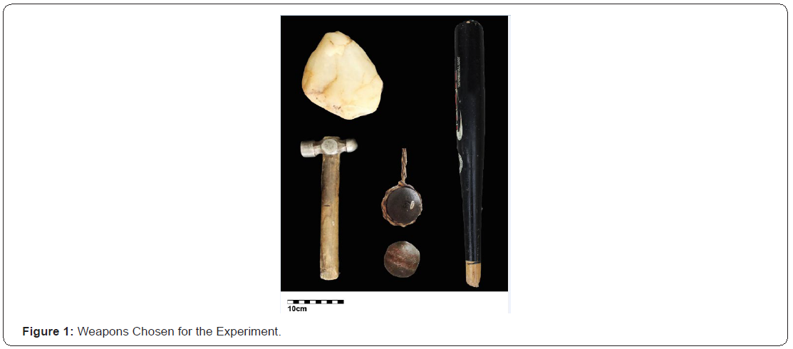
Adopting Gordon’s Model [1], the experimental research consisted in striking pig heads previously prepared and then analyzing the resulting marks on bone. A total of 21 skulls from subadult pigs (Sus scrofa domestica) of undetermined sex were used for this study. The age of death of all the pigs is similar (24 weeks (+/- 2)). The specimens were assigned a Pig Identification Number (PIN) from 0 to 20. The weapons used were metal Hammer (H), wooden club (baseball bat) (C), stone (S) and boleadora (B) (Figure 1). These objects were chosen for this experiment because they are frequently reported in forensic cases. A hammer, a stone or a wooden club can be easily used as homicidal weapons since they are common blunt objects that can be seen in every human daily situation. A boleadora is an object commonly found in rural areas of Patagonia (Argentina), which can be used as a weapon of interpersonal violence. It is a throwing hunting weapon composed of a stone ball (e.g. andesite), with a groove carved around the circumference and with a rope or strip of leather tied to the stone [18-20]. The strikes were executed without releasing the rope. Furthermore, weapons with varying characteristics (e.g. size, weight and material) were chosen to produce different injury patterns and make associations between the morphology of the injury and the instrument. The first skull (PIN 0) was impacted only to test the different instruments and experimental device (i.e. force and impact site) and ensure that the blow produced a potentially recordable effect. The other 20 skulls were divided into 4 groups of 5 animal subjects. Each group was assigned a blunt object. The pig heads were distributed randomly into the groups so that the varying characteristics of the pig skulls, such as size, did not influence the results of the weapon blows.

The skin of the pig heads was removed with knives and scalpel. A thin layer of soft tissue (i.e. muscle, fat, cartilage) was preserved to mimic the human scalp. In this way, the interface between the bone and the surface of each head was represented by 3 to 5 mm of muscle mass and connective tissue. The pig Sus scrofa domestica was chosen as a human proxy for this experiment following the criteria already established and evaluated by different authors [1,2,21-24]. In comparative terms, this species shares several physical traits such as bone and skin with Homo sapiens making it useful mainly in forensic contexts. The pig heads were placed in such a way that they kept limited mobility in relation to a vertical axis, i.e. they neither hang freely when they were impacted nor were, they completely stuck, since none of these situations resemble natural mobility resulting when striking a living individual. The device designed for this purpose consisted of a base on which the heads were placed in order to mimic the support that the spine provides to the human skull (Figure 2). Such device consisted of two buckets filled with sand placed on the floor, side by side, with one end of a wooden stick inserted into each bucket. The opposite ends were introduced into the zygomatic arches, extending to the orbital cavity. In turn, the zygomatic arches were secured to plastic pull tight seals attached to an elastic cord which dangled from a rail on the roof, thus allowing the desired mobility.
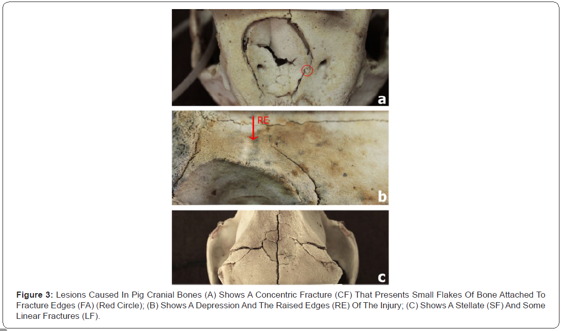
One of the authors of this study delivered all the blows with a hammer, club and stone, whereas in the case of the boleadora, an expert in the art of employing it inflicted the blows. The entire experimental process was filmed and photographed. The subsequent cleaning of the material was done by boiling the remains with detergent for domestic use, removing the remaining tissue and leaving them to dry at room temperature. In order to quantify the damage, depth (D), maximum (MD) and perpendicular (PMD) (orthogonal to the maximum diameter) diameters of each injury were measured with a digital caliper with an accuracy of 0.01 mm. Besides, the data obtained from fresh bones injuries (perimortem) was analyzed using a set of qualitative variables. Firstly, the presence of fractures was recorded. If it was positive, then fractures produced were registered as linear (LF), concentric (CF) and/or stellate (SF). Subsequently, the presence or absence of fragments (small flakes of bone) attached to Fracture edges (FA) was recorded. Furthermore, the presence or absence of Raised Edges (RE), which are produced when the bone material is plastically deformed. Finally, the presence or absence of Bone Loss (BL), i.e. the lack of anatomical units or their fragments [25-28] (Figure 3). These variables were examined with the naked eye and/ or with a 10X magnifying glass. The data were analyzed with uni-, bi-, and multivariate methods, and with parametric and nonparametric methods, depending on the nature of the data set.
Results
A total of 64 blows were delivered to 20 experimental skulls of Sus scrofa domestica, using 5 skulls per weapon: 19 blows with a hammer, 15 with a wooden club, 15 with a stone and 15 with a boleadora. Each skull was struck 3 times, except for PIN 2, 3, 4 and 5 (corresponding to the hammer), which were impacted 4 times each. However, to perform the analysis, only 61 blows (18 with hammer, 15 with club, 15 with stone and 13 with boleadora) were considered, as some of them impacted exactly on the same area, thus preventing an accurate recording of each injury. These redundant blows (RB) were counted as one provided that it was not possible to distinguish one blow trace from another. Nine of the 61 blows recorded for the analysis did not affect the bone material, i.e., they did not cause injuries. This indicates that 14.75% of the blows delivered were absorbed by the soft tissue.
Quantitative variables
For the quantitative analysis, 12 of the 61 blows were not considered relevant for this study since they affected other statistical population: two cases correspond to situations in which more than one blow impacted the same place; the other case involved one blow to the zygomatic bone, which is not included in this work. Therefore, in total, only 49 blows were considered for these analyses.
Evaluation of dependence between blows on the same skull
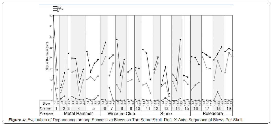
Before analyzing the variables, it was explored graphically if the application of three (or four) impacts on each experimental head (in different places) influenced the bone resistance and, therefore, the pattern of injuries produced (i.e. if each blow caused more damage than the previous one). The sizes of the injuries inflicted were illustrated in a multiple graph (the three variables plotted on the same figure) following the chronological order of the blows to each skull, separated by the weapon used. The pattern expected if each blow influenced the following one is an increase in the size of every variable in the same cranium. (Figure 4) shows that there is no pattern of increase in the size of the injury between one blow and the next, for any of the weapons. For example, blow 4.3 made with hammer decreases the size of the three variables but in Cranium 2 the opposite occurs. This indicates the independence of the blows, meaning that no relationship was found between the size of the injury and the order of the blows.
Univariate methods
Fig. 5 (A, B and C) shows the distribution of the medians for the injuries per weapon in relation to the variables MD, PMD and D, respectively. As a result of the Kruskal-Wallis test, no significant differences were observed between the weapons for the variable PMD (p= 0.089). However, there were differences for the variable’s MD (p= 0.029) and depth (p= 0.002). Therefore, a posteriori tests were carried out to identify which objects differed. In relation to MD pairs, it was observed that medians for boleadora and stone differed significantly (p= 0.024), and in relation to depth, the hammer showed significant differences with all the weapons: boleadora (p= 0.019), club (p= 0.012), and stone (p= 0.002).
Bivariate methods
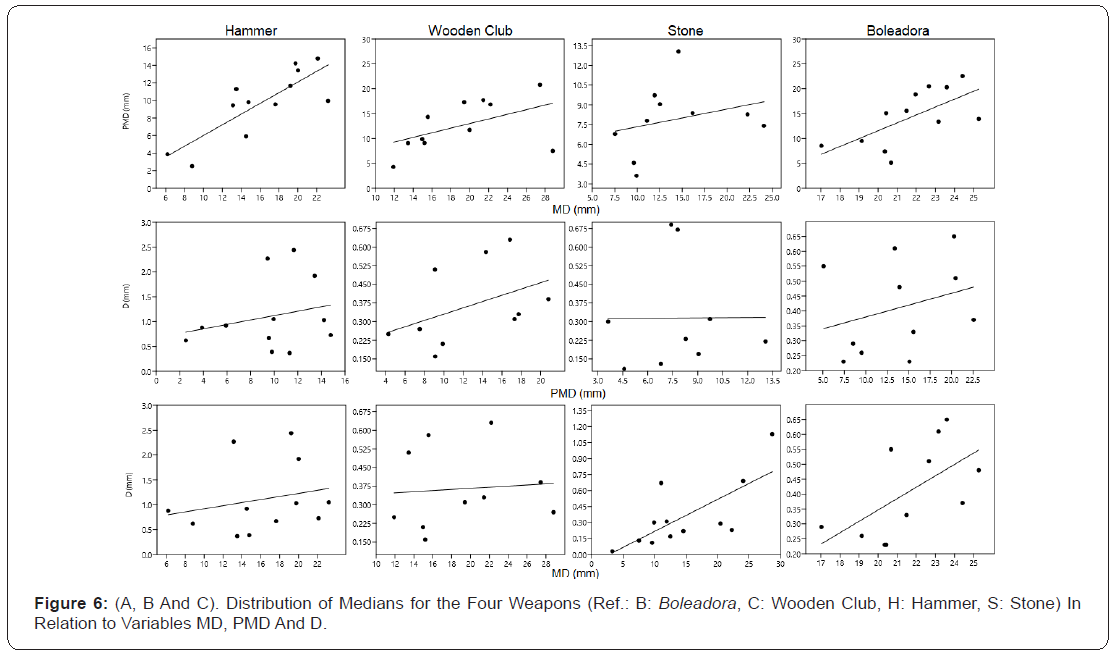
To analyze the data through bivariate methods, log(x) was applied and then the outliers were eliminated. Table 1 shows the results of the Spearman correlation coefficient among all the combinations of metric variables, for all the instruments used. Figure 6 shows regression lines of the variables MD, PMD and D for all the weapons. It was observed that for hammer (r2=0.745-p=0.008) and for boleadora (r2=0.636-p=0.026), the relationship between PMD and MD is positive. Therefore, when the maximum diameter increases, the perpendicular diameter to the maximum also increases. It was also observed that MD and D correlate with each other for the stone (r2=0.665-p=0.013) and the boleadora (r2=0.651-p=0.030).

Multivariate methods
Permutational Multivariate Analysis of Variance (PERMANOVA) was performed to compare the effects among weapons based on all metric variables. It showed significant differences between the weapons (p= 0.017). A posteriori tests for the analysis between pairs showed significant differences between boleadora-hammer (p= 0,041), and boleadora-stone (p= 0,009). In this analysis, the data was transformed into natural logarithm of each value plus one [ln (x+1)] [29].

Qualitative variables
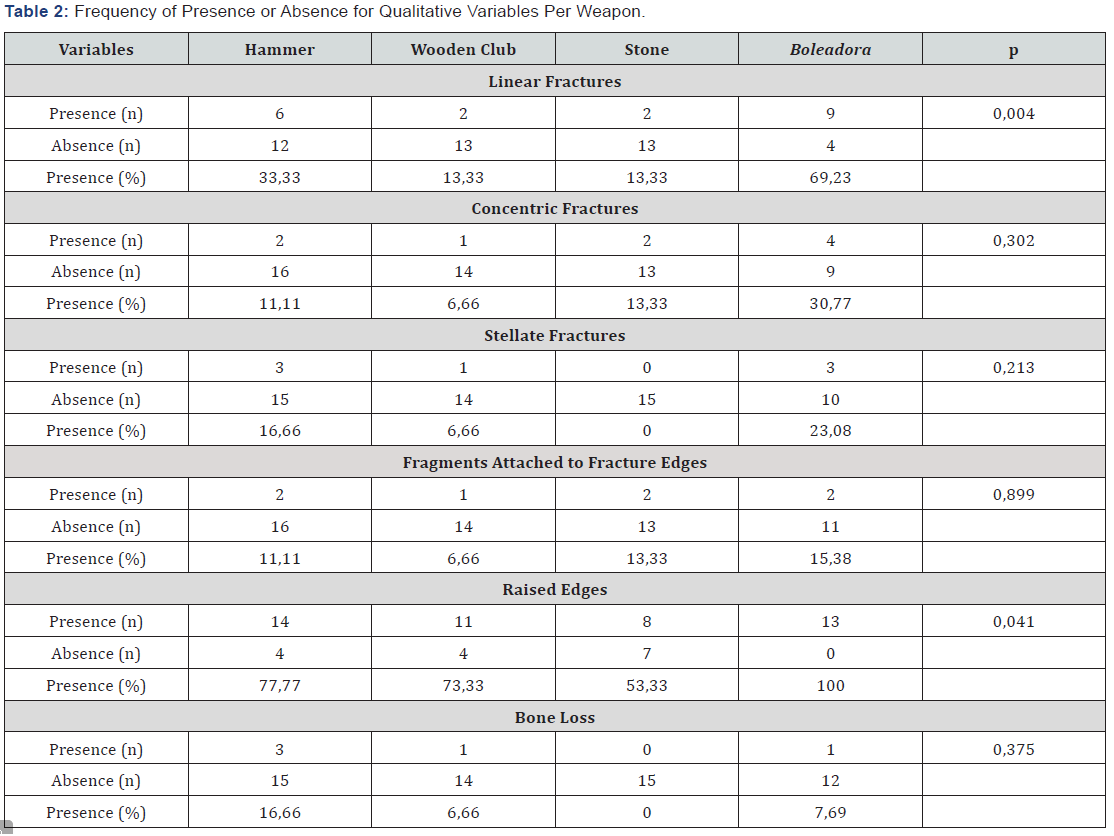
The associations between weapons and qualitative variables (i.e. LF, CF, SF, FA, RE and BL, expressed as frequencies of presences and absences) were evaluated by contingency tables (chi-square test) for each variable independently. Null hypothesis states that presence/absence ratio (P/A ratio) is homogeneous across weapons. Rejection of null hypothesis implies that at least one weapon produces a different P/A ratio. Table 2 summarizes the information collected from the experimental marks. Statistically significant differences were found for LF and RE. Conversely, the other variables did not show significant differences between the weapons as a result of the statistical tests.
Discussion & Conclusion
Pig bones are suitable analogous for experimental studies of fracture patterns [7,10] because of their similarities with human bones: both are made of external and internal tables and a diploe [28,30]. However, pigs’ skulls differ in having thinner tables, thicker diploe, and greater total thickness of the three layers if compared with human skulls. These differences might make pig cranial bones more resistant to blunt force trauma than human ones. There are also differences in the anatomy of the skulls: in the case of pigs the parietal bone is small, square and flat, while in human skulls are large and convex laterally [28]. Notwithstanding this considerations, the pigs’ skulls are still convenient proxies for human skulls, moreover in countries were laws do not allow the use of corpses with experimental purposes (e.g. body farms). In most experimental studies, only one blow is applied to each skull based on the assumption that the second blow will cause greater damage [6,28]. However, in this work evidence showed that each blow causes injuries independently of the others (a maximum of four blows). Therefore, it is possible to deliver more than one blow in different areas of a pig skull without affecting its resistance. Through the univariate analysis, the results showed that it is possible to recognize the marks on pig skulls made by the different blunt weapons using metric and categorical variables. The univariate analysis of morphometric variables of the marks is useful in finding differences between the patterns of injury produced by the different weapons. Similar results were found by Small [31], who concluded that the depth and the maximum diameter served to find differences between the instruments, although the perpendicular diameter to the maximum was not useful by itself to identify the weapon. In addition, the multivariate analysis showed significant differences between the marks associated with the different objects when analyzing the three metric variables together.
For MD, differences between B and S were identified. The scatterplot of marks by weapons (Figures 5.a) shows that B varies in a smaller range than S. This may be due to the spherical shape of B which always generates the same impact area. In contrast, S has an irregular shape, so the impact area can vary depending on the side of the impact. This observation is relevant because when the weapon is irregularly shaped it may create different marks. Therefore, an irregularly shaped weapon could be associated with a range of possibilities. In other weapons, the morphology of the injury is more limited and subject to the shape of the impact surface of the weapon. The strong correlation that exits between the diameters (e.g. MD and PMD) of B and H supports the hypothesis that the surface impacted by these weapons is always similar.
In addition, significant differences were observed between H and the other effectors (B, S and C) for the variable depth, which helps infer that the mark left by H is characterized by having a greater depth. This may be since H hits a small area and the energy of the blow is concentrated causing greater damage. Blows to a smaller focal area of bone tend to cause a high stress level in the damaged area and, conversely, as the area increases, the tension decreases [8,9]. The latter would support the fact that C and S have fewer presence observations in the variables that qualitatively account for the damage than B and H (Table 2). In addition, the hardness of the raw material directly influences the energy transfer at the time of injury production. While metal largely transfers the energy of the impact, wood tends to absorb the blow [6]. It should be noted that the depth increases when the maximum diameter of the stone and boleadora increases as well. This could be because the diameter of the weapons increases towards the center or equator. If these weapons hit with more force, they would cause a greater depression and therefore a greater area of impact. Moreover, the material of the weapon (metal, wood or rock in this case) is a factor that could be influencing this variable. However, the data obtained from this study is not enough to perform further tests which can confirm this hypothesis. Finally, with respect to the qualitative variables, it was observed that the presence/absence of linear fractures as well as raised edges would be useful by themselves to find differences between the effectors. The other variables (e.g. CF, SF, FA, BL) would not contribute individually to the identification of an effector. However, the whole data corresponding to all the qualitative variables help quantify a percentage of damage caused by each instrument. Thus, it would be possible to classify the various weapons into categories based on the damage caused.
This work represents an experimental approach to the study of injuries caused by blunt weapons. The goal is to provide forensic science with information on which to base interpretations of the weapon used to produce injuries, at the bone level, in a victim. The results have raised new questions that should be examined in depth in future studies where the hypotheses can be refined, and the number of factors involved can be reduced (for example, tests with weapons having the same characteristics, different sizes, or areas of impact, etc.). Furthermore, it is expected that further studies will determine the impact energy through the analysis of the film recorded and include other variables (e.g. area and perimeter) and/or other instruments.
Acknowledgement
We thank Fernando Archuby by for his cooperation on the statistical analysis and Antonio Flores for striking the pig heads with the boleadora. Diego and Verónica García gently donated the pig heads from their farm. This study was funded by PI- 40-A-613 UNRN grant.
References
- Krogman WM (1962) The Human Skeleton in Forensic Science. (1st ), In: Charles C Thomas, (Edt.),Spring field, USA, 17(6): 7-12.
- Rajesh Kumar Verma, Mukesh K Goyal, Shiv Kochar (2010) Age Assessment from Radiological Cranial Suture closure in Fourth to Seventh decades. J Indian Acad Forensic Med 32(2).
- Shetty U (2009) Macroscopic study of cranial suture closure at autopsy for estimation of age. Anil Aggrawal's Internet. Journal of Forensic Medicine and Toxicology 10(2).
- Dwight T (1890) The closure of the cranial sutures as a sign of age. Boston Med Surg J 122: 389-392.
- Gaur VB, Sahai VB, Singh A, Kharat A (2007) Determination of Age in living by closure of Cranial Sutures A Radiological Study. J Indian Acad Forensic Med 29: 32-34.
- Reddy SS, Kaushik A, Prakash SDR (2011) Clinical applications of reverse panoramic radiography. Dent Hypotheses 2: 190-198.
- Whaites E (2002) Dental panoramic tomography. In: Michael Parkinson (Edt.), Essentials of Dental Radiography and Radiology, (3rd ), Churchill Livingstone: An imprint of Elsevier Science Limited, pp.161-176.
- Langland OE, Langlais RP, Mc David WD, Del Balso AM (1989) Special Panoramic techniques. In: Lea Febiger (Edt.), Panoramic radiology (2ndedn). Philadelphia, Lea Febiger, USA, pp. 102-123.
- Mukherjee JB (2007) Identification. In: Textbook of Forensic Medicine and Toxicology. (3rdedn) In: Karmakar RN(Edt.), Academic publishers Kolkata, India, pp.156-157.
- Fouad AA, Metwalley HE, Seif EA, El Megid, LAM (1999) Study of skull sutures as a tool for identification. J Forensic Med Clin Toxicol 7: 57-70.
- Sabini RC, Elkowitz DE (2006) Significance of differences in patency among cranial sutures. J Am Osteopath Assoc 106:600-604.
- Todd TW, Lyon DW (1924) Endocranial suture closure, its progress and age relationship. Part I-Adult males of the while stock. Am J Phys Anthropol 7: 325-384.
- Modi J (2010) Personal identity. In: Modi’s medical jurisprudence and toxicology. (23rd). In: Mathiharan K, Patnaik KA, (Edt.),Lexis nexis, pp. 287.
- Parikh (1990) Personal identity. In: Parikh’s Textbook of Medical Jurisprudence, Forensic medicine and Toxicology (5th), CBS, p. 39-50.
- Moondra AK (2000) Age assessment from vault suture closure in elderly persons (an autopsy study in Haroti Region).
- Vyas PC, Sischer H (1996) Age estimation by closure of suture of skull in individuals of 25-45 yrs of age of Jaipur area. Dissertation for MD (Forensic Medicine) University of Rajasthan.
- Parmar P, Rathod GB (2012) Determination of age by study of skull sutures. Int J Current Res & Rev 4: 127-133.


