Emergency Perimortem Total Body CT-Scan Examination in a Child Victim of Out-of-Hospital Cardiac Arrest: A First Step Toward Finding the Truth
Sue Antunez1*, Ophélie Ferrant-Azoulay1, David Grevent2, Gilles Orliaguet2, Estelle Vergnaud3, Philippe G Meyer3 and Caroline Rey-Salmon4
1Forensic Medical Unit, Unité Médico Judiciaire des Yvelines, France
2Paediatric Radiology Department, Centre Hospitalier Universitaire Necker Enfants Malades, France
3Department of Pediatric Anesthesiology and Critical Care, Hospitalier Universitaire Necker Enfants Malades, France
4Forensic Medical Unit, Université Descartes-Paris 5, France
Submission: May 13, 2018; Published: June 19, 2019
*Corresponding author:Sue Antunez, Forensic Medical Unit, Unité Médico-Judiciaire des Yvelines, UMJ 78, Centre Hospitalier Versailles Le Chesnay, Le Chesnay, France
How to cite this article: Sue Antunez, Ophélie F A, David G, Gilles O, Estelle V, et al. Emergency Perimortem Total Body CT-Scan Examination in a Child Victim of Out-of-Hospital Cardiac Arrest: A First Step Toward Finding the Truth. J Forensic Sci & Criminal Inves. 2019; 12(1): 555826. DOI: 10.19080/JFSCI.2018.11.555826.
Keywords: Fatal child abuse; Perimortem ct scan; Forensic examination; Resuscitation; Pulmonary contusion; Traumatic; Spinal dislocation; Pediatric trauma
Case Report
We report a case of sudden out-of-hospital cardiac arrest in a 20 months-old child, where full-body contrast-enhance perimortem CT-scan, performed on an emergency basis, could rapidly oriented toward severe abdominal and spine trauma from fatal child abuse, confirmed by further forensic investigations
Case Description
A previously healthy 20 months old girl was found pulseless at home. Child’s parents only reported a recent history of upper airway infection, and a sudden syncope followed by seizures, while playing, gently throwing her up in the air. Immediate resuscitation failed to restore spontaneous cardiac rhythm, and the girl was referred, under continuing resuscitation and mechanical ventilation, to our pediatric trauma center
Upon arrival, there was no evidence of external trauma or bruising, the girl remained pulseless with fixed dilated pupils, and extreme skin pallor. Instantly measured hemoglobin was 5.5g/dl, evocating severe blood
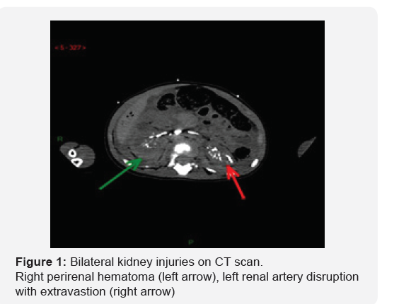
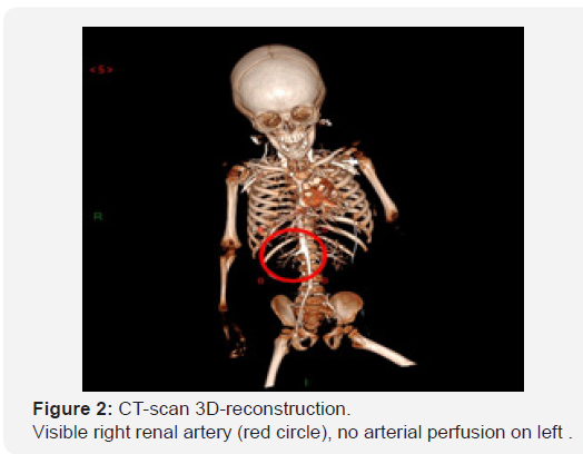
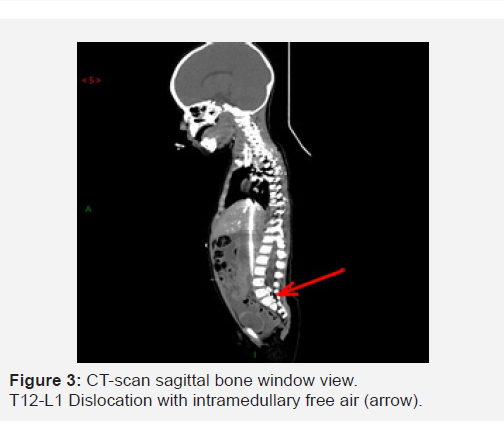
losses, and possibly inflicted trauma in the absence of reliable history. Despite massive transfusion and continuing resuscitation, the girl remained in refractory asystole. Taking account of clinical evidence of hemorrhagic shock, with no external signs of trauma, without compatible reported medical history, the girl was maintained mechanically ventilated, and transferred to the radiology ward for emergency full body CT- scanning with IV contrast injection. Perimortem emergency CT-scan depicted no major brain lesions, a bilateral mild pulmonary contusion, a large hemoperitoneum without spleen or liver laceration, and a massive retroperitoneal hemorrhage (with left renal artery disruption, right peri-renal hematoma and T12-L1 spinal dislocation (Figures 1-3).

Immediate burial certificate delivery was denied, and the State prosecutor commanded a complete forensic investigation. It revealed a complete spinal dislocation at T12-L1 level, a Keywords: Fatal child abuse; Perimortem ct scan; Forensic examination; Resuscitation; Pulmonary contusion; Traumatic; Spinal dislocation; Pediatric trauma 500ml hemoperitoneum, left renal fracture with complete arterial disruption, and right perirenal hematoma. Forensic brain examination found a bilateral, posterior mild parenchymal hematoma, and petechial lesions of the scalp. The forensic inquiry came to the final conclusions that the girl sustained an inflicted extreme hyperextension at the thoraco-lumbar junction, resulting in T12-L1 dislocation, bilateral kidney injuries, and left renal artery disruption, causing exsanguination, and death. Considering these evidences, parents were charged with inflicted injuries causing death.


Discussion
On an emergency basis, fatal abuse and neglect could be difficult to distinguish from sudden unexpected infant death in infants [1]. In many countries, fatal child abuse could be poorly investigated and identified, leading to underestimation [2,3]. In older, previously healthy children, trauma must systematically be advocated, even in the absence of reported history [4].
In our case, with a recent history of upper airway infection, without reliable history and external evidence of trauma, a sudden death resulting from medical etiology could be First suspected. However, instant measurement of blood hemoglobin rapidly oriented diagnosis toward severe blood losses of unknown, but possibly traumatic, origin
Because severe abdominal and chest lesions could directly result from cardiopulmonary resuscitation and be further difficult to distinguish from intentional lesions in young children, a precise collection of clinical and radiological data is critical before medical autopsy could be completed [5]. Postmortem CT-scan has gain rapid development, and increasing interest, as an adjunct to forensic examinations. However, performed after death in unventilated and perfused children with progressing processes of decomposition, it could be difficult to interpret by radiologists unaware of forensic radiology [6]. Performing emergency CT scan within minutes preceding or following death in mechanically ventilated and IV perfused children has the advantage of obviating the changes related to postmortem processes and is routinely used in our department [7]. In our case, CT scan rapidly eliminated lesions that could result from resuscitation and identified the cause of death: a bilateral kidney injury, left renal artery disruption and associated complete thoraco-lumbar spine dislocation. These finding were confirmed by medico-legal autopsy that depicted associated non-lethal brain lesions that could be missed by early radiological examinations. The inflicted character of the lesions with extreme thoraco-lumbar hyperextension was confirmed by further medicolegal inquiries.
Conclusion
Full body CT-scan is available on an emergency basis in most trauma center. Performed in moribund mechanically ventilated children it obviates the artefacts related to death processes that could be confounding for interpretation. It could be used as a rapid diagnostic tool, and valuable adjunct to forensic examination, especially in suspected fatal child abus
References
- Bechtel K (2012) Sudden unexpected infant deaths: differentiating natural from abusive causes in the emergency department. Pediatr Emerg Care 28(10): 1085-1089.
- Banaschak S, Janßen K, Schulte B, Rothschild MA (2015) Rate of deaths due to child abuse and neglect in children 0-3 years of age in Germany. Int J Legal Med 129(5): 1091-1096.
- Makhlouf F, Rambaud C (2014) Child homicide and neglect in France: 1991-2008. Child Abuse Negl 38(1): 37-41.
- Young KD, Gausche-Hill M, Mc Clung CD, Lewis RJ (2004) A prospective, population-based study of epidemiology and outcome of out-of-hospital pediatric cardiopulmonary arrest. Pediatrics 114(1): 157-164.
- Bush CM, Jones JS, Cohle SD, Johnson H (1996) Pediatric injuries from cardiopulmonary resuscitation. Ann Emerg Med 28(1): 40-44.
- Klein WM, Bosboom DG, Koopmanschap DH, Nievelstein RA, Van Rijn RR, et al. (2015) Normal pediatric postmortem CT appearances. Pediatr Radiol 45(4): 517-526.
- Antunez S, Grevent D, Boddaert N, Vergnaud E, Vecchione A, et al. (2019) Perimortem total body CT-scan examination in severely injured children: an informative insight into the causes of death. Int J Legal Med doi: 10.1007/s00414-019-02058-5.


