Age Estimation Based on Radiographic Visibility of Periodontal Ligament Surrounding Mandibular Third Molars- A Retrospective Study
Shalvi Vora*, Freny Karjodkar, Kaustubh Sansare and Sunita Patankar
Department of Oral and Maxillofacial Radiology, Nair Hospital Dental college, India
Submission: April 03, 2018; Published: May 03, 2019
*Corresponding author:Dr Shalvi Vora, Department of Oral and Maxillofacial Radiology, Nair Hospital Dental college, Mumbai, India
How to cite this article: Shalvi V, Freny K, Kaustubh S, Sunita P. Age Estimation Based on Radiographic Visibility of Periodontal Ligament Surrounding Mandibular Third Molars- A Retrospective Study. J Forensic Sci & Criminal Inves. 2019; 11(5): 555822. DOI: 10.19080/JFSCI.2018.11.555822.
Abstract
Objective: Aims of this study were to describe accuracy of method of age estimation given by Olze et al. [1] by using visibility of periodontal ligament surrounding fully mineralized mandibular third molars and also to check use of this study in assessing age thresholds of 18 and 21 years. Introduction: Forensic age estimation has become increasingly important now a days mainly due to problems arising from globalization. Mineralisation stages and eruption of third molars are the main criteria for Dental age estimation in adolescents. However, mineralization of root of third molars can be completed before 21 years or many a times even before 18 years of age. Olze et al. [1] described a method of dental age estimation by visualisation of the periodontal ligament. Materials and Methods: It was a retrospective study in which 800 radiographs (Digital Panoramic radiographs) from the archives of Department Oral maxillofacial Radiology, from year 2015 to 2018 were screened for the visibility of PDL surrounding mandibular third molars. Scoring was given for each mandibular third molar by three observers. Collected data was statistically analysed using Statistical package for social sciences (SPSS v 21.0, IBM). Results: Minimum Age for Score 0 in case of males and females was below 18 years. Minimum age at which score 1, 2, 3 first appeared in males and females was at or above 18 years of age
Keywords: Age estimation; Radiographic visibility; Periodontal ligament; Mandibular third molars
Abbrevations: PLV: Periodontal Ligament Visibility; ICC: Intra class correlation
Introduction
Forensic age estimation has become increasingly important now a days mainly due to problems arising from globalization. The increasing number of non-national subjects with doubtful information regarding their date of birth makes forensic age estimation necessary in several contexts, in course of criminal, civil and asylum proceedings [2]. Validation of completion of 18 years and 21 years of age is of particular importance in all these areas. For this forensic age diagnostic recommends complete physical examination with radiographic examination of left hand, Dental examination and orthopantamogram for evaluation of dentition status [3]. Mineralisation stages and eruption of third molars are the main criteria for Dental age estimation in adolescents [4,5]. However, mineralization of root of third molars can be completed before 21 years [6,7], or before the 18 years [8-10]. Olze et al. [1] described a method of dental age estimation by visualisation of the periodontal ligament. In this method visibility of periodontal ligament of the mandibular third molars was assessed on orthopantamogram.
Olze et al. [1] study reported the chronological age for each grade of periodontal ligament visibility including the minimum age. He suggested PLV (Periodontal Ligament Visibility) was useful to distinguish between individuals younger than and at least 18 and 21 years of age. Another study by Timme et al. [10] reported that the minimum age of PLV stages could not discriminate age18 year threshold but was appropriate for the 21year threshold in males but not females [10,11]. Hence there is a need for another study to rule out this discrepancy. We are reporting this study which was done first time on Indian population by using method of age estimation given by Olze et al. [1].
Aims of our study were to describe accuracy of method of age estimation given by Olze et al. [1] by using visibility of periodontal ligament surrounding fully mineralized mandibular third molars and also to check use of this study in assessing age thresholds of 18 and 21 years
Materials and Methods
It was a retrospective study in which 800 radiographs (Digital Panoramic radiographs) from the archives of Department of Oral Maxillofacial Radiology, from year 2015 to 2018 were screened regarding visibility of PDL surrounding mandibular third molars. Approval from institutional ethical committee was taken for this study.
Inclusion criteria
Digital Panoramic radiographs of patients from 15-40 years of age having fully mineralized mandibular third molars.
Exclusion criteria
a) Digital Panoramic radiographs having any systemic pathological condition that could affect the visibility of PDL. b) Periapical pathology (cyst, tumors) with respect to mandibular third molars. c) Radiographs which were not clear or having any radiographic error.
Duration of study: 6 months
Eight hundred digital panoramic radiographs were assessed for Right and / or Left mandibular third molars. Each radiograph was taken on same machine K 9000 C 3D having exposure parameters of 60-90kV and 2-15 mA and exposure time 9-10.8 sec.
Three observers were selected for screening of Radiographs, one of them was Oral Radiologist having more than 15 years of experience and remaining two were residents in department of Oral Maxillofacial Radiology. Readings were taken on standardised console from digital panoramic radiograph on HP Compaq PC with using CS 3D Imaging software. Scoring was given by three observers for each mandibular third molar. The visibility of the periodontal ligament of third molars with completed root formation including apical closure were recorded in four stages

The stages were defined as: (as shown in Figure 1) • Stage 0 = The periodontal ligament is visible along the full length of all roots. • Stage 1 = The periodontal ligament is invisible in one root from apex to more than half root. • Stage 2 = The periodontal ligament is invisible along almost the full length of one root or along part of the root in two roots or both. • Stage 3 = The periodontal ligament is invisible along almost the full length of two roots.
Statistical analysis
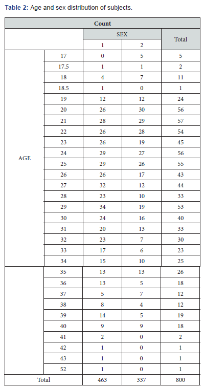
Data obtained was compiled on a MS Office Excel Sheet (v 2010). Data was subject to statistical analysis using Statistical package for social sciences (SPSS v 21.0, IBM). Reliability between the 3 observers was done using Intra class correlation (ICC) using single measures exact variables test (Table 1). Age and sex distribution of subjects has been depicted (Table 2). Descriptive statistics for both males and females for both 38 & 48 has been depicted. Using linear regression model, predication of age formula for both males and females has been generated. For all the statistical tests, p<0.05 was considered to be statistically significant, keeping α error at 5% and β error at 20%, thus giving a power to the study as 80%.


Results
Reliability between the 3 observers was done using Intra class correlation (ICC) using single measures exact variables test (Table 1). There was significant reliability between three observers. Table 3-6 shows statistical data concerning the age for both characteristics of teeth 38 and 48 per sex. For radiographic visibility of 38 in case of males showed following results- There was a statistically highly significant difference seen for the means age between the score categories (p<0.01) with mean values highest in score 3 & least in score 0. Score 0 first observed at age 18 years having mean age for score 0 was 22.23. Earliest sign of score 1 and 2 was observed at 19 years having mean ages at 25 and 25.4 respectively. Score 3 first observed at 20 years having mean age of 30.4


For radiographic visibility of 48 in case of males showed following results
There was a statistically highly significant difference seen for the means age between the score categories (p<0.01) with mean values highest in score 3 & least in score 0. Score 0 first observed at age of 17.5 years with mean age of 22 years. Score 1 and 2 first observed at age of 19 years with mean ages of 24.8 and 25.7 respectively. Earliest sign of score 3 was observed at age of 20 years with mean age of 30.5 years.
For radiographic visibility of 38 in case of females showed following results
There was a statistically highly significant difference seen for the means age between the score categories (p<0.01) with mean values highest in score 1 & least in score 0. First sign of score 0 was observed at 17 years with mean age of 21.4 years. Score 1 first appeared at age of 18 years with mean age of 23.9 years. Score 2 and 3 first observed at 20 years with mean age of 26.6 and 29.1 respectively.
For radiographic visibility of 48 in case of females showed following results
There was a statistically highly significant difference seen for the means age between the score categories (p<0.01). First appearance of score 0 was observed at 17 years with mean age 21.4 years. Score 1 first observed at 18 years with mean age of 23.7 years. Score 2 and 3 first observed at 19 and 20 years respectively with mean ages of 26.9 and 29.5 years.
Discussion
Forensic medical research mainly dealt with age estimation of living individuals. Though mineralisation stages of third molar and its eruption are main criteria in dental age estimation; they appear not be suitable for validation of completion of 18th and 21st years of age. The assessment of the periodontal ligament on radiographs of the upper jaw may generally be problematic as the maxillary wisdom teeth are often overshadowed by bone structures. Therefore, the study was restricted to lower third molars. Root development of the wisdom tooth is completed around the age of 20 years, but in a few cases, it has been shown that individuals could be under 18 years of age. Hence it is difficult to exclude this possibility in a given case.
The disappearance of the periodontal ligament is an optical phenomenon. The biological background for this may be that the membrane becomes so narrow that one cannot see it on radiographs. We Observed that radiographic image of periodontal ligament disappears some after year 20. This finding is in correlation with the finding which was observed in the study done by Olze et al. [1] in 2010. Olze et al. [1] in his study observed that score 1,2 and 3 appeared at above 18 years in both males and females. This finding is same as our study as in our study minimum age for score 1,2 and 3 were observed at or above 18 years of age in both the males and females.
Timme et al. [10] in their study observed that minimum age for scores 2 and 3 were at or above 21 years of age This finding is not correlating with the findings which were observed in Olze et al. [1] study and our study. In our study minimum ages for score 2 and 3 were 19 or 20 years. As from results of our study it was observed that score 0 first appeared below age 18 years in both males and females and score 1,2 and 3 were observed at or above 18 years of age. We can conclude that if score 1 or 2 or 3 is present for an individual; that individual should be at or above 18 years of age.
Limitations of this study
• Our study did not show any positive results for ages below or above 21 years. • This study included 800 panoramic radiographs of Indian population which were available at an institute, larger population coverage is needed for more accurate results.
References
- Olze A, Pynn BR, Kraul V, Schulz R, Heinecke A, et al. (2010) Studies on the chronology of third molar mineralization in first Nations people of Canada. Int J Legal Med 124(5): 433-437.
- Pérez Mongiovi D, Teixeira A, Caldas IM (2015) The radiographic visibility of the root pulp of the third lower molar as an age marker. Forensic Sci Med Pathol 11(3): 339-344.
- Timme M, Timme WH, Olze A, Ottow C, Ribbecke S, et al. (2017) The chronology of radiographic visibility of the periodontal ligament and the root pulp in lower third molars Sci Justice 57(4): 257-261.
- Demirjian A, Goldstein H, Tanner JM (1973) A new system of dental age assessment. Hum Biol 45(1973): 211-227.
- Gustafson G (1957) Aldersbestamningar pa tander, Odont. Tidskr 55: 556-568.
- Orhan K, Ozer L, Orhan AI, Dogan S, Paksoy CS (2007) Radiographic evaluation of third molar development in relation to chronological age among Turkish children and youth. Forensic Sci Int 165(1): 46-51.
- Prieto JL, Barbería E, Ortega R, Magaña C (2005) Evaluation of chronological age based on third molar development in the Spanish population. Int J Legal Med 119(6): 349-354.
- Caldas IM, Júlio P, Simões RJ, Matos E, Afonso A, et al. (2011) Chronological age estimation based on third molar development in a Portuguese population. Int J Legal Med 125(2): 235-243.
- Zeng DL, Wu ZL, Cui MY (2010) Chronological age estimation of third molar mineralization of Han in southern China. Int J Legal Med 124(2): 119-123.
- Ritz Timme S, Cattaneo C, Collins MJ, Waite ER, Schütz HW, et al. (2000) Age estimation: The state of the art in relation to the specific demands of forensic practise. Int J Legal Med 113(3):129-136.
- Christensen AM, Crowder CM (2009) Evidentiary standards for forensic anthropology. J Forensic Sci 54(6): 1211-1216.


