Age Estimation Using Destructive and Non- Destructive Forensic Dental Methods on an Archeological Human Sample from a 16th - 18th Century Nunnery in Brussels, Belgium
Pilar Cornejo Ulloa1, Kim Quintelier2, Wim Van Neer2,3 and Patrick Thevissen1*
1Department of Imaging and Pathology, KU Leuven, Leuven, Belgium
2Royal Belgian Institute of Natural Sciences, Belgium
3Laboratory of Biodiversity and Evolutionary Genomics, Belgium
Submission: February 07, 2019; Published: March 18, 2019
*Corresponding author:Patrick Thevissen, Department of Imaging & Pathology,Campus Saint-Raphael, Belgium
How to cite this article:Pilar Cornejo Ulloa, Kim Quintelier, Wim Van Neer, Patrick Thevissen. Age Estimation Using Destructive and Non-Destructive Forensic Dental Methods on an Archeological Human Sample from a 16th - 18th Century Nunnery in Brussels, Belgium. J Forensic Sci & Criminal Inves. 2019; 11(4): 555816. DOI: 10.19080/JFSCI.2018.11.555816.
Abstract
Introduction: Dental age estimation can be performed both in living and deceased individuals. In archeology, few studies have tested the reliability of forensic dental age estimation methods complementary to the usually applied anthropological methods. Objectives: Destructive and non-destructive forensic dental age estimations were applied on an archeological sample in order to compare their consistency with earlier obtained anthropological age estimates. Materials and Methods: One hundred and thirty four teeth from 24 archeological individuals were analyzed using Kvaal, Kvaal and Solheim, Bang and Ramm, Lamendin, Gustafson, Maples, Dalitz and Johanson’s forensic dental age estimation methods. Results: The range of the forensic dental age estimates (point predictions) was wider than the age ranges obtained by the anthropologist. Furthermore, a high variability in the forensic dental age predictions could be observed in each subject. Discussion: The wider variation observed comparing the forensic dental and the anthropological results are mainly due to the necessity to apply tooth position specific forensic dental methods in an archeological sample with incomplete dentitions and to diagenetic changes in the archeological collection.
Keywords: Forensic odontology; Dental age estimation; Archeology; Anthropologic age estimation
Abbrevations: CE: Common Era; FDI: Fédération Dentaire Internationale
Introduction
Identification of human remains and the estimation of their biological age are principal investigations in bioarchaeology and forensic sciences. In the former, the age of an individual is essential for further analysis of the demographic and adaptive characteristics of the studied population [1]. In the latter, age estimations are most important as part of human identification and in cases of living individuals without or with doubted age documentation [2]. Between 2004 and 2005, archaeological rescue excavations took place at the site of the former Poor Clare Nunnery in Brussels [3]. The nunnery was founded at the beginning of the 16th century of the Common Era (CE) and abandoned by the end of the 18th century CE. During the archaeological excavation, preceding the construction of an underground parking lot, 51 articulated skeletons were unearthed. The human remains pertaining to 48 adults (29 females, 15 males and 4 indeterminate individuals) and three sub-adults were examined using osteological sex and age estimation methods. Sex estimation was based on the morphological characteristics of the cranium, mandible and pelvis Ferembach, et al. [4] & Quintelier et al. [5]. Age estimation was primarily based on degenerative changes of the pubic symphysis and the auricular surface of the ilium Lovejoy, Suchey, Brooks, Quintelier et al. [5-8]. Dental wear, cranial suture closure, degenerative changes at the sternal end of the 4th rib were only carried out as supplementary age estimation techniques Hunger, İşcan, Quintelier et al. [7,9,10] & Maat [11]. No dental age estimation had been conducted by the anthropologists
Different forensic dental age estimation methods have been developed to estimate the age from pre-natal to adult. In children and sub-adults, dental development has been studied and documented. In adults, morphological changes in teeth were the main studied variables [12]. They have advantages in terms of simplicity, cost and time and can easily be applied on archeological samples [13-20] Kvaal et al. [21]. All existing forensic dental age estimation methods provide an estimated age (point prediction) and most of them report a related measure of uncertainty [22].
The aim of this study was to apply different forensic dental age estimation methods on the Poor Clare Nunnery collection and to compare the uniformity between the obtained dental age estimations and earlier obtained anthropological age estimates [23,24].
Materials and Methods
The skeletal remains of 24 individuals from the post-medieval Poor Clare Nunnery in Brussels were retained for study based on the presence of either premolars, canines or incisors. This material belongs to the Heritage Service of the Brussels Capital Region, where it is stored. For the data collection it was allowed to photograph and radiograph all teeth, to extract maximum seven teeth per individual and to cut one extracted tooth from each studied subject. The extracted teeth were kept in individually labeled bags and returned to the respective skeleton after examination. The present study received approval by the Ethics Committee of the University Hospitals Leuven on November 24th, 2015.
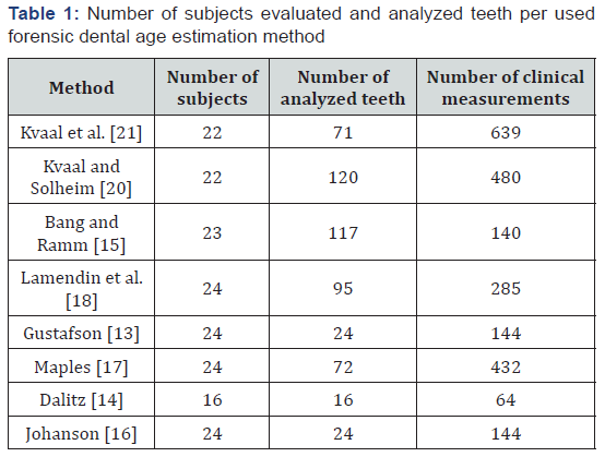
One hundred and thirty four teeth were extracted, i.e. 56 incisors, 27 canines, 23 first premolars and 28 second premolars. Teeth with extensive caries compromising the pulp or with cracks through the enamel complicating the radiographical analyses were not selected. Teeth with hypercementosis, interfering with the morphological analysis, were also excluded. On each extracted tooth, the corresponding tooth position specific forensic dental age estimation methods described in the respective following publications were applied: [13-18,20] Kvaal et al. [21] (Table 1). During the tooth selection, priority was given to upper central incisors since they were described by three different authors as better age predictors compared to the other teeth [18, 19,21].
From each extracted tooth, bucco-lingual periapical radiographs were taken using a NOMAD® (Aribex Inc., Orem, Utah, USA) portable unit. A fixed output of 60kV was used and the exposure time varied from 0.18 to 0.20 seconds, depending on the tooth type. Each tooth was attached to the Sigma M sensor (Aribex Inc., Orem, Utah, USA) using transparent tape and then radiographed mimicking the parallel technique. The images were captured and processed using Cliniview® software (Instrumentarium Dental, Tuusula, Finland) and exported in .jpg format for further analysis.
Two groups of data were collected: the first on intact and the second on sectioned teeth. On the intact teeth, radiographical and clinical measurements were established according to Kvaal et al. [21] (Figure 1). The radiographical measurements were performed after importing the radiographs in ADOBE Photoshop C5® (Adobe systems, San José, California, USA).
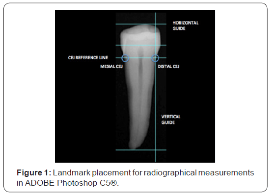
Each tooth was oriented using the cemento enamel junction (CEJ) as landmarks. A line was drawn from the mesial CEJ to the distal CEJ. The tooth was then rotated to make that line the horizontal. Subsequently, guides were dragged to the desired landmarks and the distance between them was measured. This information was recoded and integrated in the correspondent formulas proposed by Kvaal et al. [21] and Kvaal and Solheim [20,19].
The 24 sectioned teeth were cut longitudinally, using a hand piece and a dental stone grinder, described as the half tooth technique by [25]. The obtained sectioned teeth were photographed at the cut side together with a millimeter grid on a transparent layer, using a Dino‐Lite® digital microscope (AnMo Electronics Corp., New Taipei City, Taiwan).
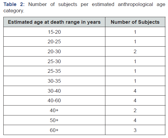
The anthropological age estimations were reported as the 5, 10, or 20 years age range or the age range older than 40, 50 or 60 years, the obtained osteological point estimates were fitting in respectively (Table 2). When an age estimation method required implementing the subjects sex the anthropological sex estimates were used.
The point estimates of the forensic dental age estimation methods and their mean were calculated and compared with the anthropological age ranges in four groups: per subject, per applied forensic dental age estimation method, per tooth position, per tooth type. Descriptive statistics were used to compare the forensic dental and anthropological age estimations.
Results
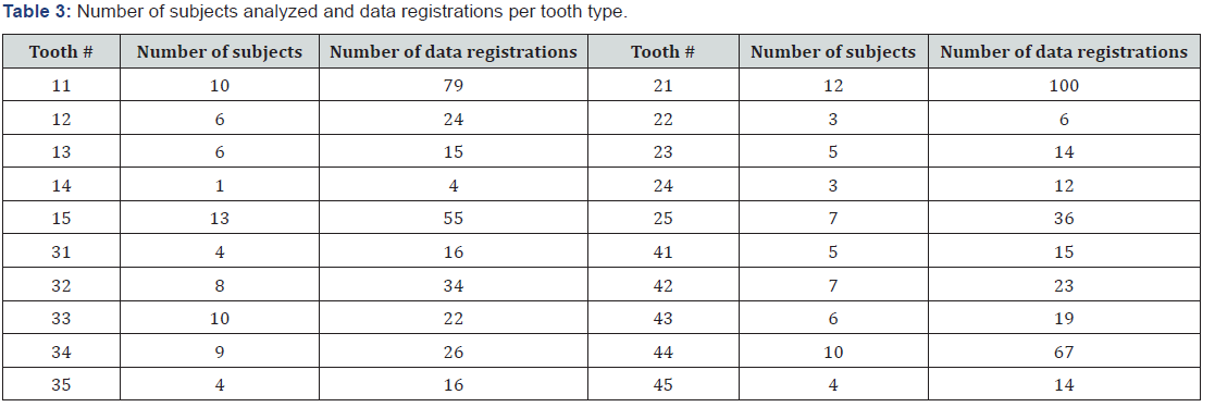
Five hundred and thirty nine data registrations were performed on 133 teeth. Not all subjects had the same number of teeth qualifying for inclusion, nor were all qualifying teeth in a condition that allowed to apply the age estimation methods. The number of teeth analyzed per applied forensic dental age estimation method was reported in (Table 1). The number of data registered on the different tooth positions (Fédération Dentaire Internationale (FDI) tooth numbering system) was shown in (Table 3).
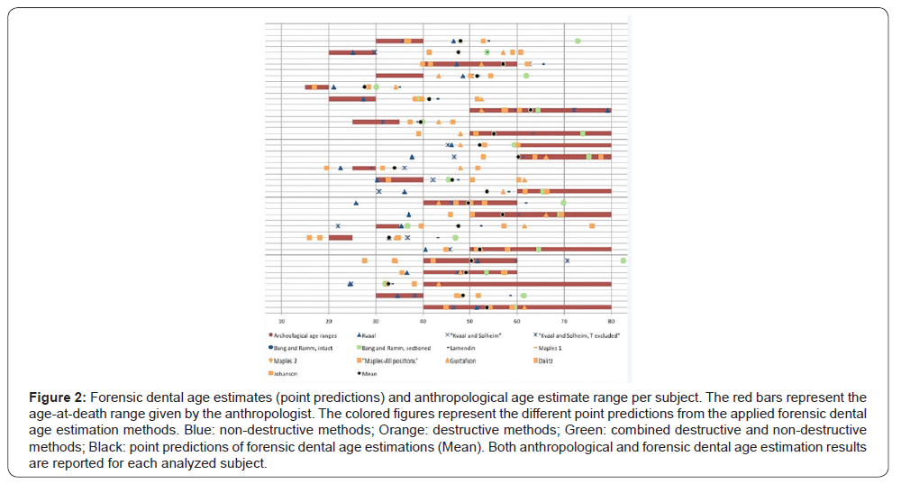
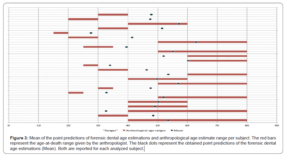
Per subject all the forensic dental age estimates were compared with the anthropological age estimates. A high variability in forensic dental age estimates was noted with the majority of them falling out of the anthropological age ranges (Figure 2). Only in six out of 24 subjects did the majority of dental age estimates fall within the anthropological age range. Furthermore only nine mean forensic dental age estimates fell in the corresponding anthropological age range (Figure 3).
Grouping the forensic dental age estimates per used method revealed a high variability between them (Figure 2). The destructive methods showed slightly higher agreement with the archeological age estimates than the non-destructive ones. [16,17] rendered the highest agreement with ten forensic dental point estimates falling within an anthropological age n range. Kvaal et al. [21] rendered the lowest agreement with eight correspondences.
Grouping the forensic dental age estimates according to tooth position, the upper central incisors (n=22) showed the highest agreement (50%) with the anthropological results. Similarly, grouping according to tooth type, the incisors (n=56) showed the highest agreement (58%). Among all the applied forensic dental methods, only [20] required the (estimated) sex for tooth 32 and 42. Seven teeth were analyzed using the equations proposed by these authors and in six cases, the obtained point estimates were older than the anthropological age range
Discussion
When performing age estimations, the gold standard to validate the prediction performances is the real age at death of the analyzed individuals. As is always the case when dealing with archeological collections, this information was lacking and the only existing age estimates eligible to be used as reference were the anthropological age estimates. Moreover, demographic information on Brussels, available in historical sources dating to the 16th century, suggests a lifespan of up to 80 years [26]. The opportunities to work with a well preserved archeological collection and even more, to obtain permission to destroy part of it, are scarce. For these reasons it was decided to carry out the study and to concentrate on the differences in results obtained with forensic methods compared to those from a classical anthropological approach.
To identify factors influencing the age estimation outcomes the obtained age estimates were analyzed in four groups: per used subjects, per age estimation method, per tooth position and per tooth type
The first group allowed for a direct comparison with the age prediction results obtained by the anthropologist. The observed high variability in the dental point predictions made it impossible to establish a narrower age range than the anthropological one. Although all forensic dental age outcomes provided measures of uncertainty related to each point estimate, performing a metaanalysis was not considered due to the already known high variability in obtained point predictions.
The second group showed a high variability between the mean age estimates per forensic dental age estimation method. This might be explained by the state of preservation of the skeletal remains. An alteration of the macro- and micro-structure due to environmental stress affects mainly the root transparency of the tooth [27,28]. This could be the cause of inaccurate measurements in forensic dental age estimation methods based on this parameter [13-18,20]. Moreover, root transparency has been documented to be prone to structural alterations due to taphonomic effects [27,29,30]. For this reason, Vlèek & Mrklas [30] suggested to either exclude or modify root transparency based methods in older samples.
On the other hand, secondary dentin based methods such as [20], avoid the influence of the age-prediction-altering taphonomic effects [27-29]. During the unearthing of the skeletal remains, it was possible to determine that the examined subjects were initially buried in wooden coffins but these disintegrated over time, leaving the skeletons in direct contact with the soil [24]. Undoubtedly the teeth were affected by diagenetic processes, altering their microstructure and, therefore, the reliability of the data registration
The third group, based on tooth position indicated that forensic dental age estimates from tooth 11 and 21 matched the anthropological results best (50% agreement). The sample size, its heterogeneity in present teeth and the restrictions of teeth allowed to be extracted or cut per subject impeded a fair comparison of results based on tooth position. Moreover, the results were certainly affected by a selection bias of extracted and cut teeth. Indeed, with the knowledge of previously mentioned restrictions and after a literature search, a priority was given to select upper central incisors. This tooth position has been demonstrated to relate best to age for the considered dental parameters [18,19,21]. The fourth group, based on tooth type, indicated that incisors hadthe highest agreement with the anthropological results (58%). However the same reflections as in the third group apply. Therefore, the fact that forensic dental age estimation methods are generally tooth-type- and even tooth-position-specific, limits their application in archeological samples with incomplete dentitions
The sex was only required on a small number of sampled teeth (n=15). Presumably, this variable did not increase errors in the forensic dental age estimates, since sex estimation methods have proved to be reliable and accurate in the hands of experienced anthropologists, especially when analyzing adults [31].
Finally it is important to note that the forensic dental age estimation methods used the present study were established on contemporary reference samples. Originally Solheim’s method was also included in the study, but unrealistic age estimates with no apparent explanation were obtained [19]. This forced its exclusion and suggested to carefully interpret the results of the included methods.
Conclusion
A wide intra-individual variation in age estimates was obtained depending on the forensic dental methods. Moreover the forensic dental and anthropological age estimations differed to a large extent. This was partly explained by the incomplete dentition, the small sample size and diagenetic alterations. Additional analyses on more extensive samples will be needed to verify whether forensic dental and anthropologicall ageing methods can reach higher agreement
Acknowledgements
1. We thank Dr. A. Degraeve, Director of the Heritage Service of the Brussels Capital Region for providing access to the collection and for permission to sample. 2. We thank Jannick De Tobel for presenting current research at the triennial IOFOS Conference Leuven, Belgium 2017.
References
- Ubelaker DH (1999) Human skeletal remains: Excavation, analysis and interpretation Taraxacum, Washington DC, USA.
- Garmus A (1996) Lithuanian forensic osteology. Vilnius: Baltic Medico-Legal Association.
- Claes B (2006) Archeologisch onderzoek ter hoogte van het voormalige Arme Klarenklooster (Br.). Archaeologia Mediaevalis 29: 28-34.
- Ferembach D, Schwidetzky I, Stloukal M (1980) Recommendations for age and sex diagnosis of skeletons. J Hum Evol 9: 517-549
- Quintelier K, Vandenbruaene M, Watzeels S (2012) A capite ad calcem. Protocol voor het macroscopisch morfologisch en metrisch onderzoek van niet-verbrand, menselijk skeletmateriaal, aangehouden binnen het agentschap Onroerend Erfgoed. Relicta 9: 263-284.
- Lovejoy CO, Meindl RS, Pryzbeck TR, Mensforth RP (1985) Chronological metamorphosis of the auricular surface of the ilium: a new method for the determination of adult skeletal age at death. Am J Phys Anthropol 68(1): 15-28.
- Brooks ST, Suchey JM (1990) Skeletal age determination based on the os pubis: a comparison of the Acsádi-Nemeskéri and Suchey-Brooks methods. Hum Evol 5(3): 227-238.
- Suchey J, Katz D (1986) Skeletal age standards derived from an extensive multiracial sample of modern Americans. Am J Phys Anthropol 69: 269.
- Hunger H, Leopold D (1978) Identifikation, Berlin.
- Işcan MY, Loth SR, Wright RK (1984) Metamorphosis at sternal rib end: a new method to estimate age at death in white males. Am J Phys Anthropol 65(2): 147-156.
- Maat GJR (2000) The impact of diet on age at death determination based on molar attrition and Forensic Odontology, Proceedings of the European IOFOS Millennium Meeting. Leuven, pp. 49-54.
- Soomer H, Ranta H, Lincoln MJ, Penttilä A, Leibur E (2003) Reliability and Validity of Eight Dental Age Estimation Methods for Adults. J Forensic Sci 48(1): 149-152.
- Gustafson G (1950) Age determination on teeth. J Am Dent Assoc 41(1): 45-54.
- Dalitz GD (1962) Age determination of human remains by teeth examination. J Forensic Sci 3(1): 11-21.
- Bang G, Ramm E (1970) Determination of age in humans from root dentin transparency. Acta Odontol Scand 28(1): 3-35.
- Johanson G (1971) Age determinations from human teeth. Odontol 21: 1-126.
- Maples WR (1978) An improved technique using dental histology for estimation of adult age. J Forensic Sci 23(4): 764-770.
- Lamendin H, Baccino E, Humbert JF, Tavernier JC, Nossintchouk RM, et al. (1992) A simple technique for age estimation in adult corpses: the two criteria dental method. J Forensic Sci 37(5): 1373-1379.
- Solheim T (1993) A new method for dental age estimation in adults. Forensic Sci Int 59(2): 137-147.
- Kvaal S, Solheim T (1994) A non-destructive dental method for age estimation. J Forensic Odontostomatol 12(1): 6-11.
- Kvaal S, Kolltvit KM, Thompson IO, Solheim T (1995) Age estimation of adults from dental radiographs. Forensic Sci Int 74(3): 175-185.
- Willems G (2001) A review of the most commonly used dental age estimation techniques. J Forensic Odontostomatol 19(1): 9-17.
- Quintelier K (2007) Antropologisch onderzoek. First progress report 2007 of the Royal Belgian Institute of Natural Sciences for the Heritage Service of the Brussels Capital Region, Brussels.
- Quintelier K (2007) Antropologisch onderzoek. Second progress report 2007 of the Royal Belgian Institute of Natural Sciences for the Heritage Service of the Brussels Capital Region, Brussels.
- Solheim T (1984) Dental age estimation. An alternative technique for sectioning teeth. Am J Forensic Med Path 2: 181-184.
- Aerts E, Daelemans F (1984) Sociaal-economische aspecten van het 16de-eeuwse Brussel. Tijdschrift voor Brusselse Geschiedenis 1-2: 5-44.
- Mandojana JM, Martin de las Heras S, Valenzuela A, Valenzuela M, Luna JD (2001) Differences in morphological age related dental changes depending on postmortem interval. J Forensic Sci 46(4): 889-892.
- Cunha E, Baccino E, Martrille L, Ramsthaler F, Prieto J, et al. (2009) The problem of aging human remains and living individuals: A review. Forensic Sci Int 193(1-3): 1-13.
- Lucy D, Pollard AM, Roberts CA (1995) A Comparison of Three Dental Techniques for Estimating Age at Death in Humans. J Archaeol Sci 22(3): 417-428.
- Vlèek E, Mrklas L (1975) Modification of the Gustafson Method of Determination of Age According to teeth on Prehistorical and Historical Osteological Material. Scripta medica 48: 203-208.
- Krishan K, Chatterjee PM, Kanchan T, Kaur, Baryah N, et al. (2016) A review of sex estimation techniques during examination of skeletal remains in forensic anthropology casework. Forensic Sci Int 261: 165.


