The Use of Mandibular and Maxillary Canine Teeth in Establishing Sexual Dimorphism in The Malaysian Population of Selangor
Yuvenya Kaeswaren* and Anita Zara Weinheimer
Department of Health and Life Sciences, Management & Science University, Malaysia
Submission: January 18, 2019; Published: March 18, 2019
*Corresponding author:Yuvenya Kaeswaren, Faculty of Health and Life Sciences, Management & Science University, Malaysia
How to cite this article:Yuvenya Kaeswaren, Anita Zara Weinheimer. The Use of Mandibular and Maxillary Canine Teeth in Establishing Sexual Dimorphism 002 in The Malaysian Population of Selangor. J Forensic Sci & Criminal Inves. 2019; 11(3): 555815. DOI: 10.19080/JFSCI.2018.11.555815.
Abstract
Sex determination is one of the major roles of forensics in establishing an individual’s identity. Teeth are excellent tools in living and non-living victim identification in the field of forensic investigations. Amongst all teeth, the mandibular and maxillary canine teeth are found to exhibit greatest sexual dimorphism. This study was conducted to investigate the effectiveness of using mandibular and maxillary canine width as well as intercanine distance in establishing sexual dimorphism in Malaysian population of Selangor. The sample comprised of 140 Malaysian individuals residing in Selangor, ages ranged between 18-30 years at a gender ratio of 1:1. The greatest mesiodistal width of the canine teeth and the intercanine distance were measured using digital vernier caliper with 0.01mm resolution. The values obtained were subjected to analysis and statistically significant sexual dimorphism was shown by the mandibular and maxillary canines. The mean values for left and right mandibular and maxillary canine widths were less for females than for males and the differences were statistically significant (P<0.01). The mean value for mandibular and maxillary intercanine distances for females were less than for males and the differences were statistically significant (P<0.01). Gender predictability using the standard canine index (SCI) and mandibular canine index (MnCI) showed acceptable results whereas the maxillary canine index (MxCI) showed poor gender predictability. Thus, the use of statistically significant sexual dimorphism in mandibular and maxillary canines in sex determination was conclusively established.
Keywords: Sexual dimorphism, Canine width, Intercanine distance, Mandibular, Maxillary, Sex determination and Canine index
Abbrevations: SCI: Standard Canine Index; MnCI: Mandibular Canine Index; MxCI: Maxillary Canine Index; mm: MilliMeter; CI: Canine Index; SD: Standard Deviation; ANOVA: Analysis Of Variance
Introduction
Odontometrics, the measurement and study of tooth size standards is most commonly used in age and sex determination as human teeth exhibit sexual dimorphism [1]. Differences in structure, size and appearance of teeth between males and females that can be applied to gender identification is referred to as “sexual dimorphism” [2]. Sex determination is also possible only with the help of teeth in situations where only fragments of jaw bones with teeth or teeth alone are found [3]. The comparison of tooth dimensions in males and females is the basis of sex determination using dental features. Canines, in particular, have the greatest degree of sexual dimorphism and are also better likely to survive severe trauma compared to the rest of the teeth [4]. Hence, odontometric sex assessment using the canine width, intercanine distance, and canine index has been proved highly valuable in gender identification
Standards of morphological and morphometric sex differences in the teeth, especially canine teeth, cannot be applied universally as it may differ with the population sample involved [5]. Cultural, environmental and racial factors are also known to influence tooth morphology [6]. To this date, there has been no literature documentation of canine width and intercanine distance for Malaysian population, specifically the population of Peninsular Malaysia. This study is therefore aimed at establishing a proof of sexual dimorphism using mandibular and maxillary canine width and intercanine distance in the Malaysian population of Selangor for forensic purposes.
Materials and Methods
Sample selection
This study consisted of 140 volunteers comprising 70 males and 70 females in the age group of 18–30 years from three large ethnic groups (Malay, Chinese & Indian) and are Malaysian citizens residing in Selangor.
Exclusion criteria
Subjects that fall under the following criteria were excluded from the study: 1. Dental caries 2. Bleeding gums 3. Occlusal abnormalities 4. Orthodontic treatment 5. Physiologic or pathologic wear and tear (attrition, abrasion, abfraction, erosion) 6. Any trauma to canine teeth 7. Crowded or excessive spacing in the anterior teeth 8. Bad oral habits 9. Persons suffering from chronic systemic diseases were also excluded
Inclusion Criteria
1. Gingiva and periodontium in a healthy state 2. Cavity free teeth. 3. Normal over jet and overbite 4. The absence of spacing in the anterior teeth. 5. Normal molar and canine relationship
Data collection
Intraoral measurements
The methodology was adapted from Kaushal and associates [1]. Study method is to measure the mandibular and maxillary canine widths and inter-canine distance of both jaws in all subjects. Measurements will be taken intraorally with the subjects in sitting position using a digital caliper with a resolution of 0.01 mm. Each measurement was measured twice and the average value was calculated. All values are in millimeter (mm) and will be rounded off to two decimal places
Width of canines
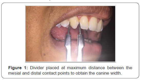
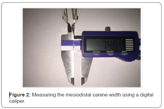
Canine width was taken as the greatest mesiodistal width which is obtained by measuring the maximum distance between the mesial and distal contact points of the canine [7] (Figures 1-2). Four readings were taken from each subject; right mandibular canine width, left mandibular canine width, right maxillary canine width and left maxillary canine width.
Intercanine distance
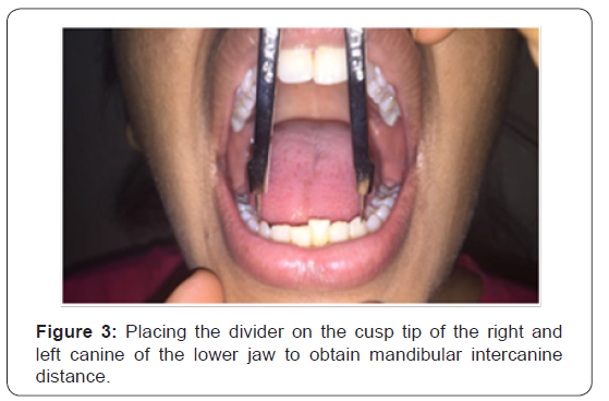
The measurement of the width from the cusp tip of the canine on the right side to the cusp tip of the canine on the left side of the jaw is known as the intercanine distance (Figure 3). Two readings were taken from each subject; mandibular intercanine distance and maxillary intercanine distance.
Canine Index
Canine index was calculated for all four canine tooth of each subject: right mandibular canine index, left mandibular canine index, right maxillary canine index and left maxillary canine index. The formula for calculating the mandibular canine index (MnCI) and maxillary canine index (MxCI) was adapted from Rao and colleagues [10] who derived it for establishing sex identity.

Antifungal resistance
The standard canine index (SCI) value was used as a cut off point to differentiate males from females. Each canine tooth will therefore have its respective SCI. It is calculated using the following formula adapted from Rao and colleagues [10]:

Where, CI = Canine index SD = Standard deviation
Sexual Dimorphism
The following formula given by Garn and associates [11] was used to calculate the sexual dimorphism of the right and left mandibular and maxillary canines:

Where, Xm = Mean value of male canine width Xf = Mean value of female canine width
Statistical Analysis
The collected data was entered in a spreadsheet (Excel 2010, Microsoft office) and is analyzed using statistical analysis software (IBM SPSS Statistics version 22). T-test and Analysis of variance (ANOVA) was done to compare the mean width of canines in males and females. Significance is set at 0.05 (P < 0.05) [Figures 1-3].
Results
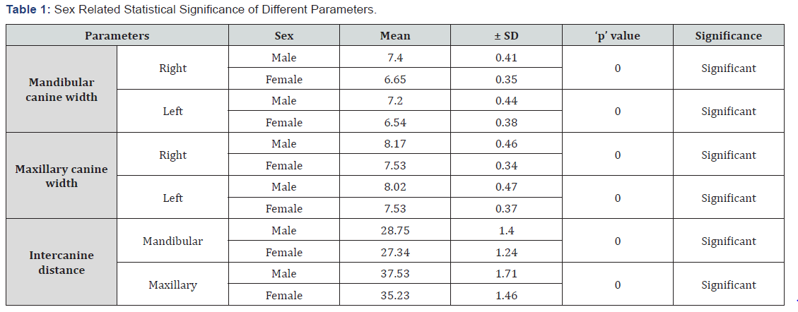
Males showed greater mean value for each canine in comparison to females in both maxillary and mandibular jaw (Table 1). Mean values of canine widths in males and females were found to be statistically significant (P < 0.01). Findings were similar for intercanine distance (p <0.01). The right mandibular canine index in males ranged from 0.2176 to 0.2940 mm with a mean of 0.2579 ± 0.0165 mm, whereas it ranged from 0.2167 to 0.2832 mm with a mean of 0.2437 ± 0.0138 mm in females (Table 2). This was statistically significant (p < 0.05). The left mandibular canine index value in males ranged from 0.2193 to 0.2923 mm with a mean of 0.2507 ± 0.0160 mm, and in females it was 0.1813 to 0.2688 mm with a mean of 0.2397 ± 0.0147 mm (Table 2). This was also statistically significant (p < 0.05).

The right maxillary canine index in males ranged from 0.1913 to 0.2457 mm with a mean of 0.2178 ± 0.0124 mm, whereas it ranged from 0.1905 to 0.2425 mm with a mean of 0.2139 ± 0.0108 mm in females (Table 2). This was not statistically significant (p > 0.05). The left maxillary canine index in males ranged from 0.1886 to 0.2482 mm with the mean of 0.2139 ± 0.0126 mm and in females it ranged from 0.1816 to 0.2407 mm with a mean of 0.2139 ± 0.0119 mm (Table 2). This was not statistically significant (p > 0.05).
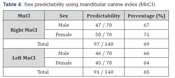
Using the right mandibular canine index (Table 3; 0.2495 mm), sex was correctly predicted only in 47 males out of 70 males and out of 70 females only 50 females were correctly predicted (Table 4). Thus, overall correct sex prediction was noted in 97 out of 140 individuals (69% accuracy). Similarly using the left mandibular canine index (Table 3; 0.2446 mm), 65% overall accuracy in sex prediction was noted with 46 out of 70 males and 45 out of 70 females correctly predicted.

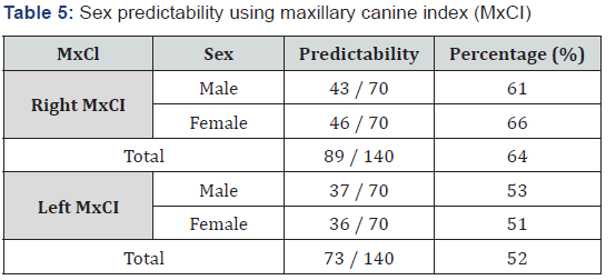
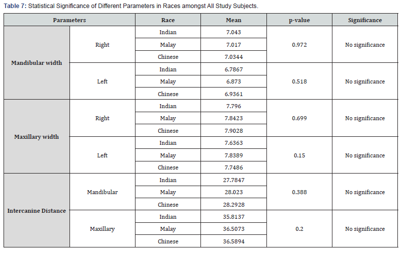
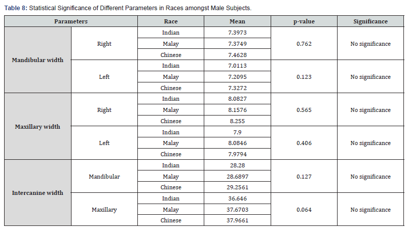
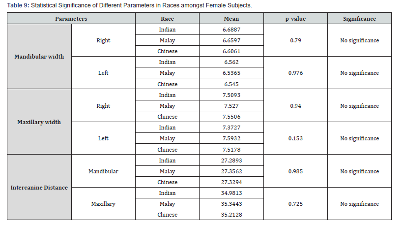
The sex predictability using right maxillary canine index (Table 3; 0.2151 mm) showed 64% overall accuracy in sex prediction with sex being correctly predicted in 89 individuals (43 males and 46 females). Using the left maxillary canine index (Table 3; 0.2135 mm), sex was correctly predicted only in 37 out of 70 males and 36 out of 70 females. Thus, overall correct sex prediction had 52% accuracy (Tables 4-6). As depicted in Table 6, the right mandibular canine exhibited the greatest percentage of sexual dimorphism of 11.28% followed by the left mandibular canine at 10.10%, right maxillary canine at 8.50% and the left maxillary canine at 6.5%. There was no statistical significance between each gender in different races (p>0.05). The canine width, canine index and intercanine distance were not influenced by race in the Malaysian population of Selangor (Tables 7-9).

Discussion
As shown in Table 1, males showed greater mean value for each canine in comparison to females in both maxillary and mandibular jaw. Mean values of canine widths in males and females on the right and left sides were compared using t-test and were found to be statistically significant (P < 0.01). Findings were similar for intercanine distance (p <0.01).
These findings were in accordance with a number of previous studies in the field. Bakkannavar and associates [8] conducted a study on the South Indian population and reported statistically significant differences in the width of the mandibular and maxillary canine teeth as well and intercanine distance between male and female (p < 0.001). They also observed that the values in males were higher than their counterparts. Al-Rifaiy and Abdullah also observed higher mean values in the width of the mandibular and maxillary canine teeth and intercanine distance amongst males than in females of the Saudi Arabian population. However, they reported no statistical significance in the canine widths but a significant difference in the intercanine distance (p < 0.0001) between male and female.
Maxillary canine index (MnCI) showed poor statistical significance whereas the mandibular canine index (MnCI) was highly significant. Moreover, sex determination using standard canine index (SCI) and mandibular canine index (MnCI) showed acceptable results (Table 4), whereas the maxillary canine index (MxCI) showed poor gender predictability (Table 5). The overall level of accuracy for sex determination using MnCI and MxCI was found to be 67% and 58% respectively.
Comparable findings were reported by a number of authors [1,9] Rai & Kaur; stating that there is an existence of a statistically significant sexual dimorphism by observing the MnCI in males and females. However in contrast with the findings in the present study, Acharya & Mainali [12] observed no statistical significant sexual dimorphism in MnCI in their study conducted on the Nepalese population. As for the right and left Maxillary Canine Index (MxCI), findings was in conformity with that of Bakkannavar and colleagues [8] and Gupta and associates who also reported no statistical significance of sexual dimorphism in MxCI values between male and female.
Amongst the four canine teeth, the right mandibular canine exhibited the highest degree of sexual dimorphism; followed by left mandibular canine, right maxillary canine and left maxillary canine (Table 6). Our findings were similar with Srivastava who reported a higher percentage dimorphism of 2.8% in right mandibular canine as compared to the left (2.3%).
There was no statistical significance between each gender in different races (p>0.05). The canine width, canine index and intercanine distance are not influenced by race in the Malaysian population of Selangor. However, this finding is not conclusive as the present study was mainly focused on determining sex. Therefore, further research can still be conducted to further validate findings focused on race.
Conclusion
Existence of significant sexual dimorphism in mandibular and maxillary canines in sex determination was conclusively established and is apparent from the study that the mandibular canine and mandibular canine index is a more reliable source for determining sex. Standard canine index is indeed very useful and would serve as a quick method for sex determination in cases when other advanced parameters for sex determination are not readily available. However, SCI is of limited value and should be used as an adjunct tool with other parameters to accurately determine sex.
References
- Kaushal S, Patnaik V, Agnihotri G (2003) . Journal of Anatomical Society of India p. 119-123.
- Kieser J A (1990) Human adult odontometrics. New York. Cambridge University Press.
- Sonika V, Harshaminder K, Madhushankari GS, Kennath JA (2011) Sexual Dimorphism in Permanent Maxillary First Molar. A Study of the Haryana Population (India). J Forensic Odontostomatol 29(1): 37-43.
- Daniel MJ, Khatri M, Srinivasan S V, Jimsha VK, Marak F (2014) Comparison of Inter-Canine and Inter-Molar Width as an Aid in Gender Determination: A Preliminary Study. Journal of Indian Academy of Forensic Medicine 168-172.
- Iscan MY, Steyn M (2013) The Human Skeleton in Forensic Medicine. Illinois: Charles C. Thomas Publisher, USA.
- Halim A (2008) Human Anatomy: Regional & Clinical for Dental Students. New Delhi: I.K. International Publishing House Pvt. Ltd, India.
- Santoro M, Ayoub ME, Pardi V, Cangialosi T (2000) Mesiodistal Crown Dimensions and Tooth Size Discrepancy of the Permanent Dentition of Dominican Americans. Angle Orthod 70(4): 303-308.
- Bakkannavar SM, Manjunath S, Nayak VC, Kumar GP (2014) Canine Index-A Tool for Sex Determination. Egyptian Journal of Forensic Sciences 5(4): 157-161.
- Reddy M V, Saxena S, Bansal (2008) Mandibular canine index as a sex determinant: A study on the population of western Uttar Pradesh. Journal of Oral and Maxillofacial Pathology 2(12): 56-59.
- Rao G N, Rao N N, Pai M L, Kotian M S (1989) Mandibular canine index - a clue for establishing sex identity. Forensic Sci Int 42(3): 249-254.
- Garn S M, Lewis A B, Swindler D R, Kerewsky R S (1967) Genetic control of sexual dimorphism in tooth size. Journal of Dental Research46(5): 963-972.
- Acharya AB, Mainali S (2009) Limitations of the mandibular canine index in sex assessment. J Forensic Leg Med 16(2): 67-69.
- Garn SM, Lewis AB, Kerewsky RS (1967) Buccolingual size asymmetry and its developmental meaning. Angle Orthodontist 37(3): 186-193.


