Lingual Morphology: A Secure Method for Forensic Identification
Madhusudan Astekar*, Shipra Saxena, Gaurav Sapra, Ashutosh Agarwal and Aditi Murari
Department of Oral and Maxillofacial Pathology, Bareilly International University, India
Submission: May 24, 2018;Published: May 30, 2018
*Corresponding author: Madhusudan Astekar, Department of Oral and Maxillofacial Pathology, Bareilly International University, India, Tel: 91 7599247899; Email: madhu.tanu@gmail.com
How to cite this article: Madhusudan A,Shipra S,Gaurav S, Ashutosh A,Aditi M. Lingual Morphology: A Secure Method for Forensic Identification. J Forensic Sci & Criminal Inves. 2018; 9(2): 555759. DOI:10.19080/JFSCI.2018.09.555759.
Abstract
Background: The Morphological aspect of the dorsal surface of the tongue is unique for each and every individual. In addition to rugoscopy and cheiloscopy, the study of lingual morphology may be one of the secure methods for identification in forensic dentistry. Hence the present study was carried out to analyze the various lingual morphological aspects like shape, colour, apex, texture, median septum and other characteristics which can aid in identification purpose.
Material and Methods: A total of 100 subjects (50 each of male and female) within an age group of 21-60 years, attending the outpatient department were included in the study. The Tongue of each subject was photographed, studied and analyzed with respect to various morphological characteristics.
Results: ‘U’ shaped tongue was identified in 71(71%) subjects, followed by ‘V’ shaped in 26(26%) and ‘W’ shaped in 3(3%) subjects. With respect to tongue color, 80(80%) subjects displayed whitish, 14 (14%) pale pink, 5(5%) yellowish pink and 1(1%) reddish pink. However, 62(62%) of the study subjects had a rough texture and the rest i.e. 38(38%) had a smooth texture. About 81(81%) subjects had rounded lingual apex, 18(18%) had pointed and 1(1%) had Septate lingual apex. Among the study subjects, median septum was visible in 42(42%) of subjects. Other morphological characteristics like whitish coat on dorsal surface and indentation on lateral border of tongue were also seen.
Conclusion: The dorsal surface of the tongue provides significant details from a morphological and structural point of view which can help in forensic identification.
Keywords: Forensics Dentistry; Unique; Forensic Human identification; Tongue Fissures; Biometric Identification.
Introduction
Forensic dentistry has turn out to be a fundamental part of forensic science. The presumption behind forensic odontology is that “no two mouths are alike” [1]. Over the years, dental or oro-facial characteristics have been serving up the judicial system in forensic identification. Dental professionals have a chief task in keeping precise dental records and providing all necessary information so that legal authorities may perhaps recognize malpractice, negligence, fraud or abuse, and identity of unknown individuals [2]. Forensic odontology involves dentist’s contribution in supporting legal and criminal issues with respect to proper handling, examination, and evaluation of dental evidence [3]. Forensic dentistry has its own limitations in human identification, when the victim is edentulous or when comparative data is inadequate in point of fact to analyze the dental arches.4 In such situation, tongue being an oral internal organ which is easy to access, is resistant to decomposition and carbonization due to its location in a humid closed cavity and thus can be of importance in post-mortem evaluation [4,5].
Despite the fact that tongue is an important organ, hitherto it has not been given equal share in the detailed anatomical studies allotted to other organs in the body [6]. As we all know that tongue is well protected in the oral cavity, it is very difficult to forge [7]. Credentials of tongue can be evaluated by visual inspection, digital photography and lingual impressions techniques [5,8]. Additional methods like biometrics validation can be used which promises to convey an intensity of distinctiveness to recognition applications, that uses tongue print, fingerprint, facial scans, iris or voice to recognize users [7,9]. Tongue print is a distinctive biometric tool which cannot be forged easily. Advantages of tongue prints over other biometric systems are genetic independence (no two tongues are similar), physical protection (well encased in the oral cavity) and its stability over time [8,9]. The analysis of the lingual morphological aspects represents criteria with evidence of uniqueness for each and every individual. Hence, the present study was carried out to analyze the various lingual morphological characteristics like tongue shape, color, texture, apex, and visibility of medial septum which can aid in identification.
Material and Methodology
A total of 100 subjects, with 50 males and females each, within the age group of 20-60 years attending the outpatient department were included in the study. The study was conducted after the approval from the Institutional ethical committee. Signed informed consent was obtained from all the individuals after explaining the study details in their vernacular language. Patients with a habit of smoking and any systemic illness were excluded out from the study. Before the clinical examination of the tongue, the patients were asked to rinse the mouth gently with water in order to remove any surface debris or food particles. The subjects were instructed to protrude their tongue, so that all the features over the dorsal surface can be appreciated. Photographs were obtained from Kodak camera (Easy share CX7310) under standard environmental, lightening condition and from a predetermined distance so as to allow the creation of a database that contains pictures of respective subject’s tongue. Various parameters such as tongue shape, colour, texture, apex, median septum and other characteristics like ankylosis, bifid tongue, microglossia, macroglossia, tongue piercing, tongue tattoos etc. (if present) were analyzed. Statistical analysis was carried out using Chi-square test to analyse prevalence of various aspects of tongue morphology with respect to age and sex of the study group.
Results
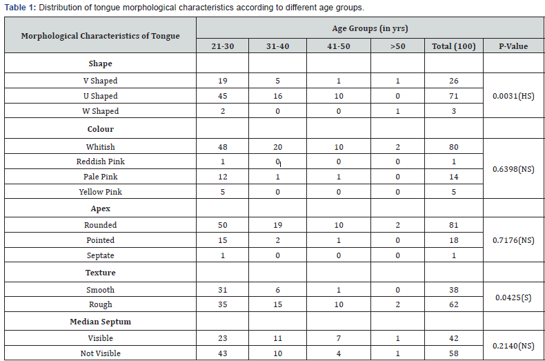
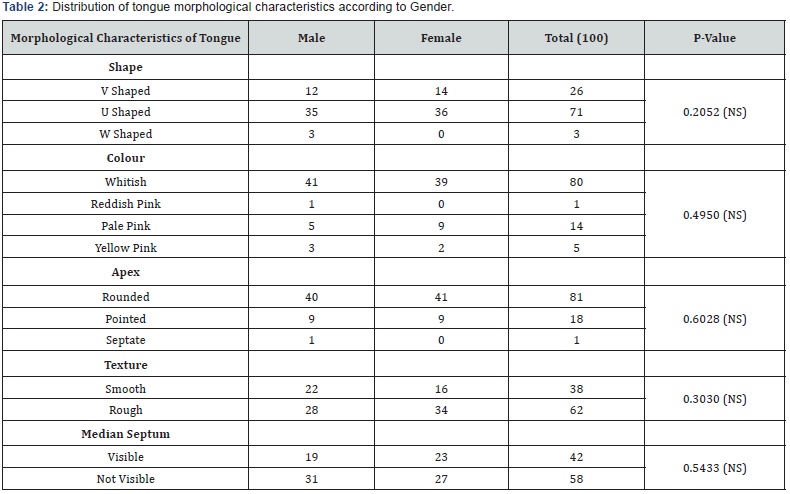
When the tongue shape (Figure 1) were analyzed ‘U’ shape (Figure 1a). was seen in 71 individuals, ‘V’ shape (Figure 1b). in 26 individuals and ‘W’ shape (Figure 1c). in 3 individuals. When the distribution of tongue shapes was statistically analyzed between the different age groups, a highly significant result was obtained with a p-value of 0.0031 (Table 1). Similarly, whereas with respect to gender, a non-significant results with a p-value of 0.205 was obtained (Table 2). When tongue color was evaluated whitish color (Figure 2a) was seen in 80 individuals, ‘Reddish pink’ (Figure 2b) in 14 individuals, ‘pale pink’ (Figure 2c) in 14 and ‘yellow pink’ (Figure 2d) in 5 individuals. When the distribution of tongue color was statistically analyzed with respect to age groups and gender, a non-significant result was obtained with a p-value of 0.6398 (Table 1) and 0.4950 (Table 2) respectively. When tongue texture was analyzed, ‘smooth’ texture (Figure 3a) was displayed by 38 individuals and rough texture (Figure 3b) by 62 individuals.


*p<0.05 consider statistically significant, HS-highly significant, NS-not significant, S-significant.

*p<0.05 consider statistically significant, HS-highly significant, NS-not significant, S-significant.
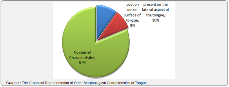
When distribution was statistically evaluated with respect to age and genders the result was significant, with a p-value of 0.0425 (Table 1). and non-significant result with a p-value of 0.3030 (Table 2) respectively. According to tongue apex, 81 individuals displayed rounded type followed by pointed by 18 and Septate by one individual. When distribution was statistically analyzed with respect to age group and gender, a non-significant result was obtained with a p-value of 0.7176 (Table 1) and 0.6028 (Table 2) respectively. The Median septum was visible (Figure 4a) in 42 study subjects and was not visible (Figure 4b) in 58 individuals. When distribution was statistically evaluated with respect to age group and gender, the result was non-significant with a p-value of 0.2140 (Table 1) and 0.5433 (Table 2) respectively. Other characteristics like a whitish coat on the dorsal surface were displayed by 9 individuals and indentation on the lateral border of the tongue (Figure 4c) by 10 of subjects (Graph 1).
Discussion
The tongue is a muscular organ situated in the oral cavity having an oral part and pharyngeal part. It has a tip, which is the anterior end of the tongue, a base forming the posterior 1/3rd of the tongue, a root which is attached to the floor of the mouth, lateral margins which are present on either side of the tongue and two surfaces i.e. dorsal and ventral. Colour of the tongue can change in numerous pathological conditions like in glossitis which is a term that is used for red and smooth tongue seen in pernicious and iron deficiency anemia. ‘Furred tongue’ presents as a partial or complete white coating on the anterior surface of the tongue. It could be due to hypertrophy of filiform papillae and hyperkeratotic as the rate of desquamation is decreased [10]. The Scalloped tongue is the term used when indentations are present on the lateral margin of the tongue due to movement of the tongue against teeth.
It could be due to tongue thrusting, bruxism, and macroglossia. Scrotal tongue or plicated tongue presents with multiple grooves and fissures that are present on the dorsal surface with a central groove present in the midline and small multiple grooves radiating from the central groove. As this condition is associated with certain syndromic cases like Down syndrome and Melkersson-Rosenthal syndrome [11], it can be helpful in individual identification. Other important clinical and morphological characteristics of tongue include a median sulcus or septum which divides the tongue centrally. It begins proximal to the apex of the tongue and ends at the foramen cecum [10]. The middle portion of the septum is the thickest and strongest. It is clearly seen in anterior two-thirds of the tongue. Its widest part is about its middle, and it diminishes gradually both anteriorly and posteriorly. It gives off attachment to the medial part of the transverse muscle [6,12]. Wide medial septum narrows towards the anterior 1/3rd, and adopts a flared shape towards the tip of the tongue [5,12].
A deep triangular septum having its tip towards the lingual apex and the base towards its “V” extending up to the anterior third of the dorsal surface can be seen. Near to the tip, a superficial, narrow and thin continuation of the septum is seen. The present study found V shape tongue as the most predominant shape among the study subjects which was consistent with the study done by Nadeem J et al., [13,14] Contrastingly comparison was not possible with Stefanescu CL et al., [15] as the shapes were classified as ovoid, ellipsoid, rectangular, pentagonal, trapezoidal to asymmetrical, the last of these being ruled out of any geometrical shapes. The present study has reported narrow, shallow and interrupted type of median septum in majority of subjects, whereas in the study by Stafenseu CL et al., [5] longitudinal median septum was barely visible with irregular bearing and shape, and small lateral grooves, followed by a discontinuity and then a small twisty area, which turns into a wide and superficial area only to change its nature by becoming narrow and deep.
The present study has encountered indentation in lateral border of tongue in 5 subjects which were inconsistent with the study done by Jeddy N [8] in 2007 who do not encounter any indentation on the lateral border. Jin-Woong park et al., [14], Manoj Diwakar [7] had carried out studies on tongue identification by Biometric analysis. The present study has tried to classify the shape of the tongue in U-Shaped, V-Shaped and W-Shaped tongue, whereas the tip of the tongue as round, pointed and Septate for ease of identification and classification. The tongue presents both geometric shape information and physiological texture information which are potentially useful for identity verification application [7,15]. The present study focuses on the importance of tongue in individual identification in the field of forensics. Furthermore, studies need to be carried out with larger sample size and different geographical distribution, to create a database.
Conclusion
The Tongue is unique to each person with respect to its shape and surface texture. Moreover, it has an advantage of being secured within the oral cavity from various environmental assaults. There are very fewer studies on tongue morphology for identification purposes in forensics. The present study is an attempt to study various morphological features of the tongue and their prevalence with age and gender which could be helpful for individual identification purposes in forensics. This study motivates to fulfill the need of more alike studies, based on morphological aspect and biometric analysis, for educational and forensic purposes. Moreover, some morphological features like color and texture could be subjective and gets altered due to various environmental, physiological and pathological causes. Thus, these factors attenuate their value for identification purposes.
References
- Dinakaran J, Dineshkumar T, Nandhini G, Priyadharshini N, Rajkumar K (2015) Gender determination using dentition. SRM Journal of Research in Dental Sciences 6: 29-34.
- Verma AK, Kumar S, Rathore S, Pandey A (2014) Role of dental expert in forensic odontology. Natl J Maxillofac Surg 5(1): 2-5.
- Singh K, Bhullar RK, Agrawal A, Chaudhary H, Anandani C, et al. (2012) Teeth and their Secrets–Forensic Dentistry. Medico-Legal Update 12: 85-89.
- Beghini M, Pereira T, Montes J, De Moura D, Dezem T (2017) Morphometric Analysis of Tongue in Individuals of European and African Ancestry. J Forensic Invest 5: 5-8.
- Stefanescu CL, Popa MF, Candea LS (2014) Preliminary study on the tongue-based forensic identification. Rom J Leg Med 22: 263-266.
- Abd-El-Malek S (1939) Observations on the morphology of the human tongue. J Anat 73: (pt2) 201-205.
- Diwakar M, Maharshi M (2013) An extraction and recognition of tongue-print images for biometrics authentication system. International Journal of Computer Applications 61(3): 36-42.
- Jeddy N, Radhika T, Nithya S (2017) Tongue prints in biometric authentication: A pilot study. J Oral Maxillofac Pathol 21: 176-179.
- Kaur G, Singh D (2015) A Novel Biometric System based on Hybrid Fusion Speech, Signature and Tongue. International Journal of Computer Applications 119: 30-39.
- Byrd JA, Bruce AJ, Rogers III RS (2003) Glossitis and other tongue disorders. Dermatol Clin 21(1): 123-34.
- Joseph BK, Savage NW (2000) Tongue pathology. Clinics in Dermatology 18: 613-618.
- Williams PL, Warwick R, Dyson M, Bannister LH (1989) Gray’s anatomy 37th edn. Churchill Livingstone, Edinburgh.
- Radhika T, Jeddy N, Nithya S (2016) Tongue prints: A novel biometric and potential forensic tool. J Forensic Dent Sci 8(3): 117-201.
- Park JW, Kang SK, Jung ST (2011) Tongue Diagnosis System Based on PCA and SVM. World Academy of Science, Engineering and Technology 5(12): 1729-1732.
- Jain AK, Bolle R, Pankanti S eds. (2006) Biometrics: personal identification in networked society Springer Science & Business Media 479.


