Sudden Death Caused by Disorders of the Cardiovascular System
Syarifah Hidayah Fatriah*
Forensic and Medicolegal, Faculty of Medicine Riau University, Indonesia
Submission: December 19, 2017; Published: January 08, 2018
*Corresponding author: Syaraifah Hidayah Fatriah Riau Univeristy, Indonesia; Email: syarifah.hf@gmail.com
How to cite this article: Syarifah H F. Sudden Death Caused by Disorders of the Cardiovascular System. J Forensic Sci & Criminal Invest. 2018; 7(1): 555704.DOI10.19080/JFSCI.2018.07.555704
Abstract
Sudden death was defined as one which has taken place within 24 hours from the onset of symptoms and signs of disease, based on World Health Organization (WHO) definition. Cardiovascular disease is the leading global cause of Sudden death and dominates as the cause of death in the future. Cardiovascular disease is a disease that involves the heart or blood vessels. There are many risk factors linked to cardiovascular disease.
Keywords: Sudden Death; Cardiovascular System; Forensic
Introduction
By the year of 2030,scientists estimates that non-contagious disease will be the cause of more than three-quarter of deaths in the world and cardiovascular disease is responsible for more number of deaths compared to contagious disease such as HIV/ AIDS, tuberculosis and malaria in developing countries, maternal and perinatal deaths and nutrition disorders. Therefore, cardiovascular disease has become the largest contributor in number of deaths in general and will continue to dominate as the cause of death in the future [1].
Discussion
Symptoms caused by heart disease appears mostly due to myocardial ischemia, myocard contraction or relaxation disorder, blood vessel obstruction or abnormal hearth rhythm/ beat frequency [2].
In general, death caused by heart disease can be classified into several types as the following :
Atherosclerosis Heart Disease
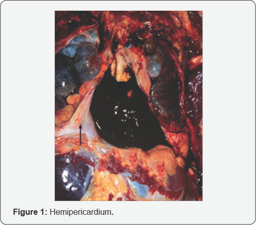
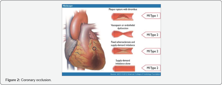
It is estimated that 75-90% of cardiac artery atherosclerosis can lead to sudden death. This incident may occur on 25-40% of individuals who previously have suffered myocardial infarction and followed by contraction or heart rhythm disorder which then leads to death. In most cases, infarct happens if an atherosclerotic plaque ruptures, forming a thrombus that may cause artery occlusion.The amount of myocard damage caused by coronary occlusion is determined by the areas supplied by the affected blood vessels, whether it is completely clogged or not. The amount of blood supplied by the collateral artery of the affected tissue, the amount of oxygen required by the myocard where blood supply suddenly becomes restricted [2]. During an autopsy, it has been discovered that approximately 75% of coronary artery constriction occurs on the branch of the left coronary artery and left ventricle rupture is the cause of most hemopericardium cases found by forensic pathologist during autopsy. Left ventricle rupture occurs approximately 3-7 days post of myocardial infraction (Figure 1)[3].

In several death cases due to atherosclerotic heart disease autopsy. Besides a constricted coronary artery, it was also where the previous history of heart disease was found to be discovered that infarct, fibrosis wth hypertrophy and left unclear, shows signs of chronic ischemia condition during ventricle dilatation has taken place (Figure 2)[3,4].

This corresponds to the literature which stated that approximately 25-40% of sudden deaths originated from coronary atherosclerosis and the only significant discovery during an autopsy was the constriction of cardiac arteries of less or more than 75% of the coronary artery lumen [5]. The estimation of cardiac artery constriction can be seen by the cross sectional area of the vessel lumen. Where the radius of artery for an amount of 50% is to be considered equal by substracting the area by 75%. An example of an autopsy case performed to a 58 year old woman who previously showed up to the emergency unit with pain complains located on the upper back area with an electrocardiography showing normal results. During an autopsy, a hemorrhage within the pericardium was discovered containing 500ml of blood clot and cardiac artery constriction. The infarction was discovered at the heart apex up to the posterior part of the left ventricle (Figures 3 & 4)[3].


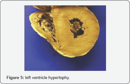
Hypertension: Blood pressure that remains uncontrolled for a long period of time can lead to many changes of the myocard and coronary artery structure and cardiac conduction system disorder. These changes may eventually induce the development of left ventricle hypertrophy, coronary artery disease and several conduction system disease, and also systolic and diastolic dysfunction of the myocard with the potentially occurring complication such as myocard infarction, cardiac arryhtmia and congestive heart failure. A macroscopic view of the heart discovered the presence of cardiac wall hypertrophy (Figure 5) [5].

Valve Disorder: The valve disorder which is oftenly associated with death case are the mitral valve prolapse, aorta valve stenosis and endocarditis.
Mitral valve stenosis: The mitral valve stenosis is the damage of heart valve characterized by the improper closing of the mitral valve. The mitral valve is a valve consisting of 2 sheaths located between the left atrium and left ventricle. This particular valve helps regulate the blood flow from the atrium towards the ventricle by opening up during the atria contraction closes during the ventricle contraction in order to prevent blood flowing back towards the atrium. In a mitral valve prolapse, the valve bulges upwards into the atrium and sometimes causing blood reentering the left atrium [6].
Aorta valve Stenosis: the aorta valve stenosis is the narrowing or occlusion of the aortal valve. The aorta valve is a valve located on the left ventricle that will open up when blood will enter the aorta in order to be distributed throughout the body. Normally, the aortal valve consist of three sheaths that will close and open, thus allowing blood to flow through.
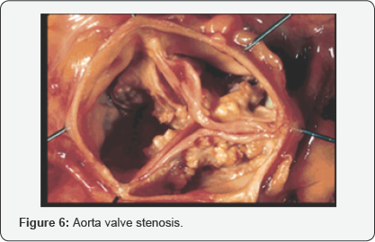
On an aorta valve stenosis, usually the valve only consist of 2 sheaths, therefore having a slightly much more narrow lumen and can potentially block blood flow. Thus, the left ventricle will have to pump stronger in order for the blood to pass the valve. Aorta valve stenosis is the result of scar tissue formation and accumulation of calcium within the valve sheath. This pasticular aorta valve stenosis only occurs after the age ofsixty but symptoms itself might only appear after the age of seventy until eighty years old. Aorta valve stenosis can also be caused by rheumatic fever on children. This condition is usually accompanied by the mitral valve disorder, either in form of stenosis, regurgitation or both. In a macroscopic view, it can be seen that the size of valve remains the same while the heart continues to expand. A valve may only consist of 2 sheaths, in which normally should be three, or having an abnormal form such as the funnel shape. The valve oftenly becomes rigid or narrows due to the accumulation of calcium precipitation (Figure 6) [7].

Endocarditis
Endocarditis is a condition characterized by the presence of infection within the endocardium. This condition is usually due to bacteria spreading from other particular parts of the body which damages the heart area. Endocarditis is a rare condition but significantly worrying due to the potential of causing heart valve destruction (Figure 7) [8].

Conclusion
Sudden death is a major source of mortality in developed countries. During an autopsy we can found hemopericardium, atherosclerosis, hypertrophy ventricel, valve stenosis or infection within the endocardium. Histopathologic examination can establish a differential diagnosis of the cause of sudden death from cardiovascular system.
References
- Valentin F and Bridget B K (2010) Promoting Cardiovascular Health in the Developing World.
- Sherwood L (2012). Fisiologi jantung dalam Fisiologi Manusia dari Sel ke Sistem. Edisi 8 EGC: Jakarta.
- Isselbacher Braundwald, Wilson, Martin, Fauci (2012) Harrison prinsip prinsip ilmu penyakit dalam Volume 3.
- 4 James JP, Jones R, Karch SB, Manlove J (2011). Simpson's Forensic Medicine 13th Edition p: 54-57.
- Dolinak D, Matshes E, Lew E (2012) Sudden Natural Death in Forensic Pathology principles and practice pp: 71-116.
- Kamran Riz (2014) hypertensive heart disease.
- Beckerman J (2013) Mitral valve prolapse.
- Michael A (2015) Stenosi Aorta.


