Ectopic Thyroid with Multinodular Goiter-Case Report
Introini Lucía1, Lago Gonzalo2, D´Albora Ricardo2 and Mintegui Gabriela1*
1Clinic of Endocrinology and Metabolism, School of Medicine, Hospital of Clinics, Uruguay
2Clinic of Otorhinolaryngology, School of Medicine, Hospital of Clinics, Uruguay
Submission: December 22, 2022; Published: January 05, 2023
*Corresponding author: Gabriela Mintegui, Clinic of Endocrinology and Metabolism, School of Medicine, Hospital of Clinics, Uruguay
How to cite this article: Introini L, Lago G, D´Albora R, Mintegui G. Ectopic Thyroid with Multinodular Goiter-Case Report. J Endocrinol Thyroid Res. 2023; 7(2): 555706. DOI:10.19080/JETR.2023.07.555706
Abstract
The ectopic thyroid gland is a rare congenital disorder. It occurs in most cases at the level of the cervical midline and rarely in lateral submandibular topography. We present the case of a 44-year-old woman who consulted for a painful, progressively growing right submandibular tumor in which a diagnosis of ectopic multinodular goiter was made and surgical treatment by resection of the thyroid tissue was decided. She always presented normal thyroid function and the reason for surgery was the neck deformity that bothered the patient and the pain. In the postoperative period, she presented TSH of 40 and levothyroxine was started. We present this case because cases of submandibular ectopic multinodular goiter as the only functioning tissue are rare.
Keywords: Ectopic thyroid; Multinodular goiter
Introduction
The ectopic thyroid gland is defined as that which isn’t in its orthotopic position in the anterior region of the neck, at the pre tracheal level between the second and fifth rings [1,2]. The thyroid gland originates from a diverticulum located in the midline between the first and second branchial arches. This begins its caudal migration along the path of the thyroglossal duct, from the foramen caecum to its final pretracheal position where it merges with the lateral primordia. The duct is reabsorbed by the sixth week.
The alteration in this migration causes ectopic topography in the midline, presenting the majority of cases (90%) at the lingual level. The distortion in the migration of the lateral primordia is posed as a possible hypothesis for the presentation of submandibular ectopic topography, being more frequent in these cases the coexistence with a normally functioning and normally positioned gland at the medial level. However, the single submandibular presentation is less common and the tissue is usually functional. Other authors suggest the existence of thyroglossal duct cysts that can move laterally adopting the submandibular topography [3,4].
Clinical Case
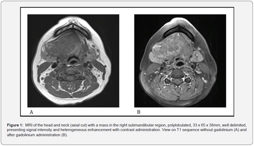
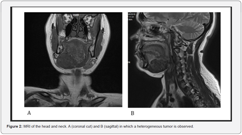
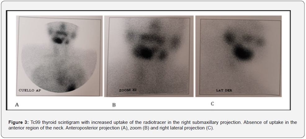
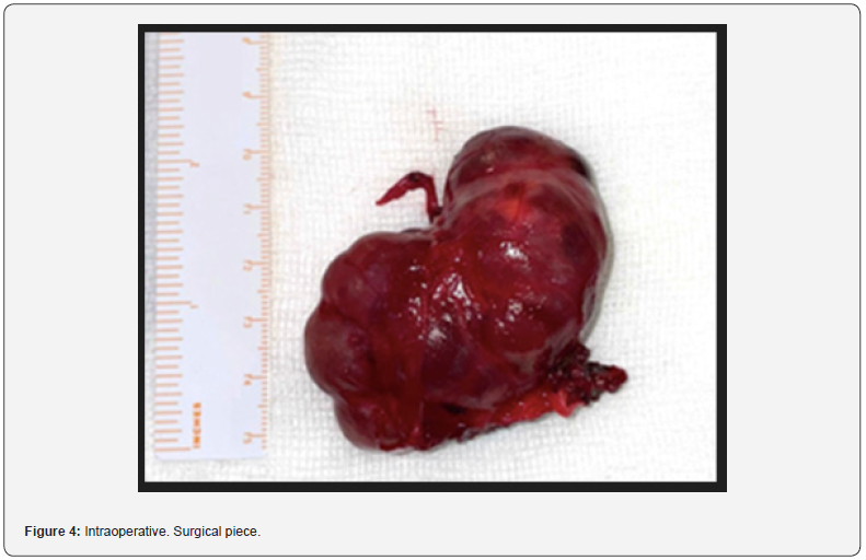
A 44-year-old woman with a history of smoking and benign breast disease. She consulted due to a right submaxillary tumor, two years old, with progressive growth, associated with pain. No dysphagia, odynophagia, hoarseness, or dyspnea. On physical examination, a tumor of 7cm in largest diameter, firm, mobile, not attached to the mandible, stood out in the neck. It wasn’t visualized in the floor of the mouth; it was painful on bimanual palpation. Imaging and cytology studies were requested for evaluation. Neck ultrasound: formation located at the level of the floor of the mouth behind and below the mylo-hyoid and genio-hyoid muscles, rounded 59 x 36 x 25mm, with mixed heterogeneous content and interior vascular signal. No alterations of the salivary glands. Magnetic resonance imaging (MRI) of the neck reported: a mass with lobulated contours, with signal intensity and heterogeneous post-gadolinium enhancement, measuring 33 x 65 x 38 mm (Figure 1&2). Functional study with thyroid scintigraphy with mTc99: increased uptake of the radiotracer in the right submaxillary projection, without uptake in the anterior region of the neck in usual thyroid’s topography (Figure 3). Fine needle aspiration (FNA) showed: numerous foamy cells with hemosiderin deposits and small, rounded cells with scant cytoplasm, isolated and arranged in conglomerates, with discrete anisokaryosis, eosinophilic substance (probably colloid) was observed; cytology that evoked the cytology of the thyroid gland. Thyroid function was always normal with TSH 2.29uUl/ml (NR: 0.27-4.2). In the postoperative period, she presented TSH of 40 and levothyroxine was started. Resection of the ectopic thyroid tissue was performed without postoperative complications with subsequent hormone replacement therapy. Pre and post-operative calcium levels remained normal without treatment. The result of the pathology showed thyroid tissue with histological changes compatible with multinodular goiter (Figure 4), without elements of malignancy. In this moment maintains normal thyroid function with levothyroxine 100mcg/day.


Discussion
The thyroid gland is normally located in the anterior region of the neck, between the second and fifth tracheal rings. It develops between the third and fourth week of gestational age, being the first endocrine gland to develop [1,2,5]. Ectopy of the thyroid gland is an infrequent clinical condition [6]. It is due to an alteration in its migration to its usual location and more frequently the ectopic tissue can be found along the path of the obliterated thyroglossal duct, from the base of the tongue to the mediastinum. It is the most frequent form of thyroid dysgenesis (48-61%), with a prevalence of 1 in 100,000-300,000 people, affecting more frequently the female sex (80%) [7].
Ectopic tissue can coexist with a normally positioned thyroid gland [6]. The lingual thyroid is the most frequent form of ectopic (90%), other positions being very infrequent. At the molecular level, genes for transcription factors such as TITF-1, TITF-2 and PAX-8 essential for the differentiation and morphogenesis of the thyroid gland have been linked. It is believed that mutations in these genes could be linked to thyroid ectopy [1,8].
The ectopic thyroid gland can be affected by any thyroid pathology that affects a gland in its normal position, such as dysfunctions, goiter, and neoplastic changes [5]. Thyroid ectopy is the most frequent cause of congenital hypothyroidism in children, although it is rarer for this to occur in adults [5]. The usual clinical presentation is asymptomatic. The symptoms are related to the size and location of the gland, as well as symptoms of dysfunction [5]. Those located in the lateral region of the neck can present as cervical tumors, which must be differentiated from salivary gland tumors, lymphadenopathy, and other subcutaneous inflammations [2,5]. There is no consensus in the literature on the assessment and management of this condition. Imaging studies (neck ultrasound, computed tomography and magnetic resonance) are the initial studies that guide the diagnosis [2,5,7].
Thyroid scintigraphy with Tc99 or I123 is crucial to determine the presence of thyroid tissue and its location, as well as to determine the presence or absence of a normally positioned thyroid gland [9], with the ectopic gland being the only functioning thyroid tissue in up to 70% of cases. the cases [10]. Likewise, PAAF is a highly sensitive procedure (95-97%) for the diagnosis of colloid tissue. Finally, the analytical study of thyroid hormones is essential to assess the presence of functional alterations of the thyroid gland [2,3,5].
Regarding the treatment of thyroid ectopy, it is recommended to take into account the patient’s age, location, regional symptoms, presence of malignancy, anesthetic-surgical risk, thyroid function. It has been proposed in the literature that in asymptomatic cases with preserved thyroid function, strict follow-up could be carried out to detect the development of complications as soon as possible. When the main discomfort is related to the size of the mass, a suppressive treatment with levothyroxine can be performed, although the result of reducing the size may be slow. Regarding surgical treatment, there is no consensus. Indications for resection of ectopic thyroid tissue generally include risk of malignancy, symptomatic patient, dysphagia, upper airway obstruction, presence of other functioning thyroid tissue, and cosmetic deformity.
It should be explained to the patient that if this is the only functioning thyroid tissue, he will need hormone replacement for the rest of his life, due to sequelae of hypothyroidism after surgery. Ablative treatment with radioiodine (I131), in those cases of patients with high surgical risk or who refuse surgery, can be considered [1,8]. The normally positioned thyroid gland is usually accompanied by four parathyroid glands. During embryonic development, the parathyroid glands originate from the third and fourth pharyngeal pouches. No associated parathyroid tissue or PTH expression has been found in ectopic thyroid tissue. This indicates their different embryonic origin [11]. In the case presented, it is evidenced by the absence of alterations in calcium levels, both preoperative and postoperative.
Conclusion
This case illustrates a rare condition of a right submandibular ectopic thyroid presenting as the only functioning thyroid tissue. These are difficult-to-managing clinical cases where the formation of multidisciplinary teams is essential to define behaviors and treatment options. This pathology should be taken into account in the differential diagnosis of oropharyngeal and cervical tumors.
References
- Guerra G, Cinelli M, Mesolella M, Tafuri D, Rocca A, et al. (2014) Morphological, diagnostic and surgical features of ectopic thyroid gland: a review of literature. Int J Surg 12(Suppl 1): S3-S11.
- Penella P, Portillo M, Álvarez R, Mesa M (2022) Bocio multinodular ectópico submandibular: a propósito de un caso y revisión de la literatura. Rev otorrinolaringol cir cabeza cuello 82(1): 65-69.
- Radkowski D, Arnold J, Healy GB, McGill T, Treves ST, et al. (1991) Thyroglossal duct remnants. Preoperative evaluation and management. Arch Otolaryngol Head Neck Surg 117(12): 1378-1381.
- Morgan NJ, Emberton P, Barton R (1995) The importance of thyroid scanning in neck lumps - a case report of ectopic tissue in the right submandibular region. J Laryngol Otol 109(7): 674-676.
- Ibrahim NA, Fadeyibi IO (2011) Ectopic thyroid: etiology, pathology and management. Hormones (Athens) 10(4): 261-269.
- Deshmukh AD, Katna R, Patil A, Chaukar DA, Basu S, et al. (2011) Ectopic thyroid masquerading as submandibular tumour: a case report. Ann R Coll Surg Engl 93(6): e77-e80.
- Santangelo G, Pellino G, De Falco N, Colella G, D’Amato S, et al. (2016) Prevalence, diagnosis and management of ectopic thyroid glands. Int J Surg 28(Suppl 1): S1-S6.
- Bersaneti J, Silva R, Ramos RR, Matsushita M, Souto L, et al. (2017) Ectopic thyroid presenting as a submental mass: A case report. Head Neck Pathol 5(1): 63-66.
- Sood A, Kumar R (2008) The ectopic thyroid gland and the role of nuclear medicine techniques in its diagnosis and management. Hell J Nucl Med 11(3): 168-171.
- Lukáš J, Drábek J, Lukáš D, Zemanová I, Rulseh A, et al. (2020) Ectopic thyroid with benign and malignant findings: A case series. Int J Surg Case Rep 66: 33-38.
- Gu T, Jiang B, Wang N, Xia F, Wang L, Gu A, et al. (2015) New insight into ectopic thyroid glands between the neck and maxillofacial region from a 42-case study. BMC Endocr Disord 15(1): 72-78.


