Visual Vignette: Adrenal Myelolipoma
Nanik Ram, Abdul Aziz and Sajjad Ali Khan*
Department of Medicine, Aga Khan University Hospital, Pakistan
Submission: November 02, 2021; Published: December 10, 2021
*Corresponding author: Sajjad Ali Khan, Department of Medicine, Section of Endocrinology, Aga Khan University Hospital, Karachi, Pakistan
How to cite this article: Nanik R, Abdul A, Sajjad A K.Visual Vignette: Adrenal Myelolipoma. J Endocrinol Thyroid Res. 2021; 6(4): 555691. DOI: 10.19080/JETR.2021.06.555691
Case Report
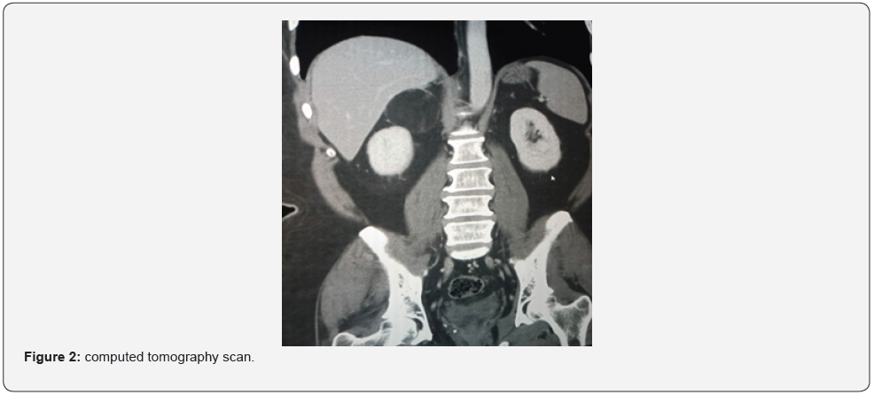
53-year male known case of ischemic heart disease s/p angioplasty (2008), incidentally found to have adrenal mass on ultrasound abdomen as part of investigation for abdominal pain. There were no symptoms of weight loss, decreased appetite, hematuria or episodic hypertension. On examination he had normal vitals and systemic examination was normal. His lab works up showed: Creatinine = 1 mg/dl, Serum sodium = 142 mmol/L, potassium = 5.5 mmol/L, VMA = 12.1 mg/24 hours (< 13.6 mg/24 hours), aldosterone = 6.60, DHEA-SO4= 153 microgram/dl (80-560), low dose overnight dexamethasone suppression test = 1.1 (normal). His CT scan is shown in Figure 1 and 2.
What is the diagnosis?
Answer: Adrenal myelolipoma: CT scan (Figure 1,2 [arrows]) showed well defined rounded mass in the right suprarenal space, arising from the right adrenal gland, which contain predominantly fat with Hounsfield unit of -95, findings are compatible with an adrenal myelolipoma measuring approximately 60 * 49.3 mm.
Left adrenal gland appear unremarkable. Myelolipoma is a rare, benign and non-functional neoplasm that primarily occurs in the adrenal gland and is made up of macroscopic fat and mature hematopoietic tissue, like bone marrow [1]. Because of the widespread use of ultrasonography, computed tomography (CT), and magnetic resonance imaging (MRI), myelolipoma has become more common, accounting for 10–15 percent of all incidental adrenal masses [2]. CT shows myelolipoma as a well-delineated mass with heterogeneous attenuation and low-density fat tissue. Management of adrenal myelolipoma should be individualized. If the size is less than 4cm, conservative treatment can be used with imaging as a follow-up. Surgery is recommended for symptomatic tumors, lesions that are rapidly expanding, or tumors that are larger than 6 cm in diameter [3]. This is to avoid the risk of life-threatening rupture and hemorrhage. The laparoscopic approach is more superior to the open method. Adrenal myelolipoma has a good prognosis, with recurrence-free survival rates of up to 12 years [3].


References
- Feng C, Jiang H, Ding Q, Wen H (2013) Adrenal myelolipoma: a mingle of progenitor cells? Med Hypotheses 80(6): 819-822.
- Wani N, Kosar T, Rawa I, Qayum A (2010) Giant adrenal myelolipoma: incidentaloma with a rare incidental association. Urol Ann 2(3): 130.
- M Ramirez, S Misra (2014) Adrenal myelolipoma: to operate or not? A case report and review of the literature. Int J Surg Case Rep 5(8): 494-496.


