




 IJCSMB Home



Pantoprazole Attenuates Cardiac Hypertrophy: Insights from Network Pharmacology, Molecular Docking, and In Vitro Validation
Anil Kumar Prajapati1,2* and Gaurang Shah2
1 Indus Institute of Pharmacy and Research, Indus University, Ahmedabad, Gujarat, India
2 Pharmacology Department, L. M. College of Pharmacy, Ahmedabad, Gujarat, India
Submission:September 18, 2026; Published:September 28, 2026
*Corresponding author: Anil Kumar Prajapati, 1Indus Institute of Pharmacy and Research, Indus University, Ahmedabad, Gujarat, India
How to cite this article: Anil Kumar P, Gaurang S. Pantoprazole Attenuates Cardiac Hypertrophy: Insights from Network Pharmacology, Molecular Docking, and In Vitro Validation. Int J Cell Sci & Mol Biol. 2026; 8(3): 555736. DOI: 10.19080/IJCSMB.2026.08.555736
Abstract
Purpose: Cardiac hypertrophy is a major pathological process contributing to the progression of heart failure and other cardiovascular complications. Inflammatory signaling pathways have been increasingly recognized as key regulators of hypertrophic remodeling. The present study aimed to investigate the potential cardioprotective effects of pantoprazole and to identify its molecular targets associated with cardiac hypertrophy using an integrated network pharmacology, molecular docking, and in vitro experimental approach.
Methods: Network pharmacology analysis was performed to identify overlapping targets between pantoprazole and cardiac hypertrophy–related genes using Gene Cards, OMIM, and Swiss Target Prediction databases. A protein–protein interaction (PPI) network was constructed using STRING and Cystoscope to determine key hub targets. Gene Ontology (GO) and KEGG pathway enrichment analyses were conducted to identify relevant biological pathways. Molecular docking was carried out using Auto Dock Vina to evaluate the binding affinity of pantoprazole with selected targets. In vitro validation was performed using H9C2 (2-1) cardiomyocytes exposed to high-salt conditions to induce hypertrophy.
Results: Network pharmacology identified 29 overlapping targets between pantoprazole and cardiac hypertrophy, with key hub genes including JAK2, CXCR2, MMP9, RAF1, and EGFR. Pathway enrichment analysis indicated significant involvement of inflammatory and chemokine signaling pathways. Molecular docking suggested favorable interactions of pantoprazole with JAK2 and CXCR2. In vitro experiments demonstrated that pantoprazole significantly reduced high salt–induced cardiomyocyte hypertrophy in H9C2 (2-1) cells. Although α-smooth muscle actin (α-SMA) levels showed a decreasing trend following pantoprazole treatment, the reduction was not statistically significant.
Conclusion: These findings suggest that pantoprazole may attenuate cardiac hypertrophy through modulation of inflammatory signaling pathways. The results provide preliminary evidence supporting the potential repurposing of pantoprazole as a therapeutic candidate for preventing pathological cardiac remodeling.
Keywords: Pantoprazole; H9C2 (2-1) cell line; Cardiac Hypertrophy; In silico studies
Abbreviations: α-SMA: Alpha-Smooth Muscle Actin; BC: Betweenness Centrality; BP: Biological Process; CC: Cellular Component / Closeness Centrality; CXCR2: C-X-C Motif Chemokine Receptor 2; DC: Disease Control; DMEM: Dulbecco’s Modified Eagle’s Medium; DMSO: Dimethyl Sulfoxide; ECM: Extracellular Matrix; EGFR: Epidermal Growth Factor Receptor; ELISA: Enzyme-Linked Immunosorbent Assay; FBS: Fetal Bovine Serum; GO: Gene Ontology; H9C2 (2-1): Rat Cardio myoblast Cell Line; HUGO: Human Genome Organization; JAK2: Janus Kinase 2; KEGG: Kyoto Encyclopedia of Genes and Genomes; MAPK1: Mitogen-Activated Protein Kinase 1; MMP9: Matrix Metalloproteinase-9; MTT: 3-(4,5-Dimethylthiazol-2-yl)-2,5-Diphenyltetrazolium Bromide; NC: Normal Control; OMIM: Online Mendelian Inheritance in Man; PARP1: Poly(ADP-ribose) Polymerase 1; PDB: Protein Data Bank; PPI: Protein–Protein Interaction; PTZ: Pantoprazole; RAF1: RAF Proto-Oncogene Serine/Threonine-Protein Kinase; SEM: Standard Error of the Mean; SMILES: Simplified Molecular Input Line Entry System; STRING: Search Tool for the Retrieval of Interacting Genes/Proteins
Introduction
Cardiac hypertrophy, defined as a prolonged increase in cardiac mass, occurs due to chronic systolic or diastolic wall stress [1]. Cardiac hypertrophy develops as a pathological response to various cardiovascular stressors, including myocardial infarction, chronic systemic hypertension, valvular heart disease, and persistent inflammatory processes. Inflammation plays a crucial role in initiating and promoting cardiac remodeling by activating multiple signaling pathways that contribute to hypertrophic growth and fibrosis. Due to the high prevalence of these conditions, there is an increasing need to develop strategies that can prevent or attenuate cardiac hypertrophy. Sustained cardiac injury and subsequent structural remodeling can lead to serious complications such as arrhythmias, heart failure, and sudden cardiac death. Therefore, targeting inflammation and inhibiting cardiac hypertrophy are important therapeutic approaches for reducing cardiovascular morbidity and mortality [2,3].
In therapeutic settings, proton pump inhibitors are frequently recommended to treat gastrointestinal disorders such as Barrett's oesophagus, Zollinger-Ellison (ZE) syndrome, and gastro-oesophageal reflux disease (GERD) [4]. Recent preclinical investigations indicate that PPIs modulate pro-inflammatory cytokines to reduce tissue inflammation [5,6]. Furthermore, studies demonstrate that proton pump inhibitors can suppress profibrotic molecules while stimulating antifibrotic pathways to diminish tissue fibrosis [7]. A prior data mining investigation identified a significant association between proton pump inhibitor utilization and enhanced outcomes in heart failure, especially in instances attributed to ischemic and non-ischemic heart disorders, encompassing cardiac hypertrophy [8]. A recent observational study provided more support for this, showing that patients with heart failure who used proton pump inhibitors had a better prognosis [9]. Additionally, there is evidence that proton pump inhibitors enhance endothelial function and control inflammatory reactions [10].
Additionally, research has revealed that the stomach isoform of H⁺/K⁺ ATPase is present in the heart, and pantoprazole has been shown to decrease cardiac contractility in both human and rabbit heart tissue [11]. These results suggest that proton pump inhibitors might directly affect heart failure. The very low pH of parietal cells is thought to be the reason for proton pump inhibitors specificity for proton pumps, as they need an acidic environment to activate. It is possible, therefore, that proton pump inhibitor prodrugs directly affect the heart without the usual activation mechanism since cardiac cells do not have a low enough pH to activate proton pump inhibitors [12]. The high-salt diet model serves as a well-established representation of salt-induced cardiac hypertrophy, providing a valuable framework for investigating the impact of excessive salt intake on cardiovascular health [13]. Also, elevated salt levels have been shown to promote tissue inflammation and contribute to the development of cardiac fibrosis [14]. Preclinical studies suggest that proton pump inhibitors modulate various pro-inflammatory cytokines to regulate tissue inflammation.
Additionally, emerging evidence indicates that proton pump inhibitors inhibit profibrotic molecules, thereby mitigating tissue fibrosis; however, the precise underlying mechanisms remain unclear. To further investigate the potential cardioprotective effects of pantoprazole, we employed network pharmacology and molecular docking approaches. Network pharmacology was utilized to systematically analyze the complex interactions between pantoprazole and multiple molecular targets involved in the pathogenesis of cardiac hypertrophy. This method integrates databases and computational tools to predict drug–target interactions and construct signaling networks. Molecular docking was performed to evaluate the binding affinity and interaction patterns of pantoprazole with the critically different molecular targets identified. Additionally, an in vitro study will be performed using the H9C2 (2-1) cardiac cell line, in which cardiac hypertrophy was induced by high-salt exposure to assess the potential cardioprotective effects of pantoprazole.
Materials and Methods
Network Pharmacology
Gene Screening for Pantoprazole-Associated Disease Targets
To determine potential disease-associated targets of pantoprazole, the compound was queried in the PubChem database using the keyword “Pantoprazole,” from which its molecular structure and SMILES representation were obtained. The retrieved SMILES code was subsequently entered into the Swiss Target Prediction platform to predict possible molecular targets.
Gene Screening for Cardiac Hypertrophy Related Targets
To identify genes associated with cardiac hypertrophy, the keyword “cardiac hypertrophy” was queried in the OMIM and Gene Cards databases. The retrieved datasets were merged, and duplicate entries were removed to compile a comprehensive list of hypertrophy-associated targets. Overlapping targets between pantoprazole and cardiac hypertrophy were then determined using the Venny tool to generate a Venn diagram. The intersecting targets were imported into the STRING database to construct a protein–protein interaction (PPI) network, restricted to Homo sapiens, with the confidence score set at 0.95, while disconnected nodes were excluded. The resulting PPI network was visualized and analyzed using Cytoscape (version 3.10.3). Further network characterization was performed with the CytoHubba plugin, which calculates topological parameters, including degree, closeness centrality (CC), and betweenness centrality (BC). Targets scoring above the mean for all three parameters were designated as hub genes and selected for subsequent analysis.
KEGG Pathway Enrichment Analysis
The previously identified differentially expressed genes were analyzed for KEGG pathway enrichment using the functional annotation tool g: Profiler, with a significance threshold set at p < 0.05. Genes associated with cardiac disease, as revealed through this enrichment analysis, were subsequently selected and visualized using an online bioinformatics visualization platform.
Molecular Docking
The 3D structures of the target proteins, EGFR (PDB ID: 8SC7) [15], JAK2 (PDB ID: 6VN8) [16], MMP9 (PDB ID: 5UE4) [17], RAF1 (PDB ID: 3OMV) [18], and CXCR2 (PDB ID: 6LFL) [19] were selected from the Protein Data Bank (PDB), a digital archive of protein structural information. Auto Dock Vina performed molecular docking studies to assess the interactions between these target proteins and Pantoprazole ligands. Before docking, the protein and ligand files were prepared in PDBQT format. Preprocessing included removing water molecules and introducing polar hydrogen atoms and Kollman charges to ensure proper charge distribution and protein polarity. A receptor grid box was generated around each macromolecule, with dimensions and center coordinates tailored for each target as follows:
1. 8SC7: Grid box size of 40 × 30 × 26 Å (x, y, z) and center coordinates at 21.849 × 0.988 × 51.037 Å.
2. 6VN8: Grid box size of 32 × 40 × 34 Å (x, y, z) and center coordinates at 53.724 × -20.314 × -11.072 Å.
3. 5UE4: Grid box size of 40 × 40 × 40 Å (x, y, z) with center coordinates at 47.44714 × 63.92154 × 42.34468 Å.
4. 3OMV: Grid box size of 40 × 46 × 40 Å (x, y, z) and center coordinates at 7.18303 × 16.88606 × 33.0137 Å.
5. 6LFL: Grid box size of 40 × 40 × 40 Å (x, y, z) and center coordinates at 47.988 × -30.32 × 187.892 Å.
The prepared protein structures in PDBQT format were then utilized for molecular docking to investigate potential binding affinities and interaction patterns between the ligands and target proteins. Molecular docking studies were conducted using the prepared protein structures in Auto Dock Vina to explore the binding affinity and interactions of the ligands with key target proteins: EGFR (PDB ID: 8SC7), JAK2 (PDB ID: 6VN8), MMP9 (PDB ID: 5UE4), RAF1 (PDB ID: 3OMV), and CXCR2 (PDB ID: 6LFL). Docking was performed within an optimized grid box, generating ten poses for each ligand. The binding energy of Pantoprazole was evaluated. The docking interactions were visualized and analyzed using Biovia Discovery Studio 2024 for a detailed understanding of ligand-protein interactions.
In Vitro Study
The H9C2 (2-1) rat cardiac myoblasts were obtained from the National Centre for Cell Science, Pune, India. Subsequently, the cells were maintained in high-glucose Dulbecco’s Modified Eagle’s Medium (DMEM; HyClone) enriched with 10% fetal bovine serum (FBS; Himedia), 100 U/mL penicillin, and 100 µg/mL streptomycin, and incubated in a humidified atmosphere at 37°C with 5% CO₂ and 95% air.
Cell Viability Analysis
The MTT (2,5-diphenyltetrazolium bromide) assay was used to determine cell viability. H9C2 (2-1) cells at passage number 20 were plated at a density of (2×10³ cells/well) and seeded into 96-well plates and incubated for 24 hours. Subsequently, the cells were exposed to Pantoprazole (PTZ) concentrations of 1 mM, 500 μM, 250 μM, and 125 μM for an additional 24 hours. Following the treatment, 5 mg/mL MTT solution was introduced to each well and incubated at 37°C for 2 hours. The medium was carefully eliminated without disrupting the produced formazan crystals, and 150 µL of Dimethyl sulfoxide (DMSO) was added to dissolve them at room temperature. The absorbance was then measured at 540 nm using a Varioskan Flash (Thermo Scientific) ELISA plate reader. Cell viability was calculated and presented as a percentage of the value observed in the control group.
High salt-induced cardiac cell hypertrophy
H9C2 (2-1) cells at passage number 25 were seeded at a density of 2 × 10⁴ cells per well and incubated for 24 hours to allow adherence and establish proper morphology. Subsequently, the cells were exposed to a 240 mM salt solution consisting of 110 mM DMEM supplemented with 130 mM NaCl to induce hypertrophy. This group was designated as the Disease Control group, while wells containing only DMEM media served as the Normal group. Treatment groups were established based on the highest cell viability observed in the MTT assay. All three groups were maintained for 48 hours. After the 48-hour incubation time, the cell area was analyzed using Q Capture Pro software to assess hypertrophic changes. Additionally, culture supernatants were collected to evaluate levels of alpha-smooth muscle actin (α-SMA), a key marker of cardiac fibrosis. Quantification of α-SMA was performed using a commercial ELISA kit (Krishgen Biosystem, India; Catalog No. KLR3435) according to the manufacturer’s protocol (n = 3).
Results
Pantoprazole and its therapeutic targets
A total of 110 main targets for Pantoprazole were obtained from the Swiss Target Prediction database. After removing duplicates, 105 unique targets were identified and considered as Potential candidates associated with Pantoprazole. For cardiac hypertrophy, 1,187 genes were retrieved from Gene Cards and 310 from OMIM, resulting in 1,497 genes after deduplication. These were standardized to 1,192 protein-coding genes using the HUGO database. The intersection of Pantoprazole targets and cardiac hypertrophy-related genes resulted in 29 shared targets (Figure 1), with their degree values summarized in Table 1. A protein–protein interaction (PPI) network for these 29 targets was constructed using Cytoscape 3.10.3, as illustrated in Figure 2.
GO enrichment and KEGG pathway analysis
Five primary targets were identified from the PPI network based on degree, betweenness centrality (BC), and closeness centrality (CC) values exceeding their respective median thresholds. Gene Ontology (GO) analysis results, covering the top 10 terms across biological processes, cellular components, and molecular functions, are presented in Figure 3. KEGG pathway enrichment analysis is presented in Figure 4, while Figure 5 illustrates the involvement of pantoprazole-associated targets in the chemokine signaling pathway (Table 2), (Figures 6-13).
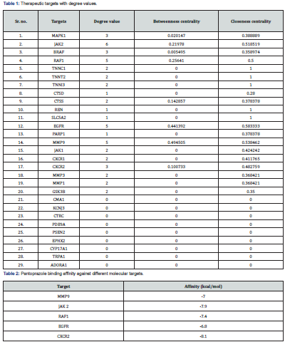
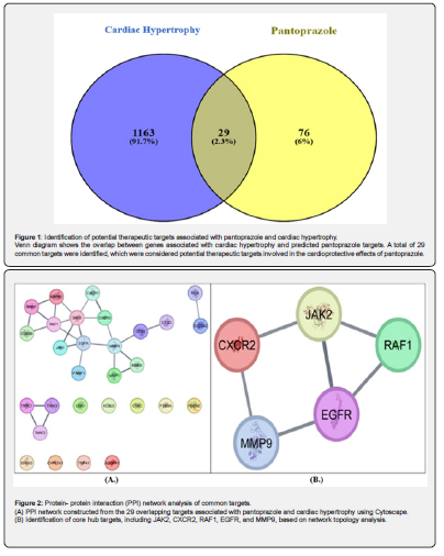
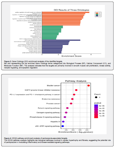
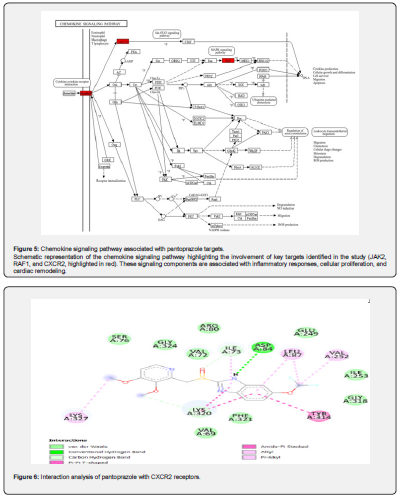
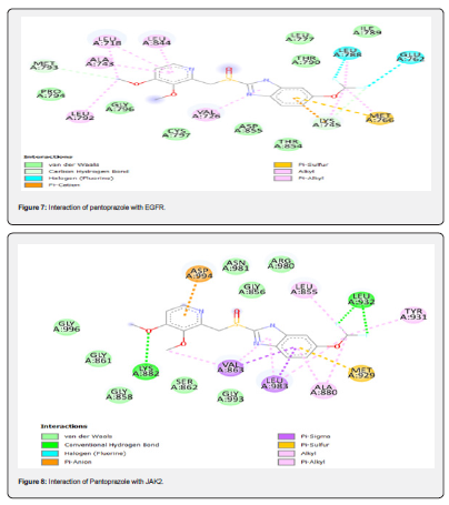
Table 2 summarizes the binding affinities of pantoprazole with selected molecular targets implicated in cardiac hypertrophy. Among the targets analyzed, pantoprazole showed the strongest affinity for CXCR2 (-8.1 kcal/mol) and JAK2 (-7.9 kcal/mol), suggesting potential modulation of inflammatory and signaling pathways critical in cardiac hypertrophy. Moderate binding affinities were observed with RAF1 (-7.4 kcal/mol) and MMP9 (-7.0 kcal/mol), indicating possible interactions with pathways involved in extracellular matrix remodeling and cellular proliferation. The weakest binding was observed for EGFR (-6.8 kcal/mol), although this still indicates a measurable interaction.
Two-dimensional interaction shows the binding interactions between pantoprazole and the CXCR2 protein. Pantoprazole forms a conventional hydrogen bond with ASP84, which contributes to ligand stabilization within the binding pocket. Additional van der Waals interactions are observed with residues such as SER76, GLY324, VAL72, ILE73, GLU249, ILE253, GLY318, VAL69 and PHE321. These interactions suggest that pantoprazole may interact favorably with CXCR2, a key mediator in inflammatory and fibrotic signaling pathways (Figure 6).
Two-dimensional representation of pantoprazole interactions with the epidermal growth factor receptor (EGFR) binding site. The ligand forms multiple van der Waals interactions with residues including MET793, PRO794, GLY796, CYS797, ASP855, THR854, LYS745, ILE789, LEU777, and THR790. These interactions may contribute to the stabilization of pantoprazole within the EGFR binding pocket (Figure 7).
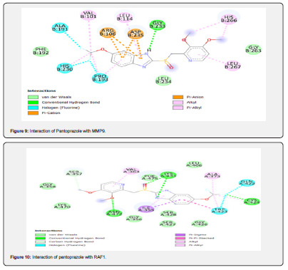
Two-dimensional docking interaction shows the binding orientation of pantoprazole within the JAK2 binding pocket. Pantoprazole forms conventional hydrogen bonds with LYS882 and LEU932, which may contribute to stabilization of the ligand within the active site. Several van der Waals interactions involving such as ASN981, ARG980, GLY856, GLY858, SER862, GLY861, and GLY993. These interactions suggest that pantoprazole may interact with JAK2, a key regulator involved in inflammatory and fibrotic signaling pathways (Figure 8).
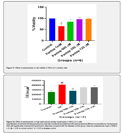
Two-dimensional docking interaction diagram showing the binding orientation of pantoprazole in the MMP9 binding site. Pantoprazole forms a conventional hydrogen bond with GLY233, along with van der Waals interactions involving residues such as PHE191, LEU234, and GLY263 (Figure 9).
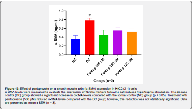
Two-dimensional interaction analysis shows that Pantoprazole forms conventional hydrogen bonds with ASN472, LYS431, and CYS424, accompanied by van der Waals interactions with residues including GLY358, LYS470, GLY356, SER427, SER428, GLY426, LEU406, and PHE475. These interactions may contribute to the stabilization of pantoprazole within the RAF1 active site (Figure 10).
Cell viability was assessed using the MTT assay after treatment with different concentrations of pantoprazole (1 mM, 500 μM, 250 μM, and 125 μM). Results are expressed as percentage cell viability relative to the control. Data represent mean ± SEM (n = 6). *p < 0.05 compared with control (Figure 11).
Quantification of cell area following exposure to high-salt conditions and treatment with pantoprazole at different concentrations. Pantoprazole (500 μM) significantly reduced the hypertrophic response compared with the disease control group. Data are presented as mean ± SEM (n = 3). #p < 0.05 vs normal control; *p < 0.05 vs disease control.
Discussion
Cardiac hypertrophy is a hallmark of pathological cardiac remodeling and represents a critical intermediate stage in the progression toward heart failure. Persistent hemodynamic stress, metabolic imbalance, and inflammatory activation collectively contribute to structural and functional alterations in cardiomyocytes, leading to myocardial hypertrophy and fibrosis. Accumulating evidence suggests that inflammatory signaling pathways and kinase-mediated regulatory networks are central drivers of hypertrophic remodeling, making them attractive therapeutic targets for preventing disease progression [20]. In recent years, system-based approaches such as network pharmacology have emerged as valuable tools for understanding complex disease mechanisms and identifying potential therapeutic targets. Unlike conventional single-target strategies, network pharmacology considers the multifactorial nature of diseases and highlights the importance of multi-target interactions within biological networks [21].
In the present study, a combined network pharmacology, molecular docking, and in vitro validation approach was applied to investigate the potential cardioprotective effects of pantoprazole in cardiac hypertrophy. Network pharmacology analysis identified several overlapping targets between pantoprazole and cardiac hypertrophy–associated genes, including JAK2, CXCR2, MMP9, RAF1, and EGFR. These proteins are widely recognized as key regulators of inflammatory signaling, extracellular matrix remodeling, and cardiomyocyte growth, suggesting that pantoprazole may influence multiple pathways involved in pathological cardiac remodeling. Among these targets, JAK2 plays a crucial role in the JAK/STAT signaling pathway, which has been extensively implicated in myocardial hypertrophy and fibrosis. Activation of the JAK2/STAT3 axis promotes fibroblast proliferation and enhances the transcription of profibrotic genes such as collagen type I and α-smooth muscle actin (α-SMA), thereby contributing to extracellular matrix deposition and myocardial stiffening [22]. Experimental studies have shown that excessive activation of JAK2 signaling accelerates pathological cardiac remodeling, whereas pharmacological inhibition of JAK2 can attenuate hypertrophic growth and reduce myocardial fibrosis [23]. In the present study, molecular docking analysis suggested that pantoprazole may interact with the binding region of JAK2, providing a potential mechanistic explanation for its predicted role in modulating hypertrophic signaling. Another important target identified in this study is CXCR2, a chemokine receptor that plays a key role in inflammatory responses and oxidative stress–mediated tissue injury. Chemokine signaling has been increasingly recognized as a central driver of cardiac remodeling, particularly through the recruitment of inflammatory cells [24]. Previous experimental studies have demonstrated that inhibition of CXCR2 signaling can significantly reduce myocardial inflammation and attenuate pathological cardiac remodeling following cardiac injury [25].
The predicted interaction between pantoprazole and CXCR2 observed in this study, therefore, suggests a possible role for pantoprazole in modulating chemokine-mediated inflammatory signaling pathways involved in cardiac hypertrophy. KEGG pathway enrichment analysis revealed significant enrichment of pantoprazole-associated targets within the chemokine signaling pathway. Chemokines are critical mediators of immune cell migration and inflammatory signaling during cardiac injury and remodeling. Persistent activation of chemokine pathways promotes inflammatory cell infiltration and stimulates fibroblast activation within the myocardium, thereby contributing to the development of myocardial fibrosis and hypertrophic remodeling [26]. The experimental findings obtained in this study further support the computational predictions. Exposure of H9C2 (2-1) cardiomyocytes to high-salt conditions induced significant cellular enlargement, reflecting hypertrophic remodeling under pathological stress. Pantoprazole treatment attenuated this hypertrophic response, suggesting that pantoprazole may exert protective effects against stress-induced cardiomyocyte growth. In addition to cardiomyocyte hypertrophy, activation of fibroblast-related markers represents another hallmark of pathological cardiac remodeling.
The increased α-SMA level observed in the disease control group reflects the activation of myofibroblasts and the initiation of fibrotic remodeling processes within the myocardium. Although pantoprazole treatment showed a tendency to reduce α-SMA level, the reduction was not statistically significant under the experimental conditions used in this study. This observation suggests that pantoprazole may primarily influence hypertrophic signaling rather than directly suppress fibrotic marker expression. The findings of this study suggest that pantoprazole may exert cardioprotective effects through multi-target modulation of signaling pathways associated with cardiac hypertrophy and inflammatory remodeling. By potentially influencing key regulators such as JAK2 and CXCR2 and modulating chemokine-mediated inflammatory signaling, pantoprazole may contribute to the attenuation of pathological cardiac remodeling. Importantly, the computational predictions presented in this study should be interpreted cautiously, as molecular docking provides preliminary insights into potential ligand–protein interactions rather than definitive evidence of molecular inhibition. Further mechanistic investigations and in vivo studies will be necessary to validate these proposed pathways and to clarify the therapeutic potential of pantoprazole in the prevention of cardiac hypertrophy and associated cardiovascular complications.
Study Limitations
Despite providing important insights into the potential cardioprotective effects of pantoprazole, several limitations should be acknowledged. First, the molecular docking results represent computational predictions and should be interpreted as hypothesis-generating rather than definitive evidence of direct molecular interactions. Second, the in vitro experiments were performed using the H9C2 (2-1) cell line, which, although widely used as a cardiomyocyte model, may not fully replicate the complexity of in vivo cardiac physiology. Third, only one of the fibrotic markers was evaluated in this study to understand the antifibrotic effects of pantoprazole. Finally, in vivo studies are required to further validate the cardioprotective potential of pantoprazole and clarify its precise molecular mechanisms in pathological cardiac remodeling.
Conclusion
This study demonstrates that pantoprazole may have potential cardioprotective effects against pathological cardiac hypertrophy. Using an integrated network pharmacology, molecular docking, and in vitro approach, pantoprazole was found to influence key signaling targets associated with inflammatory and hypertrophic pathways. The experimental findings further suggest that pantoprazole can attenuate salt-induced cardiomyocyte hypertrophy in H9C2 (2-1) cells. Collectively, these results highlight the potential of pantoprazole as a repurposed therapeutic candidate for mitigating cardiac hypertrophy and remodeling. However, further mechanistic investigations and in vivo studies are required to validate these findings and clarify the underlying molecular mechanisms.
References
- Chowdhury D, Tangutur AD, Khatua TN (2013) A proteomic view of isoproterenol induced cardiac hypertrophy: Prohibitin identified as a potential biomarker in rats. J Transl Med 11: 130.
- Prajapati AK, Shah G (2024) Exploring in vivo and in vitro models for heart failure with biomarker insights: a review. The Egyptian Heart Journal 76: 141.
- Hok KD, Rich HE, Shadid A (2025) Functional Roles of the Complement Immune System in Cardiac Inflammation and Hypertrophy. Int J Mol Sci 26(20): 9931.
- Ebrahimpour A, Ahir M, Wang M (2022) Combination of esomeprazole and pirfenidone enhances antifibrotic efficacy in vitro and in a mouse model of TGFβ-induced lung fibrosis. Sci Rep 12: 20668.
- Sayed AH, Mahmoud NS, Mohawed OAM, Ahmed HH (2024) Combined effect of pantoprazole and mesenchymal stem cells on experimentally induced gastric ulcer: implication of oxidative stress, inflammation and apoptosis pathways. Inflammopharmacology 32: 1961-1982.
- Kedika RR, Souza RF, Spechler SJ (2009) Potential Anti-inflammatory Effects of Proton Pump Inhibitors: A Review and Discussion of the Clinical Implications. Dig Dis Sci 54: 2312-2317.
- Ghebre YT, Raghu G (2016) Idiopathic Pulmonary Fibrosis: Novel Concepts of Proton Pump Inhibitors as Antifibrotic Drugs. Am J Respir Crit Care Med 193(12): 1345-1352.
- Kim J, Washio T, Yamagishi M (2004) A Novel Data Mining Approach to the Identification of Effective Drugs or Combinations for Targeted Endpoints? Application to Chronic Heart Failure as a New Form of Evidence-based Medicine. Cardiovasc Drugs Ther 18: 483-489.
- Yoshihisa A, Takiguchi M, Kanno Y (2017) Associations of Acid Suppressive Therapy with Cardiac Mortality in Heart Failure Patients. J Am Heart Assoc 6(5).
- Onda K, Tong S, Beard S (2017) Proton Pump Inhibitors Decrease Soluble fms- Like Tyrosine Kinase-1 and Soluble Endoglin Secretion, Decrease Hypertension, and Rescue Endothelial Dysfunction. Hypertension 69(3): 457-468.
- Schillinger W, Teucher N, Sossalla S (2007) Negative Inotropy of the Gastric Proton Pump Inhibitor Pantoprazole in Myocardium from Humans and Rabbits. Circulation 116(1): 57-66.
- Lin H, Li Y, Zhu H (2020) Lansoprazole alleviates pressure overload-induced cardiac hypertrophy and heart failure in mice by blocking the activation of β-catenin. Cardiovasc Res 116(1): 101-113.
- Pap D, Pajtók C, Veres SA (2023) High Salt Promotes Inflammatory and Fibrotic Response in Peritoneal Cells. Int J Mol Sci 24(18): 13765.
- Wenzel UO, Bode M, Kurts C, Ehmke H (2019) Salt, inflammation, IL‐17 and hypertension. Br J Pharmacol 176(12): 1853-1863.
- Whitehead CE, Ziemke EK, Frankowski MCL (2024) A first-in-class selective inhibitor of EGFR and PI3K offers a single-molecule approach to targeting adaptive resistance. Nat Cancer 5: 1250-1266.
- Davis RR, Li B, Yun SY (2021) Structural Insights into JAK2 Inhibition by Ruxolitinib, Fedratinib, and Derivatives Thereof. J Med Chem 64(4): 2228-2241.
- Scannevin RH, Alexander R, Haarlander TM (2017) Discovery of a highly selective chemical inhibitor of matrix metalloproteinase-9 (MMP-9) that allosterically inhibits zymogen activation. Journal of Biological Chemistry 292(43): 17963-17974.
- Hatzivassiliou G, Song K, Yen I (2010) RAF inhibitors prime wild-type RAF to activate the MAPK pathway and enhance growth. Nature 464: 431-435.
- Liu K, Wu L, Yuan S (2020) Structural basis of CXC chemokine receptor 2 activation and signaling. Nature 585: 135-140.
- Frey N, Olson EN (2003) Cardiac Hypertrophy: The Good, the Bad, and the Ugly. Annu Rev Physiol 65: 45-79.
- Hopkins AL (2008) Network pharmacology: the next paradigm in drug discovery. Nat Chem Biol 4: 682-690.
- Jiang H, Yang J, Li T (2024) JAK/STAT3 signaling in cardiac fibrosis: a promising therapeutic target. Front Pharmacol 15: 1336102.
- Chen Z, Zhou H, Huang X (2022) Pirfenidone attenuates cardiac hypertrophy against isoproterenol by inhibiting activation of the janus tyrosine kinase-2/signal transducer and activator of transcription 3 (JAK-2/STAT3) signaling pathway. Bioengineered 13(5): 12772-12782.
- Oliveira VLS, Proost P, Struyf S (2025) Chemokines in the resolution of inflammation: key players and targets for therapeutic modulation. Front Immunol 16: 1717666.
- Zhang YL, Geng C, Yang J (2019) Chronic inhibition of chemokine receptor CXCR2 attenuates cardiac remodeling and dysfunction in spontaneously hypertensive rats. Biochimica et Biophysica Acta (BBA) - Molecular Basis of Disease 1865(12): 165551.
- Li R, Frangogiannis NG (2021) Chemokines in cardiac fibrosis. Curr Opin Physiol 19: 80-91.

























